Implications
Herbivores are distinguished from other mammals by their ability to obtain energy from cellulose, the primary polysaccharide in plants. This ability is the result of microbial activity in the rumen, predominantly bacterial. Other than a recent study with ionized calcium, the role of cations in the enzymatic degradation of cellulose has not been investigated. This study demonstrates that Mg is important for both growth and cellulose digestion by three major cellulolytic species of rumen bacteria.
Introduction
Bryant et al. (Reference Bryant, Robinson and Chu1959) studied the mineral requirements of Bacteroides succinogenes strain S85 (now Fibrobacter succinogenes) for growth, and demonstrated the need for PO4 −3, NH4 +, Mg, Ca, K and Na in a purified medium. When PO4 −3, K, Mg, NH4 + or Ca were deleted singly, little or no growth occurred. For Mg, they reported a requirement for maximum growth of 0.01 mg/5 ml of medium. This converts to a requirement of 0.082 mM of total Mg.
Gong and Forsberg (Reference Gong and Forsberg1989) reported that F. succinogenes requires either Ca or Mg for adhesion to cellulose and its subsequent degradation. In a later study, Roger et al. (Reference Roger, Fonty, Komisarczuk-Bondy and Gouet1990) found that Ruminococcus flavefaciens requires both Ca and Mg for the cellulose adhesion process. Also, newer information on the molecular aspects of cellulosome structure, a cellulose-degrading nanomachine described in cellulolityc bacteria with protein–protein interactions, involves cations such as Ca and Mg as their structural and functional components (Aurilia et al., Reference Aurilia, Martin, Munro, Mercer and Flint2000; Fontes and Gilbert, Reference Fontes and Gilbert2010).
Morales and Dehority (Reference Morales and Dehority2009) investigated the ionized calcium requirements for two strains of each of the three main cellulolytic rumen bacteria (F. succinogenes, R. flavefaciens and Ruminococcus albus). All cultures except F. succinogenes A3c grew normally with a soluble substrate when sequentially transferred in an ionized calcium (Ca+2)-free medium. With cellulose as the only substrate, both strains of F. succinogenes had an absolute requirement for ionized Ca. Although not an absolute requirement, all strains of the ruminococci responded to increasing concentrations of ionized calcium when cellulose was the only substrate. Based on these results, it became of interest to investigate whether the cellulolytic rumen bacteria had any requirements for other divalent cations such as Fe, Mg, Mn and Zn.
Material and methods
Bacterial species and strains utilized were: F. succinogenes strains S85 (Bryant and Doetsch, Reference Bryant and Doetsch1954) and A3c (Dehority, Reference Dehority1963); R. albus strains 7 (Bryant et al., Reference Bryant, Small, Bouma and Robinson1958) and 8 (Hungate and Stack, Reference Hungate and Stack1982); and R. flavefaciens strains B34b (Dehority, Reference Dehority1963) and C94 (Bryant et al., Reference Bryant, Small, Bouma and Robinson1958). R. albus 8 was obtained from the Rumen Microbiology Lab, Department of Animal Sciences, The Ohio State University. The other cultures were obtained from rumen–glucose–cellobiose–agar slants (Bryant and Burkey, Reference Bryant and Burkey1953) stored at −60°C in this lab. Transfers were made to fresh slants and the cultures were transferred several times until they showed active growth. At the time that strains were being studied, cultures were transferred daily. Those strains not in use were maintained at 5°C and transferred every 3 to 4 days. Preparation of media, transfers and inoculations were carried out under anaerobic conditions, using commercial CO2 that was passed through a heated glass column filled with copper turnings. All cultures were incubated at 39ºC.
All chemicals used were reagent grade; however, in media without added Mg (Mg-free medium), the Mg concentrations ranged from 0.0032 to 0.014 mM. This resulted from trace amounts of Mg contained in the other reagent grade mineral sources included in the basal medium. Water was deionized in the laboratory (Nanopure DiamondTM Life Science (UV/UF), Ultrapure water system, Barnstead International, Dubuque, IA, USA).
The complete anaerobic medium of Scott and Dehority (Reference Scott and Dehority1965) was used as the control, and contained 0.41mMMg. Cellobiose (0.5%) or cellulose (0.75%) (ball-milled Sigmacell 20) (Sigmacell Cellulose, type 20; Sigma-Aldrich, St. Louis, MO, USA) served as energy substrates. Growth was monitored by measuring absorbance at 600 nm with a Spectronic 20® spectrophotometer (Bausch and Lomb, Rochester, NY, USA). Cellulose degradation was measured using the procedure described by Hiltner and Dehority (Reference Hiltner and Dehority1983).
Mg concentration of the different media was determined by Atomic Absorption Spectrophotometry (Varian Spectr AA200, Palo Alto, CA, USA). To remove all other possible mineral contamination, glassware was washed, soaked in 20% HCl for 24 h, then rinsed three times with deionized water.
Inoculum preparation was started by transferring a loop full of culture from a slant to cellobiose liquid divalent-cation-deficient medium (1/8 of the cation concentrations in normal medium). The culture was incubated overnight at 39°C and the next morning 1.0 ml was transferred into complete cellobiose medium that was free of divalent cations. Cultures were incubated until they reached an OD between 0.6 and 0.7, depending on the strain involved. Between 0.6 and 0.9 ml of the culture was then added to either cellobiose complete liquid divalent-cation-free medium or Mg-free medium (depending on the requirement being investigated) to attain 0.1OD. Using this as an inoculum, 0.1 ml was added to culture tubes containing 7 ml of liquid medium.
Preparation of inocula for NH3-free cellulose medium was similar, with a slight variation: inocula (at least eight tubes) was initially grown in a Mg-limited medium (0.05 mM). When OD reached 0.6 to 0.7, the tubes were centrifuged (15 to 20 min, 900×g), and the supernatant was decanted and discarded. Phosphate buffer was added to re-suspend the bacterial cells, the culture was mixed, OD determined and enough buffer was added to reach 0.7 to 0.75 OD. Then 1 ml was used to inoculate each culture tube of the different treatments studied in NH3-free cellulose media.
After the lag period on cellobiose medium, growth was monitored by optical density at 600 nm (Spectronic 20® spectrophotometer). Readings were taken every 1 or 2 h, depending on growth of the bacteria, until they reached maximum growth. Blank tubes (non-inoculated) were incubated simultaneously. Each experiment was run in duplicate and replicated once.
To determine Mg requirements for growth in cellobiose liquid medium, concentrations of 0, 0.05, 0.10, 0.20, and 0.82 mM of Mg were used. The complete medium of Scott and Dehority (Reference Scott and Dehority1965) served as the control (0.41mMMg). Mg was supplied from MgSO4. Inoculum preparation was similar to the other experiments; however, in this experiment, cellobiose Mg-deficient liquid medium (0.05mMMg) was used to prepare the pre-inoculum and cellobiose Mg-free liquid medium to prepare the inoculum. Incubation and monitoring of growth was similar to the other growth experiments. Each experiment was run in duplicate and replicated once (n=4).
To determine which mineral, ionized calcium or Mg, is required for cellulose degradation by R. flavefaciens, strains B34b and C94, an NH3-free cellulose medium was used. The control contained all the divalent cations, including both Ca+2 and Mg. Treatments were: Ca+2-free, Mg-free and Ca+2–Mg-free. The procedure to prepare the pre-inoculum was similar to the cellulose degradation experiment with F. succinogenes in the earlier study by Morales and Dehority (Reference Morales and Dehority2009). Samples were taken, at 0 h and at 60 h, when no additional visible disappearance of cellulose was observed. Two replicates were run, each containing four tubes per treatment. Blanks (tubes not-inoculated) were included and incubated.
Experimental design
For Mg growth requirements, a complete factorial design was used, 2(3)×6; factors were: strains (2), within species (3) and Mg concentrations (6). The two strains of each species were run simultaneously, but each species was run at different times; then the replications were considered as Block 1 and Block 2. Tube 1 and Tube 2 were the experimental units, because the OD was measured on each tube. The experimental design to evaluate Mg requirements for cellulose degradation in NH3-free cellulose media for R. flavefaciens was a factorial design 2 ×4 (2 strains and 4 treatments). The experiment was run in duplicate and replicated one time.
Statistical analysis
Growth data were fitted mathematically with a logistic model (Zwietering et al., Reference Zwietering, Jongenburger, Rombouts and Van’t Riet1990): Y=A/(1+exp{[(4×B)/A]×(C−time)+2}), where Y represents growth, using non-linear regression analysis (NLIN PROC; SAS Institute, 1999) to determine A (maximum growth (maximum absorbance)); B (growth rate (absorbance units/h)); and C, the lag time (h). In general, statistical significance was accepted at P⩽0.05; and a tendency was accepted when 0.05>P⩽0.10. Maximum growth, weighted by the reciprocal of its standard error, was used for ANOVA analysis, as described below.
Growth data
Least squares means (LSM) were compared with linear and quadratic contrasts (PROC MIXED, SAS Institute, 1999). Fixed effects were [Mg], species, strain within species and the interaction of strain within species×[Mg]. Random effects were block within species and tube within block. The extended and reduced models were compared and, because they were different (P<0.05), the extended model was used. Homogeneity of variance evaluation showed that an unstructured variance test for analysis of maximum growth response should be used. When the linear effect for any of these was significant, it was not possible to estimate a requirement because an end-point was not attained. When a quadratic effect was significant, non-linear analysis was used to define the break-point and the plateau of the function (PROC NLIN, SAS Institute, 1999). When this method did not have a solution, the maximum of the first derivative of the quadratic function (PROC REG, SAS Institute, 1999) was used to estimate requirements. The extended model used for growth studies was Y=[Mg]+species+strain(species)+block+tube(block)+[Mg]×strain(species)+error.
Cellulose degradation, NH3-free cellulose media
In this experiment no degradation occurred with Ca+2–Mg-free and Mg-free media and these treatments were not included in the overall analysis; thus only the treatments with Ca+2+Mg (control) and Ca+2-free, were analyzed; then the extent of degradation was estimated and the data for this parameter were analyzed by ANOVA, obtaining LSM per strain×cation treatment interaction (PROC GLM, SAS Institute, 1999). No mean separation was made, because no differences were found between treatments within strain (P>0.05).
The statistical model used was: Y=cation treatment+strain+strain×cation treatment+error.
Results
Cations required for bacterial growth with specific attention to Mg requirements
In preliminary experiments, the requirement of R. flavefaciens B34b, R. albus 7 and F. succinogenes A3c and S85 for Ca+2, Mg, Mn, Zn and Fe was studied qualitatively in cellobiose liquid medium (Scott and Dehority, Reference Scott and Dehority1965) with single deletion and single addition experiments (data not shown). Concentrations for each cation were the same as those used in the Scott and Dehority (Reference Scott and Dehority1965) medium. R. flavefaciens B34b had an absolute requirement for Mg, since any combinations of divalent cations without Mg, did not support growth. Maximum growth of R. albus 7 and both strains of F. succinogenes was decreased when Mg was not present in the medium. Deletion of Mn, Fe or Zn did not appear to effect growth of the four strains. Thus, based on these qualitative results, the Mg requirement for growth in cellobiose liquid medium was investigated with all strains. Treatments evaluated only total concentration of Mg, because an instrument to measure ionized Mg concentrations was not available.
Growth, measured as OD, was characterized by a logistic function. The statistical analysis of the parameters of the growth curves (A, B and C) showed significant main effects for species, strain within species, [Mg] and for the interactions: species×concentration and strain within species×concentrations (P<0.05). Since R. flavefaciens B34b and R. flavefaciens C94 did not grow when they were cultured in Mg-free media, the data from 0 [Mg] were not included in the statistical analysis.
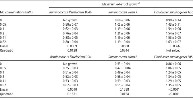
As can be seen in Table 1, neither R. flavefaciens B34b or R. flavefaciens C94 grew in Mg-free medium. Maximum growth for R. flavefaciens B34b, evaluated using A from the logistic function, responded to both linear and quadratic contrasts (P=0.0009 and 0.0138, respectively). R. flavefaciens C94 responded linearly, with an increasing maximum OD (P=0.0010) to increasing [Mg]. Calculating the maximum of the quadratic function, the Mg requirement can be estimated for R. flavefaciens B34b as 0.54 mM of Mg. Since R. flavefaciens C94 showed an increased linear response to increased [Mg], a requirement could not be determined because an end-point or plateau was not attained.
Effect of different Mg concentrations (mM) on growth of several cellulolytic rumen bacteria in liquid media with cellobiose as the energy sourceFootnote 1

1 For each strain, maximum extent of growth was estimated by linear and quadratic contrasts, P-values are presented.
2 OD values measured at 600 nm.
Maximum extent of growth for R. albus 7 (Table 1) was reduced when Mg was not present in the media and it responded linearly (P=0.0568) to increasing [Mg], with a tendency for a quadratic response (P=0.0744). R. albus 8 (Table 1), showed a quadratic response (P=0.0154) for maximum growth (maximum OD) as [Mg] increased. The Mg requirement for maximum growth was estimated as 0.51mMMg. The linear response to [Mg] shown by R. albus 7, suggests it has a higher Mg requirement for maximum growth than R. albus 8.
Both strains of F. succinogenes responded in maximum growth to increased [Mg] (Table 1). F. succinogenes A3c had a positive linear response (P=0.0366) to increased [Mg], whereas F. succinogenes S85 showed both increasing maximum growth in linear and quadratic ways to increased [Mg] (P<0.0001, respectively). Mg requirement for F. succinogenes A3c cannot be defined from the data available and the method used for its estimation. For F. succinogenes S85, the Mg requirement for maximum growth can be estimated as 0.56 mM of Mg from the quadratic function. Apparently, F. succinogenes A3c has a higher requirement for Mg than F. succinogenes S85, considering that F. succinogenes A3c had a linear response to [Mg]. The requirement for F. succinogenes S85 attained in this study is higher than the 0.082 mM requirement reported by Bryant et al. (Reference Bryant, Robinson and Chu1959) for the same strain. Thus, only the two strains of R. flavefaciens have an absolute requirement for Mg, while the remaining species and strains have different Mg requirements for optimum growth.
Cellulose degradation by R. flavefaciens in NH3-free media
R. flavefaciens had a high maximum growth and rate of growth in Ca+2-free and low Ca+2 media (Morales and Dehority, Reference Morales and Dehority2009). In contrast, as described above, R. flavefaciens has an absolute growth requirement for Mg. When the Ca+2 requirement for cellulose degradation by R. flavefaciens was studied, an increased Ca+2 concentration resulted in only a small increment in cellulose degradation, characterized as a linear response (P<0.0001; Morales and Dehority, Reference Morales and Dehority2009). To separate the potentially confounding effect of growth on cellulose degradation, due to Ca+2 or Mg, an experiment was carried out with R. flavefaciens in NH3-free media. Treatments were: Ca+2 and Mg at normal control concentrations (Scott and Dehority, Reference Scott and Dehority1965), Ca+2-free, Mg-free, and Ca+2 and Mg-free. The results are shown in Table 2. For both strains of R. flavefaciens, the extent of degradation after 60 h incubation with Ca+2 and Mg was similar to Ca+2-free (P>0.05). Neither Mg-free nor Ca+2 and Mg-free showed any cellulose degradation. Thus, when Mg was not present in the NH3-free medium, no cellulose degradation occurred. No interaction between strain and cation treatment was observed (P>0.05). A strain effect was observed (P<0.05), R. flavefaciens C94 having less cellulose degradation. These results suggest that R. flavefaciens requires Mg to degrade cellulose, and may have no requirement or require very low concentrations of Ca+2 for cellulose degradation.
Extent of cellulose degradation (mg degraded) after 60 h incubation with Ruminococcus flavefaciens B34b and C94 in the presence or absence of Ca+2 and/or Mg in NH3-free cellulose liquid mediumFootnote 1

x,yValues in the same column followed by different superscripts differ at P<0.05.
1 Milligrams of cellulose degraded.
Discussion
Several authors (Bryant et al., Reference Bryant, Robinson and Chu1959; Scott and Dehority, Reference Scott and Dehority1965; Caldwell et al., Reference Caldwell, Keeney, Barton and Keller1973; Caldwell and Arcand, Reference Caldwell and Arcand1974) have used maximum extent of growth (maximum OD) as the indicator to determine nutrient requirements. Because Mg is involved in so many different processes in the cell (Jasper and Silver, Reference Jasper and Silver1977; Durand and Kawashima, Reference Durand and Kawashima1980; Smith and Maguire, Reference Smith and Maguire1998), maximum growth may be a good indicator of Mg requirements. As a limiting nutrient it can affect both rate and extent of bacterial growth.
Considering the role of Mg in cell wall synthesis (Durand and Kawashima, Reference Durand and Kawashima1980), it would be logical to expect a high Mg requirement for Gram-positive bacteria. This is supported by the results obtained with R. flavefaciens in this study, as it has an absolute requirement for Mg. Although R. albus showed similar Mg requirements for maximum growth to R. flavefaciens, R. albus can grow without Mg in the medium, indicating the Mg requirement is not absolute for this species. This may be owing to differences in metabolic demands, different capacity to control intracellular concentrations or Mg channels may be different between the two species. Unfortunately, these explanations are only speculative, due to the lack of information related to this topic in rumen bacteria. Information from Salmonella, Escherichia coli and other pathogens shows differences in resistance to Mg starvation, which may be owing to different channel systems and variations in how different species manage their Mg status (Jasper and Silver, Reference Jasper and Silver1977; Smith and Maguire, Reference Smith and Maguire1998; Moncrief and Maguire, Reference Moncrief and Maguire1999; Chamnongpol and Groisman, Reference Chamnongpol and Groisman2002) and the complexity of Mg channels and how they are regulated and affected by [Mg] and other cations (Moomaw and Maguire, Reference Moomaw and Maguire2008).
Beveridge and Murray (Reference Beveridge and Murray1980), Beveridge et al. (Reference Beveridge, Forsberg and Doyle1982) and Beveridge (Reference Beveridge1990) reported differences in cell wall composition between two species of Bacillus, B. subtilis and B. licheniformis. These differences consisted of changes in the amount and structural disposition of teichoic, lipoteichoic and teicuronic acid in the cell wall matrix, which altered the capacity of these bacteria to bind different ions and in different proportions. The differences in Mg affinity observed for the predominant rumen cellulolytic bacteria may be related with differences in their cell wall composition, but the information available about cell wall composition for these bacteria is not enough to establish this different capacity to bind Mg (Dehority, Reference Dehority1977; Vinogradov et al., Reference Vinogradov, Egbosimba, Perry, Lam and Forsberg2001).
In earlier research, the lack of growth response from R. flavefaciens to increasing Ca+2 concentrations (Morales and Dehority, Reference Morales and Dehority2009) was not expected, based on the information reported by Durand and Kawashima (Reference Durand and Kawashima1980), who suggested that these organisms have a high requirement for Ca and Mg because these elements are needed for synthesis of the cell walls by gram-positive bacteria. In general, Mg, as a cofactor, is involved in many different steps in the synthesis of the cell walls (Ghuysen and Shockman, Reference Ghuysen and Shockman1973), both Ca+2 and Mg are part of the cell wall structure, owing to chemical interactions between compounds at the bacterial cell wall and divalent cations in the medium. Thus, teichoic and/or teichuronic acids present at the matrix of the peptidoglycan layer of gram-positive bacteria can bind Ca+2 and Mg from the medium and the amount bound will vary as a function of the cell wall composition (Beveridge and Murray, Reference Beveridge and Murray1980; Beveridge et al., Reference Beveridge, Forsberg and Doyle1982; Beveridge, Reference Beveridge1990). It is necessary to consider that these cations, while they are bound to the cell wall, are not available for exchange with the medium or other cells. If the bacteria die or the pH conditions became acidic, these cations become available in the medium (Durand and Kawashima, Reference Durand and Kawashima1980). Thus, a kind of mineral recycling can happen. But in the present study, despite the incubation conditions, this situation may not apply, since the size of the inoculum utilized was very small (0.1 ml of 0.1 OD culture) and probably would not be a source of such cations for the bacteria.
When growth was inhibited in an NH3-free medium, neither R. flavefaciens B34b or R. flavefaciens C94 degraded cellulose when Mg was deleted from the medium. No effect on cellulose degradation was observed when Ca+2 was omitted. Differences were observed between the two R. flavefaciens strains, R. flavefaciens B34b degraded more cellulose than R. flavefaciens C94 (P<0.05) when Mg was present in the medium at 0.41 mM.
A number of reports on Mg concentration in rumen contents have been published, with values ranging from 1.0 to 10.10 mM (Poutiainen, Reference Poutiainen1970; Bennink et al., Reference Bennink, Tyler, Ward and Johnson1978; Mackie and Therion, Reference Mackie and Therion1984; Scandolo et al., Reference Scandolo, Noro, Böhmwald, Contreras and Wittwer2007; Sepulveda et al., Reference Sepulveda, Wittwer, Böhmwald, Pulido and Noro2011). Considering this information and the results obtained in the present study, it may be rare to find negative effects on rumen bacteria owing to low [Mg]. However, it is possible that in certain areas where Mg deficiency is found, rumen bacteria could be affected.
The role of Mg in cellulose degradation by R. flavefaciens needs to be studied further. In this study when Mg was absent in NH3-free cellulose media, no degradation occurred, despite the normal concentration of other mineral nutrients. Variations in cellulose degradation to increasing Ca+2 concentrations were observed, which may be a reflection of Mg content in those media, and not the direct effect of Ca+2 concentration (Morales and Dehority, Reference Morales and Dehority2009). Roger et al. (Reference Roger, Fonty, Komisarczuk-Bondy and Gouet1990) found that R. flavefaciens requires both Ca and Mg for the cellulose adhesion process, thus a possible interaction between the two cations needs to be evaluated. These are questions that deserve more attention and research.
Conclusions
With cellobiose as a substrate, growth of all the species and strains studied, F. succinogenes (A3c and S85), R. albus (7 and 8) and R. flavefaciens (B34b and C94) responded to increasing Mg concentrations. The Mg requirement for both strains of R. flavefaciens was absolute. When R. flavefaciens was grown in an NH3-free medium with cellulose as the sole substrate, no degradation was observed. This suggests that Mg has a dual function for R. flavefaciens, one in growth and one in cellulose degradation.
Acknowledgements
Salary and research support were provided by state and federal funds appropriated to the Ohio Agricultural Research and Development Center, The Ohio State University.