Case report
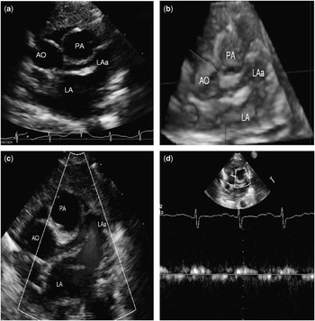
A male neonate, weighing 3.5 kg, underwent an echocardiogram to evaluate an incidental murmur found on the routine perinatal physical examination. This study suggested that there was an elevation of the right ventricular pressure associated with mild dilatation and hypertrophy of the right ventricle. The short-axis view at the level of the aortic and pulmonary roots revealed what appeared to be a vascular structure that arose from the aortic root, but was also in luminal continuity with the left atrial appendage (Fig 1a and b). A low-velocity flow, identical to that in the left atrial appendage, was sampled within this structure (Fig 1c and d). On the same short-axis view, at a slightly lower plane, a normal origin, calibre, and flow in both the right and left coronary arteries was demonstrated though, at times, the images of the left coronary artery and of the unusual vascular-like structure were super-imposed. On the basis of the echocardiographic findings, a left atrial appendage diverticulum with fistulisation to the adjacent left main coronary artery could not be excluded.
( a ) Two-dimensional and ( b ) three-dimensional echocardiogram with short-axis view showing the aortic and pulmonary roots, the left atrium, the left atrial appendage, and the diverticulum. ( c and d ) colour and power-doppler flow within the left atrial appendage and the diverticulum. Ao=aorta; LA=atrium; LAa=left atrial appendage; PA=pulmonary artery.

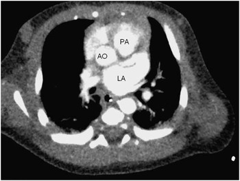
To exclude the presence of a fistulous connection, a CT scan was performed that, indeed, confirmed the presence of a left atrial appendage diverticulum. This diverticulum, resembling a “seahorse” in appearance, was located posterior to the pulmonary artery near the aortic root in close proximity to the left main coronary artery (Fig 2).
( a ) CT axial view showing the aortic and pulmonary root and the left atrium with a left atrial appendage diverticulum resembling a “seahorse”.

Left atrial appendage diverticula are common findings in healthy patientsReference Victor and Nayak 1 – Reference Peng, Yu and Yang 4 as well as in those with atrial fibrillation.Reference Lazoura, Reddy and Shriharan 2 , Reference Peng, Yu and Yang 4 Aneurysms of the left atrial appendage have been also described,Reference Victor and Nayak 1 but they are usually directed downward to the left ventricle.
Acknowledgement
None.
Financial Support
This research received no specific grant from any funding agency, commercial, or not-for-profit sectors.
Conflicts of Interest
None.
Ethical Standards
The authors assert that all procedures contributing to this work comply with the ethical standards of the relevant national guidelines on human experimentation and with the Helsinki Declaration of 1975, as revised in 2008, and has been approved by the institutional committees.