Introduction
There is no effective vaccine to prevent human leishmaniasis and the drugs available to chemotherapy have several limitations. Thus, the development of new drugs is a challenge because of the variety of Leishmania species, different clinical forms of the disease and the profile of the immune response. Therefore, new therapeutic alternatives are urgently needed and, in this context, natural products are an important source of potential molecules.
Plants from the genus Hypericum (Hypericaceae) presented many biological actions, including antiparasitic (Cargnin et al. Reference Cargnin, Vieira Pde, Cibulski, Cassel, Vargas, Montanha, Roehe, Tasca and von Poser2013), antifungal (Fenner et al. Reference Fenner, Sortino, Rates, Dall'Agnol, Ferraz, Bernardi, Albring, Nor, von Poser, Schapoval and Zacchino2005) and antiviral activities (Fritz et al. Reference Fritz, Venturi, Cargnin, Schripsema, Roehe, Montanha and von Poser2007). Considering that some phloroglucinol derivatives, including acylphloroglucinols and benzophenones, have shown leishmanicidal activity (Bharate and Singh, Reference Bharate and Singh2011; Sidana et al. Reference Sidana, Singh, Arora, Foley and Singh2011; Socolsky et al. Reference Socolsky, Salamanca, Gimenez, Borkosky and Bardon2016) and that Hypericum species are a rich source of these compounds (Ccana-Ccapatinta et al. Reference Ccana-Ccapatinta, Barros, Bridi and von Poser2015), in a previous study, our group investigated and demonstrated that the lipophilic extracts of Hypericum polyanthemum Klotzsch ex Reichardt, Hypericum carinatum Griseb. and Hypericum linoides A. St.-Hil. containing phloroglucinols were able to kill infective forms of Leishmania amazonensis (Dagnino et al. Reference Dagnino, Barros, Ccana-Ccapatinta, Prophiro, Poser and Romao2015). The purpose of this study was to evaluate the anti-Leishmania action of two acylphloroglucinols and a benzophenone isolated from Hypericum species against promastigote and amastigote forms of L. amazonensis. Our data exhibit for the first time that isouliginosin B, uliginosin B and cariphenone A isolated from Hypericum species present potent activity against both promastigote and amastigote forms of L. amazonensis.
Materials and methods
Plant material
Flowering aerial parts of H. carinatum Griseb were collected in the State of Rio Grande do Sul, in Southern Brazil, between October and December 2011. Flowering aerial parts of H. andinum Gleason were collected in the State of Cuzco, in Southern Peru, in May 2008. The plants were identified by Dr Sérgio Bordignon (UNILASSALE, Brazil) and voucher specimens were deposited in the UFRGS herbarium (ICN) (Bordignon and Ferraz 2309, and Ccana-Ccapatinta et al. 05 respectively). The collection of plants was authorized by IBAMA (no. 003/2008; Protocol 02000.001717/2008-60) and Dirección General Forestal y de Fauna Silvestre of the Republic of Peru (0147-2010-AG-DGFFS-DGEFFS).
Isolation procedures of Hypericum compounds
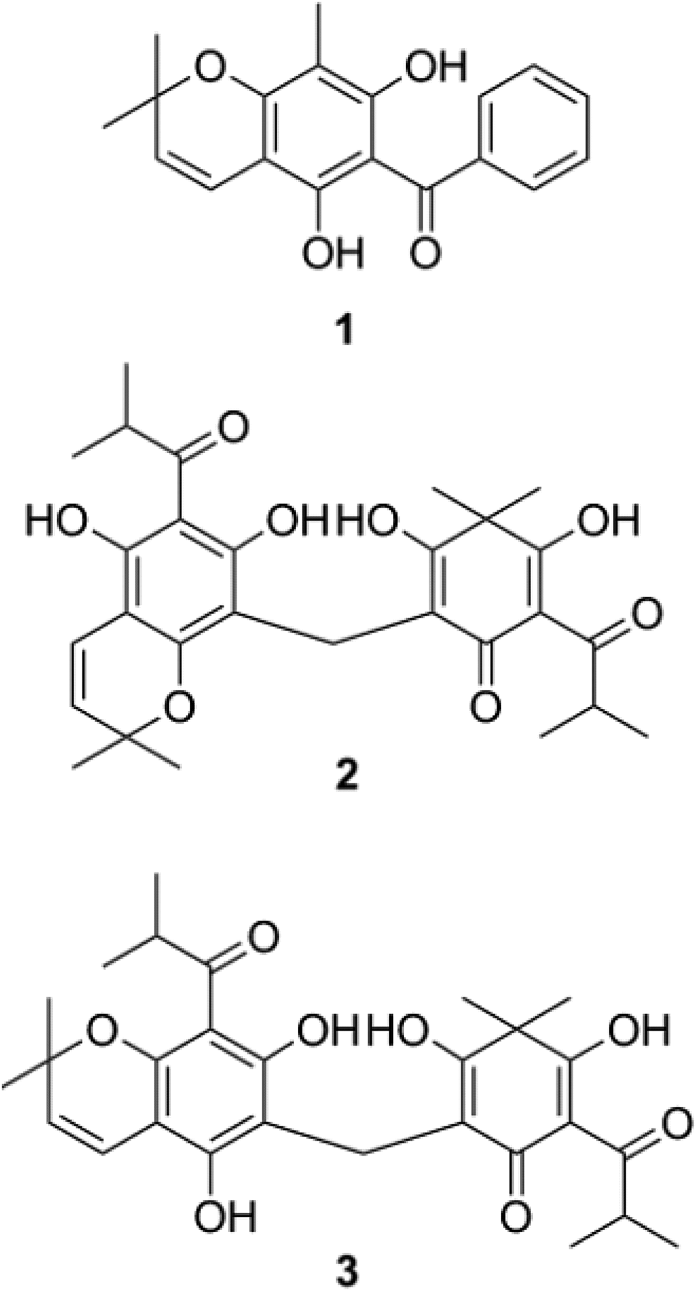
The air-dried and powdered plant materials were subjected to extraction with n-hexane over 24 h (plant-solvent ratio 1:5) until exhaustion. The n-hexane extracts were pooled and evaporated to dryness under reduced pressure and then treated with cold acetone to obtain extracts without fatty residues. The obtained extracts were submitted to chromatographic procedures, as described previously, to isolate the phloroglucinol derivatives 1–3 (Fig. 1). Cariphenone A (1) and uliginosin B (3) were isolated from H. carinatum as described by Bernardi et al. (Reference Bernardi, Ferraz, Albring, Bordignon, Schripsema, Bridi, Dutra-Filho, Henriques and von Poser2005). Isouliginosin B (2) was isolated from H. andinum as described by Ccana-Ccapatinta et al. (Reference Ccana-Ccapatinta, Stolz, da Costa, Rates and von Poser2014, Reference Ccana-Ccapatinta, Barros, Bridi and von Poser2015). The identity and purity of the isolated compounds were confirmed by 1H and 13C nuclear magnetic resonance spectroscopy.
Chemical structures of the phloroglucinol derivatives screened for anti-L. amazonensis activity.

L. (Leishmania) amazonensis culture
The MHOM/BR/73/m2269 strain of Leishmania (Leishmania) amazonensis was cultured as previously described elsewhere (Dagnino et al. Reference Dagnino, Barros, Ccana-Ccapatinta, Prophiro, Poser and Romao2015). All experimental procedures were performed in accordance with the guidelines of the National Institute of Health and the Brazilian Society for Science on Laboratory Animals, with the approval of the local Ethics Committee from Federal University of Health Sciences of Porto Alegre (process number 11-061).
Effects of isolated compounds of Hypericum on L. amazonensis viability in vitro
Promastigotes of L. amazonensis (3 × 10⁶ on stationary phase) were plated in 96 well microplates and incubated with M199 medium with 10% fetal bovine serum (FBS) in the presence of the isolated compounds 1–3 at concentrations of 0 to 100 µ m for 48 h. The activity of each compound was compared with control samples incubated with M199 medium (100% of viability) or with amphotericin B (Sigma, USA) at 1 µ m as a standard antileishmanial drug (100% of mortality). Control cells were incubated with M199 medium containing less than 0.05% of polysorbate 80 (Dagnino et al. Reference Dagnino, Barros, Ccana-Ccapatinta, Prophiro, Poser and Romao2015). To determinate the kinetics of the leishmanicidal activity, the viability of parasites treated with compounds at 50 µ m was evaluated at different time points (6, 12, 24 and 48 h) by counting the viable promastigotes using a hemocytometer (Antonello et al. Reference Antonello, Sartori, Folmer Correa, Brandelli, Heermann, Rodrigues Júnior, Peres, Romão and Silva2017). Survival rate was calculated according to the formula: percentage of survival = (average number of viable parasites in treated group/average number of viable untreated parasites) × 100. The 50% inhibitory concentration (IC50) value for each isolated compound was determined by nonlinear regression analysis using the GraphPad Software.
Field emission scanning electron microscopy
To evaluate parasite ultrastructural alterations by scanning electron microscopy (s.e.m.), L. amazonensis promastigotes were incubated with M199, M199 containing Hypericum compounds (IC50 value) or treated with amphotericin B at 0.13 µ m (IC50) for 6 h, washed with phosphate buffered saline (PBS) and fixed with 25% glutaraldehyde, distilled water in 0.2 M phosphate buffer, pH 7.4. After being washed three times in the same buffer, the parasites were adhered to glass slides previously coated with 0.1% aqueous poly-L-lysine for 30 min at room temperature. Subsequently, the slides were washed three times with 0.2 M phosphate buffer pH 7.4, post-fixed in a solution of 2% OsO4 with 0.2 M phosphate buffer (1:1) for 45 min at room temperature. All samples were dehydrated in a graded series of acetone (30–100%), critical point dried using CO2, mounted on metal stubs, and coated with gold for observation in a Field Emission Scanning Electron Microscope (Inspect F50, FEI).
Evaluation of cell membrane integrity
Leishmania amazonensis promastigote (3 × 10⁶) were untreated or treated with 25 µ m of cariphenone A, isouliginosin B and uliginosin B for 12 h. Hydrogen peroxide (2 mm) and amphotericin B (0.20 µ m) were used as positive control. After that, the parasites were harvested and washed twice with PBS, resuspended and incubated with 100 µL of propidium iodide (PI–Sigma–Aldrich®) at 50 µg mL−1 for 15 min at 25 °C. Following incubation, each sample was completed with 400 µL of PBS. A total of 20 000 events were acquired and analysed on BD FACScalibur flow cytometer and CellQuest Pro software. PI fluorescence intensity was quantified as the percentage of the fluorescence compared with the untreated promastigotes. Results were obtained from three independent experiments performed in triplicate.
Leishmania DNA fragmentation analysis
Qualitative analysis of DNA fragmentation was performed by agarose gel electrophoresis of DNA extracted from 5 × 10⁷ promastigotes of L. amazonensis (Sereno et al. Reference Sereno, Holzmuller, Mangot, Cuny, Ouaissi and Lemesre2001; Rotureau et al. Reference Rotureau, Gego and Carme2005). Leishmania amazonensis promastigote was untreated or treated with 25 µ m of cariphenone A, isouliginosin B, uliginosin B or amphotericin B at 0.2 µ m for 24 h. Cell pellets were incubated in 200 µL lysis buffer [10 mm Tris, 5 mm EDTA, 0.5% SDS (sodium dodecyl sulfate), 200 mm NaCl, 100 µL mL−1 proteinase K pH 8] for 1 h at 65 °C. DNA was precipitated with 400 µL absolute ethanol. After a centrifugation step, supernatants were discarded and the pellets were allowed to dry for 30 min at 50 °C followed by resuspension in 20 µL recovery buffer (10 mm Tris, 0.1 mm EDTA pH 7.5). Following this, 10 µg of DNA from each sample was submitted to electrophoresis in the presence of migration buffer [40 mm Tris, 20 mm sodium acetate, 1 mm EDTA, pH 8.5 (Tris-borate-EDTA), 50% glycerol] on a 2% agarose gel in Tris-borate-EDTA buffer for 2.5 h at 120 V. DNA was then visualized under UV light after gel staining with ethidium bromide.
Determination of the mitochondrial membrane potential (ΔΨm)
The mitochondrial membrane potential was quantified according to Ferlini and Scambia (Reference Ferlini and Scambia2007) using rhodamine 123 (Rh 123, R8004, Sigma–Aldrich, St. Louis, MO, USA). Promastigotes of L. amazonensis (3 × 10⁶ on log phase) were either treated or not with isolated compounds at 25 and 50 µ m (about 2.5- and 5-fold the IC50 value found at 48 h of incubation) for 1, 6 or 12 h. Then the cells were washed with PBS and incubated with rhodamine 123 1 µg mL−1 for 10 min at 37°C. Hydrogen peroxide (H2O2 2 mm) and amphotericin B (0.2 µ m) were used as positive control. A total of 20 000 events were acquired on BD FACScalibur flow cytometer (Becton–Dickinson, Rutherford, NJ, USA) and analysis was performed using CellQuest Pro software (Joseph Trotter, Scripps Research Institute, La Jolla, CA, USA). Alterations in Rh 123 fluorescence were quantified using an index of variation (IV) obtained from the equation IV = (Mt – Mc)/Mc, in which Mt is the median fluorescence for the treated parasites, and Mc is the median fluorescence for the untreated parasites. Negative IV values correspond to depolarization and positive values, hyperpolarization of the mitochondrial membrane.
Determination of intracellular reactive oxygen species (ROS) production
ROS production was evaluated using the reagent 2′,7′dichlorofluorescein diacetate (DCF-DA), that becomes fluorescent when oxidated by ROS (Wu and Yotnda, Reference Wu and Yotnda2011). Promastigotes of L. amazonensis (3 × 10⁶ on log phase) were either treated or not with Hypericum compounds at 25 and 50 µ m for 1 or 6 h. Then the cells were centrifuged and incubated with 10 µ m DCF-DA for 30 min at 37 °C, followed by two washes with PBS. H2O2 2 mm and amphotericin B (0.2 µ m) were used as positive controls. A total of 20 000 events were acquired on BD FACScalibur flow cytometer (Becton–Dickinson, Rutherford, NJ, USA) and the fluorescence of the probe, indicating ROS levels were analysed using a CellQuestPro softwar e. Data are demonstrated in percentage relative to control.
Macrophage viability
Macrophage viability was measured using 3-[4,5-dimethylthiazol-2-yl]-2,5-diphenyltetrazolium bromide (MTT). RAW 264.7 cell line macrophages were plated at 1 × 10⁵ cells per well (in 96-well microplate) in DMEM plus 10% FBS and were incubated overnight at 37 °C and 5% CO2. Then, the plates were washed with PBS and cultured for 44 h in DMEM or medium plus different isolated compounds (100–1000 µ m). Subsequently, MTT was added and incubations were continued for 4 h after that. The purple formazan was solubilized and the absorbance at 570 nm determined using a Spectramax M2 microplate reader (Molecular Devices). The concentration of isolated compounds that causes 50% of macrophage cytotoxicity (CC50) was determined by nonlinear regression analysis. The selectivity index (SI) of compounds was determined by the equation: CC50 against mammalian cells/IC50 against L. amazonensis.
Hemolytic assay
The hemolytic assay was performed according to Gauthier et al. (Reference Gauthier, Legault, Girard-Lalancette, Mshvildadze and Pichette2009) with some modifications. Fresh human blood was obtained from healthy voluntary donors and the human erythrocytes were washed three times with PBS pH 7.0 and resuspended to obtain a 1% (v/v) erythrocytic suspension. The erythrocytes were incubated into 96-well microplates with Hypericum isolated compounds at concentrations ranging from 125 to 2000 µ m. The microplate was incubated at 37 °C under agitation (90 rpm) for 60 min and the absorbance of the supernatant was measured at 540 nm using a Spectramax M2 microplate reader (Molecular Devices). SDS 0.01% (100% haemolysis) and PBS were used as positive and negative controls, respectively. Each experiment was performed in quadruplicate and repeated three times.
Leishmanicidal activity of macrophages treated with isolated compounds of Hypericum and nitric oxide determination
The anti-amastigote assay was performed as previously described (Antonello et al. Reference Antonello, Sartori, Folmer Correa, Brandelli, Heermann, Rodrigues Júnior, Peres, Romão and Silva2017). RAW 264.7 cell line macrophages (1 × 10⁵ cells per well in 96 well microplates) were cultivated for 12–16 h at 37 °C and atmosphere of 5% CO2. Non-adherent cells were removed and the adherent cells were washed three-times with previously warmed DMEM medium. Then, the macrophages were infected with L. amazonensis promastigotes (5 parasites per cell) and 4 h later washed with PBS to remove not internalized parasites. Then, macrophages were incubated with medium, Hypericum compounds at 100 µ m or with lipopolysaccharide (LPS, 10 ng mL−1) plus interferon-gamma (IFN-γ, 1 ng mL−1) in the presence or absence of aminoguanidine 300 µ m, cariphenone A, isouliginosin B or uliginosin B for 48 h. Initially, we used Hypericum compounds at 100 µ m since this concentration did not cause any cytotoxicity to macrophages or erythrocytes (data not shown). Following that, the supernatant was removed for nitric oxide (NO) determination and adherent cells incubated with 100 µL of SDS solution 0.01% (w/v) in M199 serum-free medium for 20 min. Then, the cells were supplemented with 100 µL of M199 30% FBS and incubated at 26 °C until parasite releasing to determine the number of promastigotes recovered once only viable amastigotes are capable to differentiate into motile forms. The leishmanicidal activity of macrophages was analysed by determining the number of viable parasites using a hemocytometer.
The concentration of NO in the supernatants of L. amazonensis-infected macrophages was quantified using Griess's reaction (Romao et al. Reference Romao, Fonseca, Hothersall, Noronha-Dutra, Ferreira and Cunha1999). Briefly, Griess reagent [NEED (0.1% w/v) plus sulphanilamide (1% w/v in H3PO4 5% v/v)] at room temperature (100 µL) was incubated with an equal volume of supernatants. The absorbance at 540 nm was measured and NO2− calculated from a standard curve of 1–200 µ m NaNO2.
In another experimental set, we evaluated the activity of Hypericum compounds against intramacrophage amastigotes using concentrations ranging from 1 to 20 µ m. The IC50 value for each isolated compound was determined after 48 h incubation as described before for the anti-promastigote activity. The activity of each compound was compared with M199 control (100% of viability) and with amphotericin B at 0.2 µ m as a standard antileishmanial drug (100% of mortality).
Assessment of the interaction between Hypericum compounds and amphotericin B on L. amazonensis
The leishmanicidal activity of combinations of Hypericum compounds was determined as described above. The interaction between compounds was evaluated by the fractional inhibitory concentration index (FIC) method and isobologram construction (Seifert and Croft, Reference Seifert and Croft2006; Wagenpfeil et al. Reference Wagenpfeil, Treiber and Lehmer2006). For this, promastigotes (3 × 10⁶ on stationary phase) were exposed to five different concentrations of isouliginosin B (1–20 µ m) plus cariphenone A or uliginosin B, both at 1 or 5 µ m. In another experimental set, parasites were exposed to uliginosin B (1–20 µ m) plus cariphenone A (1 or 5 µ m) or amphotericin B at concentrations equivalent to −5 or −10-fold the IC50 value, i.e. 0.026 or 0.013 µM. On the other hand, infected macrophages were exposed to 4 different concentrations of isouliginosin B (1–20 µ m) plus cariphenone A or uliginosin B, both at 5 µ m. In yet another experimental set, parasites were exposed to uliginosin B (1–20 µ m) plus cariphenone A at 5 µ m or amphotericin B at concentrations equivalent to −10-fold the IC50 value, i.e. 0.013.
Each point in the isobologram (a, b) indicates the IC50 value against promastigote obtained for the combination. The FIC of each isolated compounds was calculated according to Equation: FIC = (IC50 of combination)/(IC50 of isolated compound). The sum FICs (ΣFICs) were calculated as FIC of isouliginosin B + FIC of cariphenone A, as FIC of isouliginosin B + FIC of uliginosin B, FIC of cariphenone A + FIC of uliginosin B, and FIC of uliginosin B + FIC of amphotericin B. The mean ΣFIC was calculated for each combination and then compared with the reference values and reported as synergistic interactions when ΣFIC ⩽ 0.5, indifferent when 0.5 < ΣFIC < 4.0 or antagonistic when ΣFIC ⩾ 4 (Seifert and Croft, Reference Seifert and Croft2006). The interaction of the isolated compounds was also analysed by constructing an isobologram using the IC50 values of compounds isolated, which were plotted on the x- and y-axes. The line joining these two points is the additivity line. They were subsequently plotted the concentrations of associated compounds required to cause 50% death of the parasites (IC50 combinations). Antagonistic interactions were defined as points above the additivity line and as synergistic when the points below the line (Wagenpfeil et al. Reference Wagenpfeil, Treiber and Lehmer2006).
Effect of Hypericum compounds on the expression of CD80 and CD86 costimulatory molecules on macrophages
To better investigate the immunomodulatory activity of Hypericum compounds on macrophages we assessed the expression of surface costimulatory molecules CD80 and CD86 on L. amazonensis-infected macrophages. For this, BALB/c mice (n = 3) were euthanized under lidocaine (10 mg kg−1 i.p.) and thiopental (100 mg kg−1 i.p.) and the peritoneal cells were harvested by washing the peritoneal cavity with 3 mL of PBS. Cells (2 × 10⁵ per well) were distributed in 96-well microplate and incubated overnight in RPMI medium at 37 °C in an atmosphere of 5% CO2. Then, the cells were washed with PBS to remove non-adherent cells, and adherent macrophages were infected with L. amazonensis (5 parasites per cell). Four hours later, the cultures were washed to remove not internalized Leishmania and cells were incubated with RPMI medium or with cariphenone A, isouliginosin B or uliginosin B at concentration of 10 µ m. After that, cells were stained with monoclonal antibodies conjugated with anti-mouse CD80-fluorescein isothiocyanate (FITC) (clone 16-10A1; BIOGEMS, USA) or anti-mouse CD86-phycoerythrin (PE) (clone GL1; BIOGEMS, USA), anti-CD14-FITC (clone Sa2-8; eBioscience). Thirty minutes after incubation, cells were resuspended in 0.4 mL of 1% BSA (bovine serum albumin) in PBS and analysed by flow cytometry. Fluorescent signals were collected in logarithmic mode (six-decade logarithmic amplifier). Macrophages were identified and gated according to their forward scatter (FSC) and side scatter (SSC) profiles related to the CD14 expression. The expression of CD80 and CD86 were evaluated in CD14 + macrophages, based on fluorescence-1 (FL1-FITC) vs fluorescence 2 (FL2-PE) dot plots. A minimal of 20 000 events of gated cells were acquired for analysis using FACScalibur flow cytometer (Becton-Dickson, Rutherford, NJ, USA). The analysis was performed using software FlowJo 7.6.3 (TreeStar, Ashland, Oregon, USA).
Ethical considerations
All of the experimental procedures with mice were performed in accordance with the guidelines of the National Institute of Health and the Brazilian Society for Science on Laboratory Animals with the approval of local Ethics Committee (CEUA UFCSPA number 505/17). The hemolytic assay using human blood erythrocytes obtained from healthy voluntary donors was approved by the Research Ethical Committee from the Federal University of Health Sciences of Porto Alegre (project under authorization CAAE 63282416.6.0000.5345).
Statistical analysis
The results were expressed as mean ± s.e. of the mean from the average of four replicates in each experiment. Comparisons of parameters between different experimental groups were performed by one-way ANOVA test followed by Bonferroni's post-test. Differences were considered statistically significant when P < 0.05 (*) and P < 0.01 (**). All statistical tests were performed using GraphPad Prism 6 (GraphPad Software, San Diego, CA, USA).
Results
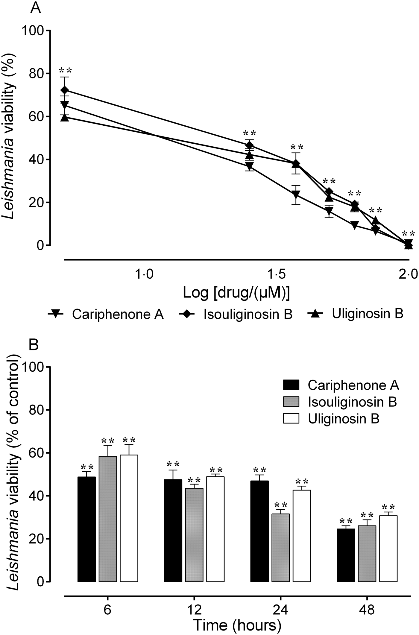
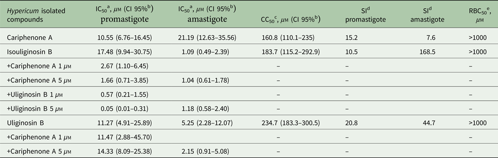
Dimeric acylphloroglucinols and benzophenone derivatives (Fig. 1) isolated from Hypericum species killed the promastigote forms of L. amazonensis in a concentration and time-dependent manner (Fig. 2A and B). When parasites were incubated with cariphenone A, isouliginosin B and uliginosin B for 48 h, all tested concentrations caused significant mortality (almost 100% of viability inhibition at 100 µ m) (Fig. 2A). Moreover, significant differences were observed in the viability of parasites treated at 50 µ m in all time points evaluated, indicating that all compounds act early, reducing the viability already in the first 6 h of exposure (Fig. 2B). The IC50 values of cariphenone A, isouliginosin B and uliginosin B on promastigote forms of L. amazonensis were calculated to be 10.55, 17.48 and 11.27 µ m, respectively (Table 1), while the reference antileishmanial drug, amphotericin B presented an IC50 48 h = 0.13 µ m (data not shown).
Leishmanicidal activity of isolated compounds of Hypericum on promastigote forms of L. amazonensis. (A) Promastigote forms (3 × 10⁶) in M199 medium were incubated with cariphenone A, isouliginosin B or uliginosin B at concentrations of 5–100 µ m for 48 h and the viability determined using a hemocytometer, (B) Time course effect of Hypericum compounds at 50 µ m on L. amazonensis. Data are expressed as means ± s.e.m. of four replicates and are representative of three independent experiments. **P < 0.01 compared with control group (M199 = 100% viability).

Leishmanicidal activity, macrophage cytotoxicity and haemolytic effect of Hypericum compounds at 48 h

a IC50: concentration of the isolated compound that causes 50% of mortality of L. amazonensis.
b CI 95%: 95% confidence interval.
c CC50: concentration of the isolated compound that causes 50% of macrophage cytotoxicity.
d SI: selectivity index, calculated as ratio of CC50 for macrophage/IC50 for L. amazonensis.
e RBC50: concentration of the isolated compound that causes 50% of red blood cell haemolysis.
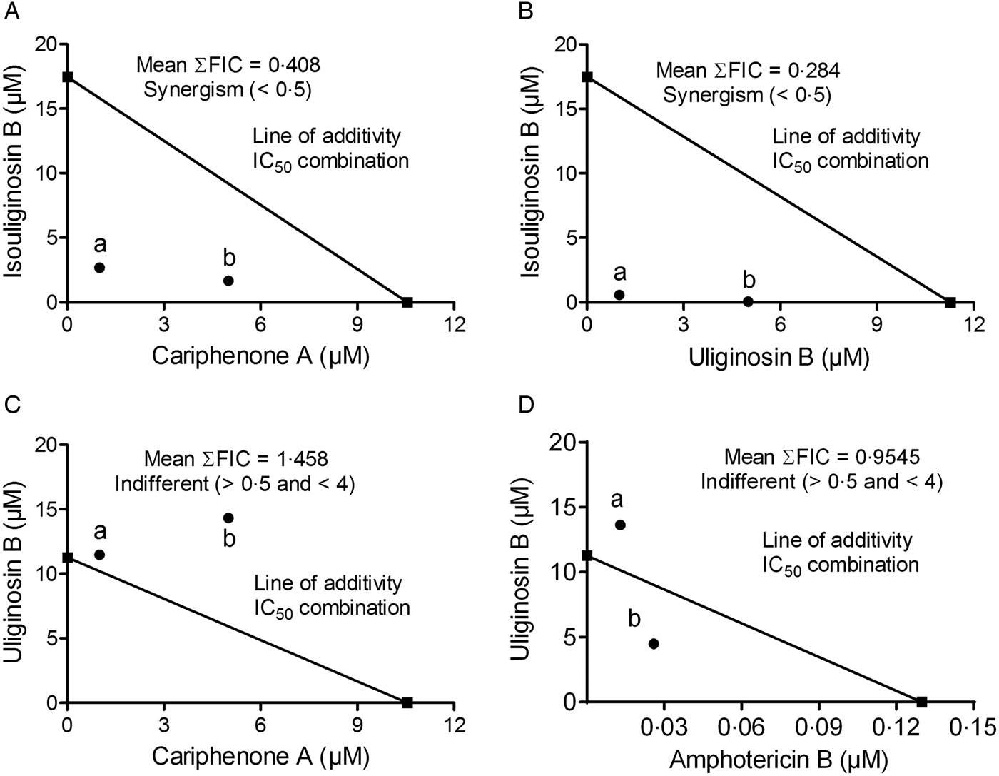
One strategy for the optimization of therapies, aiming for greater therapeutic efficacy, is the combination of two or more drugs. The isobologram analysis demonstrated that the association between different concentrations of isouliginosin B and uliginosin B at 1 µ m or 5 µ m (Fig. 3B) was the most promising association with an overall mean ΣFIC value of 0.284, when compared with the association between isouliginosin B and cariphenone A with mean ΣFIC value = 0.408 (Fig. 3A), indicating synergistic effects. However, the association between uliginosin B (1–20 µ m) and cariphenone A at 1 µ m or 5 µ m (Fig. 3C) did not decrease the IC50 values calculated to the individual compounds, generating a mean ΣFIC value = 1.458 (Fig. 3C), suggesting an antagonistic effect. With respect the association between uliginosin B (1–20 µ m) and amphotericin B at 0.013 µ m or 0.026 µ m, the isobologram analysis demonstrated an indifferent effect (ΣFIC value = 0.9545) (Fig. 3D).
Effects of combinations of isolated compounds of Hypericum on the viability of L. amazonensis promastigotes (A–D). Percentage of promastigote killing was determined by counting the number of viable parasites after 48 h of incubation as described in materials and methods. (A) Isobolograms for associations between isouliginosin B + cariphenone A, (B) isouliginosin B + uliginosin B, (C) uliginosin B + cariphenone A and (D) association between uliginosin B and amphotericin B. The points (a and b) refer to IC50 of compounds in combination (a and b). The line indicates synergy, additivity or antagonism when the points are located below, on or above the line, respectively. FIC, fractional inhibitory concentration. Experiments were performed in four replicates and are representative of two independent experiments.

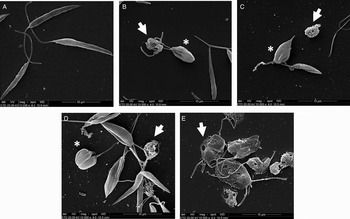
As illustrated in Fig. 4, when promastigotes were treated with IC50 Hypericum compounds values, the parasites showed striking morphological alterations. While untreated parasites exhibited elongated body and an unique flagellum, promastigotes treated with all Hypericum compounds evidenced the presence of membrane holes, flagellum duplication, rounded shape and multi-septation of the cell body.
Scanning electron micrographs of L. amazonensis promastigotes exposed to the isolated compounds of Hypericum for 6 h. (A) untreated parasites showing the typical elongated shape (parasite body and anterior flagella), (B) parasites treated with cariphenone A (IC50 = 10.55 µ m), (C) isouliginosin B (IC50 = 17.48 µ m), (D) uliginosin B (IC50 = 11.27 µ m) or (E) amphotericin B (IC50 = 0.13 µ m) showing morphological alterations such as membrane ruffles, membrane holes, flagellum loss, round-shaped forms and complete loss of typical promastigote morphology after 6 h of treatment. Note membrane disruption (arrows) and multi-septation of the cell body (asterisks). Bars: (A–E) 10 µm.

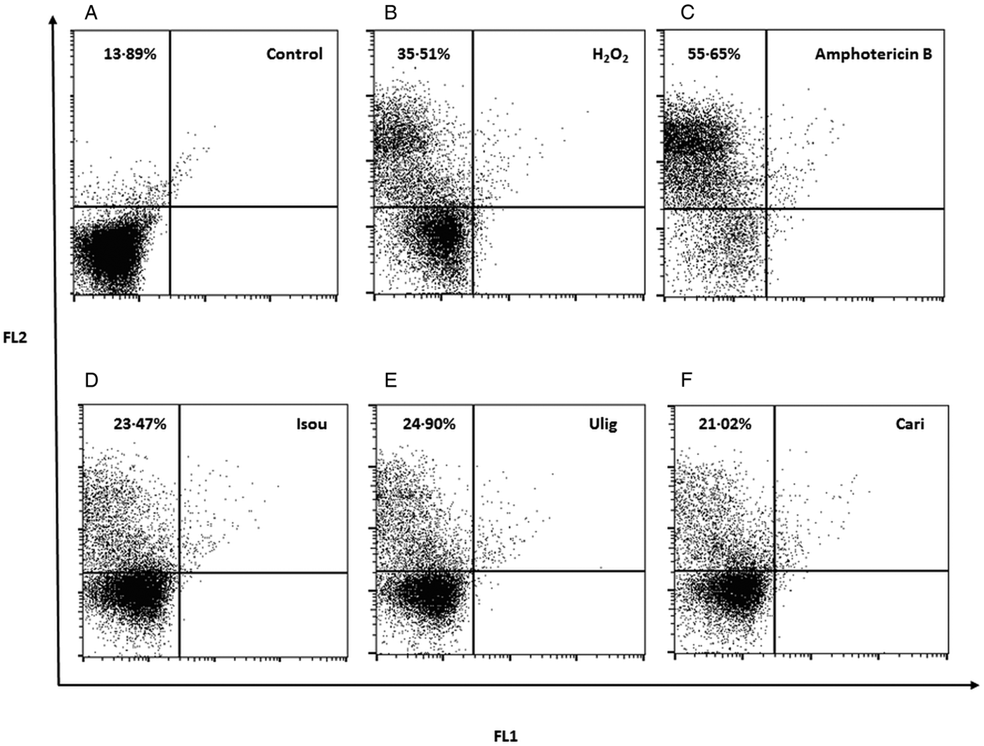
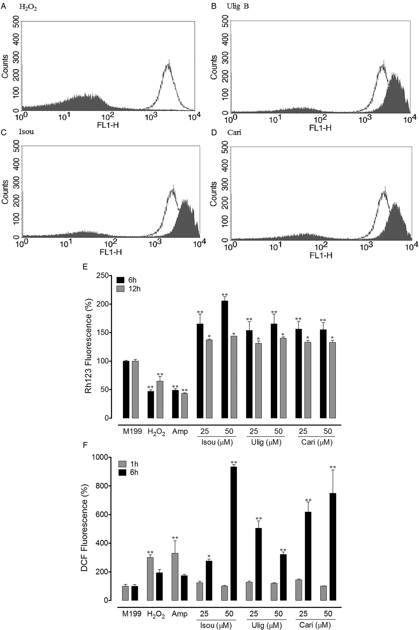
To investigate whether the leishmanicidal activity of Hypericum compounds involves the necrotic death, we evaluated the plasma membrane integrity of promastigotes treated with compounds. It was observed that compared with untreated control, PI fluorescence was significantly increased in parasites treated with 25 µ m of cariphenone A, isouliginosin B or uliginosin, as well as in parasites treated with H2O2 and amphotericin B (Fig. 5A–F). In addition, these compounds also induced DNA fragmentation after 24 h of incubation (data not shown). In relation to the mitochondrial effect, we observed that promastigotes treated with cariphenone A, isouliginosin B and uliginosin B at 25 µ m showed a marked increase in Rh 123 fluorescence after 12 h of incubation compared with the control group, indicating mitochondrial hyperpolarization [Fig. 6B–D). Moreover, according to the flowing formula IV = (Mt – Mc)/Mc], the Rh 123 fluorescence variation between treated and untreated parasites found positive IV values (cariphenone A: IV = 0.70, isouliginosin B: IV = 0.82 and uliginosin B, IV = 0.69) confirming ΔΨm hyperpolarization. Similar patterns of hyperpolarization were detected after 6 h of incubation with Hypericum compounds at 25 and 50 µ m (Fig. 6E). However, no mitochondrial membrane potential alterations were triggered by the compounds at 25 and 50 µ m after 1 h of incubation (data not shown). As expected H2O2 induced depolarization of ΔΨm (IV = −0,16) (Fig. 6A and E). Moreover, promastigotes treated with cariphenone A, isouliginosin B and uliginosin B at 25 and 50 µ m for 6 h, but not 1 h, presented significant increase in ROS production (Fig. 6F). The incubation of L. amazonensis with H2O2 and amphotericin B induced significant increase in ROS at 1 h of treatment.
Determination of membrane integrity of L. amazonensis using propidium iodide-PI. Acquisition dot plot of control promastigotes (A), promastigotes in the presence of H2O2 2 mm (B), in the presence of amphotericin B 0.20 µ m (C), in the presence of isouliginosin B 25 µ m (D), in the presence of uliginosin B 25 µ m (E) and in the presence of cariphenone A 25 µ m for 12 h (F). Upper left = PI stained cells. Experiments were performed in three replicates and are representative of two independent experiments.

Mitochondrial membrane potencial (ΔΨm) and intracellular reactive oxygen species (ROS) in promastigotes of L. amazonensis exposed to Hypericum compounds. L. amazonensis promastigotes (1 × 10⁶) were treated with H2O2 2 mm, amphotericin B 0.2 µ m, cariphenone A, isouliginosin B or uliginosin B (25 and 50 µ m for 1, 6 or 12 h) to ΔΨm or by 1 or 6 h for ROS evaluation. ΔΨm was determined using rhodamine 123 (A– E) and ROS using 2′,7′dichlorofluorescein diacetate (DCFH-DA) (F). (A–D) Demonstrative histograms of ΔΨm of L. amazonensis untreated control (panel A white), with H2O2 (panel A filled), uliginosin B (panel B filled), isouliginosin B (panel C filled) or cariphnone A (panel D filled). (E) Graphic representation of Rh 123 mean fluorescence intensity in percentage relative to control of different treatment groups. (F) Graphic representation of DCF mean fluorescence intensity in percentage relative to control of different treatment groups. Data represent mean ± s.e.m. of four replicates and are representative of three independent experiments. *P < 0.05 vs M199; **P < 0.01 vs M199.

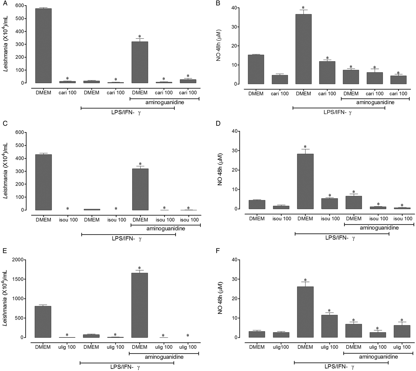
The effect of Hypericum compounds on intracellular amastigotes was first evaluated using compounds at 100 µ m. It was found that unlike the leishmanicidal activity of macrophages stimulated by LPS plus IFN-γ, cariphenone A, isouliginosin B and uliginosin B drastically reduced the viability of intracellular amastigotes by a mechanism independent of NO production (Fig. 7). Moreover, the anti-amastigote activity induced by Hypericum compounds was not associated with the increase on costimulatory CD80 (control macrophages: mean fluorescence intensity = 691.3 ± 4.43; L. amazonensis infected-macrophages: MFI = 689.7 ± 7.57; L. amazonensis-infected macrophages treated with cariphenone A: MFI = 705.33 ± 6.42; L. amazonensis-infected macrophages treated with isouliginosin B: MFI = 694.66 ± 5.50; L. amazonensis-infected macrophages treated with uliginosin B: MFI = 689.33 ± 1.15), and CD86 (control macrophages: MFI = 696.10 ± 8.71; L. amazonensis infected-macrophages: MFI = 690.33 ± 6.62; L. amazonensis-infected macrophages treated with cariphenone A: MFI = 693.93 ± 7.23; L. amazonensis-infected macrophages treated with isouliginosin B: MFI = 691.33 ± 6.02; L. amazonensis-infected macrophages treated with uliginosin B: MFI = 695.66 ± 0.57).
Effects of isolated compounds of Hypericum on the viability of amastigotes of L. amazonensis and nitric oxide production in vitro. Macrophages RAW 264.7 strain were incubated at 1 × 10⁵ cells/well and infected with L. amazonensis promastigotes. Subsequently, they were treated with cariphenone A (A and B), isouliginosin B (C and D) and uliginosin B (E and F) at 100 µ m in the presence or absence of LPS (10 ng mL−1) plus IFN-γ (1 ng mL−1) or 300 µ m of aminoguanidine. Data are expressed as means ± s.e.m. of four replicates and are representative of three independent experiments. *P < 0.05, **P < 0.01 compared with control group (macrophages incubated with DMEM).

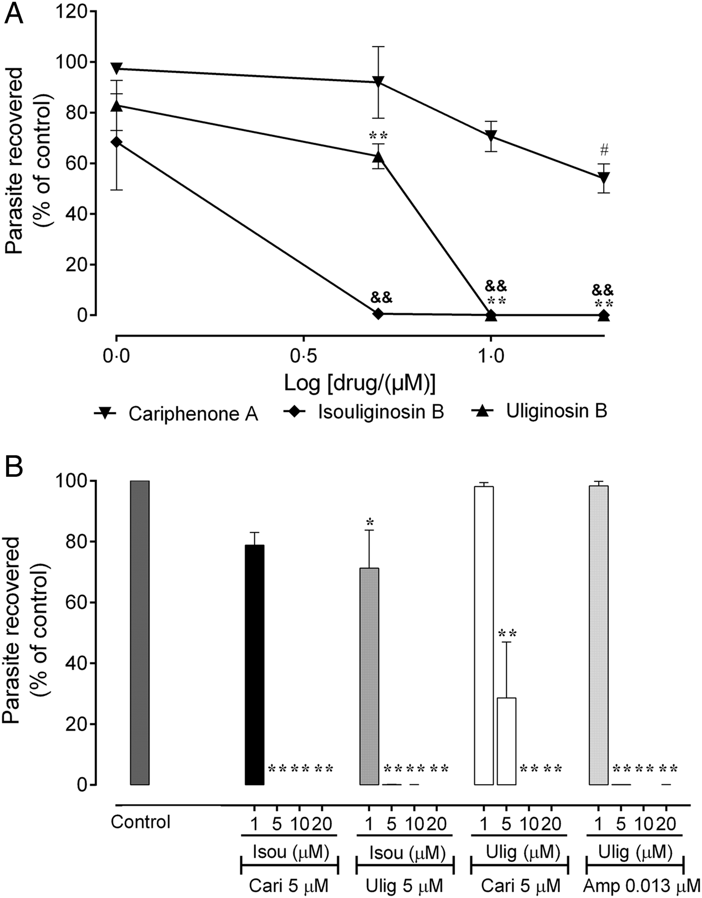
In another set of experiments, it was observed that, even at low concentrations, Hypericum compounds caused potent decrease in the intramacrophage amastigote viability (isouliginosin B: 100% amastigote mortality in concentrations ⩾5 µ m, uliginosin B: 100% mortality in concentrations ⩾10 µ m and cariphenone A: 46% mortality at 20 µ m) (Fig. 8A). The IC50 values calculated for these compounds against amastigotes indicated better or similar activities compared with promastigote activity (Table 1).
Effects of individual or combinations of isolated Hypericum compounds on intracellular amastigote forms of L. amazonensis. Macrophages 1 × 10⁵ cells per well were infected with L. amazonensis promastigotes (five parasites per cell) and incubated with DMEM medium, cariphenone A, isouliginosin B, uliginosin B (1 to 20 µ m) for 48 h. The leishmanicidal activity of treated-macrophages was determined as described in materials and methods (A). Effects of associations between isouliginosin B + cariphenone A, isouliginosin B + uliginosin B, uliginosin B + cariphenone A, and uliginosin B + amphotericin B (B). Data are expressed as means ± s.e.m. of four replicates and are representative of two independent experiments. #P < 0.05 vs M199; **P < 0.01 vs M199; &&P < 0.01 vs M199.

Regarding the effect of Hypericum compounds and amphotericin B combinations on amastigotes, it was observed that the association between isouliginosin B plus cariphenone A, as well as isouliginosin B plus uliginosin B both at 5 µ m was able to caused 100% of amastigote mortality (Fig. 8B). In addition the combination between uliginosin B plus cariphenone A at the same concentration of 5 µ m caused 72% of amastigote mortality (Fig. 8B). Moreover, the association between uliginosin B at 5 µ m and amphotericin B at 0.013 μ m resulted in 100% amastigote mortality (Fig. 8B). All combinations were more potent when compared with the effect of individual drugs.
In relation to cytotoxic effect of Hypericum compounds, cariphenone A, isouliginosin B and uliginosin B showed low rate of haemolysis (Table 1). Moreover, they also present low cytotoxicity against macrophages and high selectivity toward parasites (Table 1).
Discussion
To the best of our knowledge, this is the first study reporting the leishmanicidal activity of phloroglucinol derivatives from Hypericum species against L. amazonensis. Our data showed that these compounds presented potent leishmanicidal activity against both promastigote and amastigote stages of parasites and synergistic interactions when tested in association (isouliginosin B plus cariphenone A and isouliginosin B plus uliginosin B) against promastigotes. Moreover, these Hypericum compounds induced ROS production and mitochondrial dysfunction in promastigotes of L. amazonensis, as well as presented low toxicity towards macrophages and red blood human cells, high selectivity index and killed the amastigote intracellular forms by a mechanism independent of nitric oxide.
In a previous study, our group has demonstrated that the lipophilic extracts of H. polyanthemum, H. carinatum and H. linoides containing phloroglucinols were able to kill infective forms of L. amazonensis (Dagnino et al. Reference Dagnino, Barros, Ccana-Ccapatinta, Prophiro, Poser and Romao2015). Regarding the antiprotozoal activity of phloroglucinol derivatives, it was demonstrated that uliginosin B, and also isouliginosin B decreased the viability of trophozoites of Trichomonas vaginalis with IC50 values of 121.96 and 59 µ m, respectively (Cargnin et al. Reference Cargnin, Vieira Pde, Cibulski, Cassel, Vargas, Montanha, Roehe, Tasca and von Poser2013; Menezes et al. Reference Menezes, Rigo, Bridi, da Silva Trentin, Macedo, Von Poser and Tasca2017). In our study, the IC50 values obtained for uliginosin B, and isouliginosin B against L. amazonensis promastigotes were 11.27 and 17.48 µ m, respectively. Moreover, uliginosin B showed antimicrobial activity against Staphylococcus aureus and Staphylococcus epidermidis with minimum inhibitory concentration (MIC) of 3.0 µg mL−1, while isouliginosin B presented MIC value of 1.5 and 3.0 µg mL−1 for S. aureus and S. epidermidis, respectively (França et al. Reference França, Kuster, Rito, Oliveira, Teixeira and Rocha2009).
The leishmanicidal activity of other phloroglucinols has been reported. Formylated phloroglucinols presented activity against promastigotes of L. donovani (IC50 = 88.24 to 244 μ m) (Sidana et al. Reference Sidana, Singh, Arora, Foley and Singh2011), prenylated acylphloroglucinols lindbergins E – I against promastigotes of L. braziliensis (IC50 = 13.6–87.7 µ m) and L. amazonensis (IC50 = 16.3–151.3 µ m) (Socolsky et al. Reference Socolsky, Salamanca, Gimenez, Borkosky and Bardon2016) and phloroglucinol-terpene adducts against L. donovani promastigotes (IC50 = 0.62–7.68 µ m) (Bharate and Singh, Reference Bharate and Singh2011). In addition, nemorosone and guttiferone A induced significant mortality of L. tarentolae promastigotes with IC50 values of 0.67 and 6.2 µ m, respectively (Monzote et al. Reference Monzote, Lackova, Staniek, Cuesta-Rubio and Gille2015). Moreover, prenylated benzophenone garcinielliptone FC from Platonia insignis showed an IC50 value of 42.8 µ m against L. amazonensis promastigotes (Costa Junior et al. Reference Costa Junior, de Almeida, de Barros Falcao Ferraz, Rossatto, Silva, Silva, Militao, das Gracas Lopes Cito, Santana, de Amorim Carvalho and Freitas2013). In agreement, it was shown that benzophenone-derived bisphosphonium salts with intermediate hydrophobicity inhibited Leishmania proliferation at low micromolar concentrations (Luque-Ortega et al. Reference Luque-Ortega, Reuther, Rivas and Dardonville2010). In fact, synthetic benzophenones containing more lipophilic radicals are studied in order to increase their anti-Leishmania activity (Maciel-Rezende et al. Reference Maciel-Rezende, de Almeida, Costa, Pires, Alves, Viegas, Dias, Doriguetto, Marques and dos Santos2013).
Here, the s.e.m. observations revealed that the treatment with Hypericum compounds induces swelling and overall rounding, multi-septation of the cell body, with significant loss in body length and flagellum, corroborating evidences of the action of Hypericum extracts and isolated compounds from different plant species on L. amazonensis (Brenzan et al. Reference Brenzan, Santos, Nakamura, Filho, Ueda-Nakamura, Young, Correa, Junior, Morgado-Diaz and Cortez2012; Dagnino et al. Reference Dagnino, Barros, Ccana-Ccapatinta, Prophiro, Poser and Romao2015; da Silva et al. Reference da Silva, da Silva, Rodrigues, Farias, da Silva, Alves, Bastos, do Nascimento and Silva2015). On the other hand, it was demonstrated that the treatment of promastigotes with Hypericum compounds led to significant increase in PI staining in parasites, indicating the loss of membrane integrity. It is known that necrosis process is characterized by swelling of organelles, volume augments of the cell and membrane rupture (Laster et al. Reference Laster, Wood and Gooding1988). Although we did not deeply evaluate apoptosis, the DNA analysis by agarose gel electrophoresis revealed DNA fragmentation in parasites treated with Hypericum compounds as well as with amphotericin B or H2O2, suggesting induction of apoptosis. Further analysis should clarify this point.
Mitochondria play a pivotal role for parasite viability in Leishmania, since they are the essential source for ATP production during the mitochondrial respiratory chain (Van Hellemond and Tielens, Reference Van Hellemond and Tielens1997; Van Hellemond et al. Reference Van Hellemond, Van der Meer and Tielens1997). Then, irreversible damage and dysfunction of this vital organelle caused by drugs will have disastrous consequences on parasites. In this context, it was demonstrated that the antileishmanial drug pentamidine accumulates in the Leishmania mitochondrion and that the resistance phenotype is accompanied by lack of drug mitochondrial accumulation and its exclusion from the parasites (Basselin et al. Reference Basselin, Denise, Coombs and Barrett2002; Mukherjee et al. Reference Mukherjee, Padmanabhan, Sahani, Barrett and Madhubala2006). On the other hand, amphotericin B causes a significant decrease in mitochondrial membrane potential (Lee et al. Reference Lee, Bender and Kadenbach2002). In our study, cariphenone A, isouliginosin B and uliginosin B induced hyperpolarization of the mitochondrial transmembrane potential of L. amazonensis. Hyperpolarization is considered to be less common than mitochondrial depolarization, arising mainly from dysfunctions in Fo/F1 ATPase, and it is considered a preapoptotic event (Sen et al. Reference Sen, Das, Ganguly, Mukherjee, Tripathi, Bandyopadhyay, Rakshit, Sen and Majumder2004). Indeed, both hyperpolarization and loss of ΔΨm can result in apoptotic death of promastigotes forms, demonstrating that an unstable mitochondrial membrane potential and redox transitions can have negative consequences for mitochondrial integrity and parasite survival.
Based on that, intracellular oxidative stress was investigated in L. amazonensis promastigotes exposed to the different Hypericum compounds. In Leishmania spp., the deviation of electrons from the mitochondrial complexes is the primary source of endogenous ROS (Gille and Nohl, Reference Gille and Nohl2001). In our study, we observed a significant increase in ROS production in promastigotes treated with cariphenone A, isouliginosin B and uliginosin B at the same time of mitochondrial membrane hyperpolarization was detected. Similarly to our results, Leishmania donovani promastigotes treated with anthraquinone derivative obtained from Hypericum perforatum called hypericin, also triggered ROS production (Singh et al. Reference Singh, Sarma, Katiyar, Das, Bhardwaj, Sundar and Dubey2015).
Because Leishmania possesses a single mitochondrion, mitochondrial dysfunction and energetic metabolic plasticity in Leishmania are more limited than in other eukaryotic cells with multiple mitochondria, such as macrophages. In this respect, Hypericum compounds presented low cytotoxicity against macrophages. Thus, uliginosin B exhibited greater selectivity (SI) and best biological profile, with a SI = 20.8, followed by cariphenone A (SI = 15.2) and isouliginosin B (SI = 10.5). Literature data refer that the effectiveness of a compound is indicated by selectivity indices ⩾10 (Nava-Zuazo et al. Reference Nava-Zuazo, Estrada-Soto, Guerrero-Alvarez, Leon-Rivera, Molina-Salinas, Said-Fernandez, Chan-Bacab, Cedillo-Rivera, Moo-Puc, Miron-Lopez and Navarrete-Vazquez2010). Moreover, SI > 20 is considered adequate for subsequent in vivo studies following the drug discovery pipeline (Don and Ioset, Reference Don and Ioset2014).
In our study, the treatment with cariphenone A, isouliginosin B and uliginosin B drastically reduced the viability of intracellular amastigotes by a mechanism independent of NO, suggesting the involvement of other mechanisms including the interference with essential metabolic pathways of parasites or indirectly by the induction of other oxidants products such as oxygen reactive species that was not determined in this study. For example, de Almeida et al. (Reference de Almeida, Alves, Maciel-Rezende, Jesus Lde, Pires, Junior, Izidoro, Judice, dos Santos and Marques2015) showed that the synthetic benzophenone 2-hydroxy-4-O-(3,3-dimethyl)-allylbenzophenone presented anti-amastigote activity (IC50 L. amazonensis = 74.4 µ m) and caused a potent inhibitory effect on trypanosomatids cysteine proteases. On the other hand, hypericin isolated from H. perforatum inhibited the spermidine synthase in L. donovani promastigotes, leading to spermidine starvation and parasite death (Singh et al. Reference Singh, Sarma, Katiyar, Das, Bhardwaj, Sundar and Dubey2015).
Our data exhibit for the first time that benzophenone cariphenone A and acylphloroglucinols isouliginosin B and uliginosin B isolated from Hypericum species (alone or in association) present potent activity against both promastigote (infective forms) and amastigote forms (intracellular stage) of L. amazonensis by a mechanism dependent on ROS generation and mitochondrial dysfunction. These results indicate that cariphenone A, isouliginosin B and uliginosin B are promising candidates for the development of drugs against L. amazonensis.
Financial support
The authors are grateful to the Brazilian agencies Coordenação de Aperfeiçoamento de Pessoal de Nível Superior (CAPES, CAPES-PNPD 23038007200/11-08), Conselho Nacional de Desenvolvimento Cientifico e Tecnológico (CNPq – 307277/2013-5) and Fundação de Amparo à Pesquisa do Estado do Rio Grande do Sul (FAPERGS 1017479) for financial support and by fellowships from CAPES (APD, FMCB, VONT, GDP) and CNPq (MCM, GLvP and PRTR).
Conflict of interest
The authors declare that this article content has no conflict of interest.