Post-infective pseudoaneurysm of the left ventricle in children is extremely rare, with only five cases reported in English medical literature so far.Reference Moraes, Ferreira and Gomes Ferreira 1 – Reference Vinitha, Dhananjay and Kothari 5 Untreated, the pathology has a fatal outcome. Timely surgical intervention has generally a good outcome. We present a case of post-infective pseudoaneurysm in a 2-year-old girl with a review of literature.
Case report
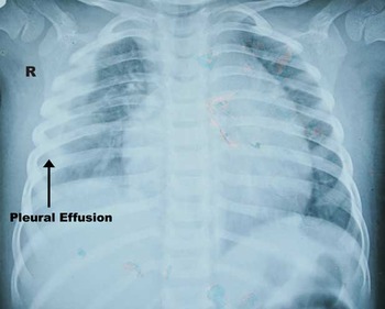
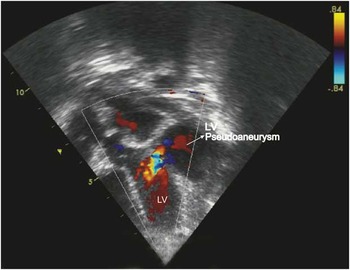
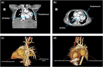
Our patient was a 2-year-old girl weighing 5.4 kg; she presented to us with a history of high-grade intermittent fever since 1 month. The fever was associated with dry cough and macular rashes. Few days later, the patient developed multiple skin abscesses and swelling of the left hip joint with painful movement. The patient was admitted at a rural centre when she started having respiratory distress 1 week after onset of fever. Investigations revealed minimal pericardial effusion and mild right pleural effusion. She was given intravenous antibiotics, but as her symptoms worsened she was referred to our centre. Hemogram showed leucocytosis with neutrophilia and increased levels of C-reactive protein. Her chest X-ray now showed moderate cardiomegaly with pleural effusion (Fig 1); however, two-dimensional echocardiography at this time showed a mild pericardial effusion. Pleural fluid examination showed evidence of bacterial infection – high protein, low sugar, and total count 5000/cm3 with 80% neutrophils. Cultures of blood and pleural fluid were, however, negative. The patient was treated with intravenous Vancomycin for 1 week. Respiratory symptoms and white blood cell counts improved, but the patient continued to have fever. Nearly 1 month after onset of symptoms, repeat two-dimensional echocardiography now showed a large left ventricular pseudoaneurysm arising from lateral wall with neck in the submitral region (Fig 2). A contrast cardiac computed tomography angiography confirmed the diagnosis (Fig 3).
Chest X-ray posterioranterior view showing pleural effusion on the right side.

Apical five-chamber view of two-dimensional echocardiography with colour Doppler showing a pseudoaneurysm communicating with the left ventricle (LV).

( a ) Coronal view of CT angiogram showing the pseudoaneurysm. ( b ) Axial view of CT angiogram showing the pseudoaneurysm in transverse dimension. ( c ) Three-dimensional VRT reconstruction of CT angiogram showing the relation of the pseudoaneurysm to the left pulmonary artery. ( d ) Three-dimensional VRT reconstruction of CT angiogram showing the pseudoaneurysm communicating with the left ventricle through a small neck (arrow head) close to the AV groove. ( d ) Three-dimensional VRT reconstruction of CT pulmonary angiogram showing the pseudoaneurysm communicating with the left ventricle through a small neck (arrow head) close to the AV groove. CT=computed tomography; VRT=volume rendering technique.

The patient was taken for emergency surgery. After midline sternotomy, the pseudoaneurysm ruptured and emergency cardiopulmonary bypass was commenced. The core temperature was dropped to 20°C and flows were reduced in order to provide neuroprotection considering the brief period of severe hypotension. Under cardioplegic arrest, the neck of the pseudoaneurysm was closed with a Goretex™ (ePTFE, Gor-Tex Patch W.L. Gore and Associates, Flagstaff, Arizona, United States of America) patch. The pseudoaneurysm wall was removed piecemeal and pericardial cavity was washed thoroughly. Gradual rewarming was done and the patient weaned of extracorporeal support. The patient was extubated on post-operative day two and showed no neurological deficit. Post-operative two-dimensional echocardiography showed mild left ventricular dysfunction with pseudoaneurysm closed off. Cultures of the operative specimen were negative. Although cultures of our patient were never found to be positive for any organism, we think the causative organism in our case must have been staphyloccous on account of classical presentation with boils and suppurative arthritis. The cultures of our patient were sterile probably because of antibiotics received priorly at the rural centre.
Discussion
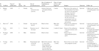
A pseudoaneurysm is when there is a breach in the vessel wall such that blood leaks through the wall but is contained by the adventitia or surrounding soft tissue. After the first report of left ventricular pseudoaneurysm developing post infective pericarditis in children by Moraes et al,Reference Moraes, Ferreira and Gomes Ferreira 1 only a few cases have been published in world literature (Table 1). There are certain common factors among these cases. First, all of them were associated with staphylococcus infection, which was isolated from different sites. Second, there was rapid progression from a generalised staphylococcal infection to development of pseudoaneurysm and ranged from 2 to 4 weeks. Third, the extent of staphylococcal infection was quite widespread, with most having septicaemia and all of them having pericarditis. Fourth, none of the cases had cultured staphylococcus in their operative specimens. Fifth, all of them had received antistaphylococcal antibiotics before development of pseudoaneurysm. Various theories have been proposed for the pathology arising after staphylococcal infection. Boer et alReference Boer, Elzenga, Boer and Meuzeleer 2 proposed that there seems to be contained rupture of ventricular wall at a site of weakening produced either by direct spread of infection from pericardium or by intramyocardial abscesses. Juneja et alReference Juneja, Sharma and Kothari 4 suspected streptokinase, given intrapericardially for better drainage, to be responsible for left ventricular pseudoaneurysm. We think staphylokinase produced by the organism might be contributing to the pathology. Staphylokinase converts inactive plasminogen to plasmin, which in turn breaks down collagen. The fact that staphylococcus has not been isolated from operative specimens and the pseudoaneurysms occurred even after antibiotics supports our theory. Quantitative staphylokinase assays might help us to further investigate this theory. As to why this is so common in the posterolateral submitral region remains to be answered. It may be due to congenital weakness in the region. In general, post- infective pseudoaneurysm present only a few weeks after the onset of staphylococcal septicaemia and pericarditis. Emergency surgery with closure of the submitral aneurysm either directly or with a patch generally has a good outlook.
Comparative analysis of post infective LV pseudoaneurysm reported so far