Implications
The understanding of the physiological, cellular and molecular mechanisms involved in milk production, particularly in the mammary gland, is required to manage dairy production. Data from literature suggest that ovarian steroids, estradiol and progesterone, positively affect mammogenesis and negatively affect milk production and lactation persistency. The objective of this review is to investigate the effect of ovarian steroids on the molecular and cellular dynamics of mammary epithelial cells, and their implications in mammogenesis and in lactation persistency in dairy cow.
Introduction
The lactation persistency defines the rate of decline in milk production after the peak of lactation. This persistency depends on maintaining the number and the activity of mammary epithelial cells (MECs) and the organization of secretory tissue in the mammary gland. It can be modulated by various factors such as milking frequency, feeding plan, hormonal status or animal health. All of these factors directly or indirectly influence the proliferation/apoptosis balance of MEC and tissue remodeling. Ovarian steroids (estradiol and progesterone) play an important role in the mammary gland development and function. During phases of mammogenesis at puberty and during pregnancy, ovarian steroids stimulate cell proliferation and promote the development of secretory structures. However, they seem to have a negative effect on the lactating mammary gland. Several studies in cattle have shown that administration of estradiol with or without progesterone for lactation induced a rapid decrease in milk yield (MY) and accelerated the process of involution of the mammary gland. Through this review, we will analyze how ovarian steroids induce mammogenesis and influence lactation persistency and by which molecular mechanisms they act on mammary gland and MECs.
Ovarian steroids
Estradiol and progesterone synthesis and modes of action
Synthesis of ovarian steroids
Estradiol and progesterone are two steroid hormones that are synthesized from cholesterol. The incorporation of cholesterol in theca cells occurs either via membrane vesicles or through specific transporters such as steroidogenic acute regulatory protein. These proteins enable the delivery of cholesterol to the mitochondria, where it undergoes its first transformation through the action of cytochrome P450scc. This enzyme is responsible for the conversion of cholesterol to pregnenolone by cleavage of the side chain of cholesterol, and this first step is a key step in the regulation of steroidogenesis (Miller, Reference Miller2007). Pregnenolone is the last common precursor for progesterone and androgens. Indeed, pregnenolone is converted to progesterone by 3β-hydroxysteroid dehydrogenase (3β-HSD), while the production of androstenedione requires successive interventions by cytochrome P45017α and 3β-HSD, with intermediate production of dehydroepiandrosterone (DHEA). After the transfer of androgens from theca cells into granulosa cells, estrogen is produced via the aromatization of androgens by cytochrome P450 aromatase, which converts androstenedione to estrone. Estrone is subsequently subjected to the action of 17β-HSD to produce estradiol. The aromatization of androgens to estrogens is the second important step in the regulation of steroidogenesis. Fadrozole, an aromatase inhibitor used to treat some cancers, including breast cancer in women (Dutta and Pant, Reference Dutta and Pant2008), prevents the synthesis of estradiol by ovarian cells in vitro and is responsible for decreases in the concentration of plasmatic estradiol and disruption of ovarian cyclicity in vivo in sheep (Benoit et al., Reference Benoit, Inskeep and Dailey1992). The regulation of estradiol synthesis thus requires modulation of aromatase activity, in addition to modulation during the first stage of steroidogenesis, which occurs at the mitochondrial level.
Ovarian steroids receptivity
The actions of estradiol and progesterone mainly occur through genomic pathways via their nuclear receptors, which act as transcription factors. The free forms of these receptors are found in the cytoplasm close to the nucleus. The binding of the hormone to its receptor induces a conformational change that allows the coupled hormone/receptor to be transferred into the nucleus and form a dimer with another coupled hormone/receptor. This dimer binds to specific DNA sequences (hormone-response elements) located in the promoter regions of target genes, where it will recruit either co-activators or co-repressors to stimulate or repress transcription of target genes. Microarray analysis identified 124 genes whose transcription is under the control of estrogen in the mammary gland, with only 3% of the genes repressed by estrogen. Among these 124 genes, several are involved in the activation of cell proliferation, such as insulin-like growth factor-1 (IGF-1), epidermal growth factor and cyclin D1 (Connor et al., Reference Connor, Meyer, Li, Van Amburgh, Boisclair and Capuco2007). Estradiol also activates transcription of the progesterone receptor (PR) (Petz et al., Reference Petz, Ziegler, Schultz, Kim, Kemper and Nardulli2004). In the mammary gland, progesterone binds to its receptor to activate Wnt-4 gene transcription, a paracrine mediator of progesterone in cell proliferation (Brisken and O’Malley, Reference Brisken and O’Malley2010), as well as transforming growth factor-β (TGF-β), Wnt-5b and insulin-like growth factor binding protein-5 (IGFBP-5), which are involved in the inhibition of milk secretion in late pregnancy (Connor et al., Reference Connor, Meyer, Li, Van Amburgh, Boisclair and Capuco2007).
Steroid nuclear receptors are a family characterized by a protein structure composed of six functional domains named A to F. Domain A/B, located at the N-terminus of the protein, is the area that has the highest variability and that differs between receptor isoforms for the same hormone. Domain C is the most conserved domain in this family of receptors and permits binding to the DNA sequence. Domain D is a fairly conserved domain that allows binding between the C domain and the hormone-specific binding domain, the E domain. Finally, the F domain is involved in the regulation of hormone-binding domain E (Connor et al., Reference Connor, Meyer, Li, Van Amburgh, Boisclair and Capuco2007). There are different isoforms of the nuclear receptors for estradiol and progesterone. The distribution of the different isoforms is tissue-specific and allows variability in the responses to the same hormone.
Changes in levels of estradiol and progesterone
Ovarian cyclicity
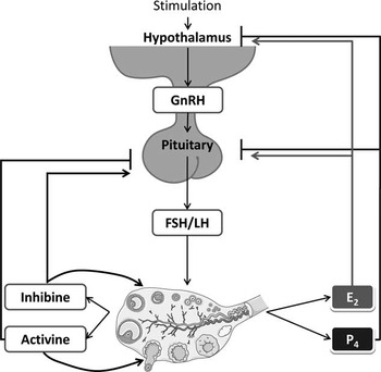
In female mammals, the ovary is the organ that ensures the release of oocytes, the female gametes. The stock of oocytes is created in the ovaries during the embryonic stage of life, and these oocytes are released regularly in females from puberty until the stock runs out. Each oocyte is surrounded by the cells of the theca and granulosa, which form a primordial follicle and ensure the maturation of the oocyte until ovulation. The cyclic activity of the ovaries begins at puberty with the initiation of the pulsatile secretion of gonadotropin-releasing hormone (GnRH). GnRH is a peptide hormone secreted by neurons in the hypothalamus (Figure 1). Its secretion is regulated by many endogenous and exogenous factors, and it mainly controls the secretion of two hormones secreted by the anterior pituitary: FSH and LH. The ovarian cycle consists of two phases. The first is the follicular phase, which corresponds to a period of ovarian follicle growth and oocyte maturation and ends with ovulation. Second is the luteal phase, which follows ovulation and is characterized by the formation of a corpus luteum. The average duration of a cycle is 21 days in cows (Wiltbank et al., Reference Wiltbank, Lopez, Sartori, Sangsritavong and Gumen2006) and goats (Baril et al., Reference Baril, Leboeuf and Saumande1993) and 17 days in sheep (Bartlewski et al., Reference Bartlewski, Baby and Giffin2011). The majority of the ovarian cycle is devoted to the luteal phase. In cows, for example, the follicular phase lasts ∼6 days, while the luteal phase lasts ∼15 days (Forde et al., Reference Forde, Beltman, Lonergan, Diskin, Roche and Crowe2011). The ovaries are the main source of estradiol and progesterone in non-pregnant female mammals. There are, however, other localized sources of steroid hormones that can still strongly influence the concentrations of circulating estradiol and progesterone.
Schematic representation of the hormonal regulation of ovarian activity. E2=estradiol, P4 progesterone →: stimulation; ┤ inhibition.

The fetoplacental unit
In pregnant females, the fetoplacental unit has been identified as a major endocrine gland. During pregnancy, many hormones are produced and finely regulated to optimize fetal development. Estradiol and progesterone are the two major hormones of pregnancy and parturition. After ovulation and during the first days of pregnancy, the corpus luteum ensures the progesterone production that is necessary for the initiation of pregnancy and implantation of the conceptus. Around the second week of gestation in cattle, the trophoblast secretes prostaglandins, testosterone, progesterone and small amounts of estrogen. The placenta synthesizes steroid hormones from cholesterol circulating in maternal blood. The concentration of progesterone in maternal cattle blood is ∼10 ng/ml on the 18th day of gestation and remains stable almost until parturition (Patel et al., Reference Patel, Takenouchi, Takahashi, Hirako, Sasaki and Domeki1999). A peak of estrogen production takes place just before parturition, resulting in a level of estradiol in maternal blood up to ∼900 pg/ml at 24 h before parturition (Patel et al., Reference Patel, Takenouchi, Takahashi, Hirako, Sasaki and Domeki1999). In contrast, estradiol is not the major estrogen of pregnancy in ruminants. Indeed, estrone obtained by the aromatization of DHEA is secreted by the placenta in significantly higher amounts (Bazer and First, Reference Bazer and First1983). The plasma level of estrone is between 70 and 140 pg/ml in mid-gestation and 600 and 1200 pg/ml at the end of gestation, reaching up to 4500 pg/ml by 24 h before parturition (Patel et al., Reference Patel, Takenouchi, Takahashi, Hirako, Sasaki and Domeki1999).
The adrenal glands
The adrenal glands are, like the ovaries in the female mammal, the seat of an important steroidogenesis process. The steroidogenic biosynthetic pathways activated in the adrenal glands involve the production of precursors for the synthesis of estradiol and progesterone. Steroid synthesis occurs specifically in tissue of mesodermal origin located at the periphery of the adrenal gland. Steroidogenesis in the adrenal cortex is stimulated by a pituitary hormone, ACTH, and allows for secretion of mineralocorticoids (aldosterone), glucocorticoids (cortisol and corticosterone) and androgens (DHEA, androstenedione and testosterone).
The mammary gland
Estrogen synthesis in the mammary gland has been demonstrated in several species of mammals in healthy and cancerous tissues (Simpson, Reference Simpson2000; Janowski et al., Reference Janowski, Zdunczyk, Malecki-Tepicht, Baranski and Ras2002; Lonning, Reference Lonning2004). In women, the estradiol concentration in breast tissue is 10 to 20 times higher than its plasma concentration after menopause (Lonning, Reference Lonning2004). Similarly, Janowski et al. (Reference Janowski, Zdunczyk, Malecki-Tepicht, Baranski and Ras2002) measured plasma estradiol in cows and found that it was significantly higher in the mammary vein than in the aorta or the uterine vessels (Janowski et al., Reference Janowski, Zdunczyk, Malecki-Tepicht, Baranski and Ras2002).
Local production of estrogen in the mammary gland seems to have systemic impact via paracrine actions. By this mode of action, estrogen produced in the mammary gland plays an important role in the development of breast cancer in postmenopausal women (Simpson, Reference Simpson2000). In the mammary gland, estrogen synthesis is particularly ensured by adipocytes that express aromatase, an enzyme necessary for the conversion of androgens to estrogens. There is also a positive correlation between the mass of adipose tissue and the risk of developing breast cancer in women (Simpson, Reference Simpson2000). The main substrate of aromatase is androstenedione, which is converted to estrone and is itself converted to estradiol by 17β-HSD. Culture of bovine mammary gland homogenates (taken a few days before parturition) shows that the bovine mammary gland can convert androstenedione to estradiol with a yield of 37% (Janowski et al., Reference Janowski, Zdunczyk, Malecki-Tepicht, Baranski and Ras2002). In women, the use of aromatase inhibitors in treatment against some breast cancers effectively reduces aromatase activity and plasma estradiol level (Lonning, Reference Lonning2004).
The mammary gland: anatomy and development
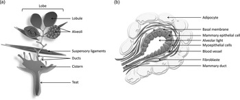
The mammary gland is the organ that produces milk and is specially organized for optimal function in milk synthesis and ejection. The secretory tissue is located in the distal regions of the udder relative to the position of the teats and gland cistern. It is composed of cells grouped into lobules, which are themselves divided into lobes. The secretory tissue is drained by a network of ducts, which opens into a cistern in ruminants (Figure 2a). In ruminants, milk is stored in the cistern and in the alveolar lumen (see below) before being discharged through the teat canal during milking or suckling. The alveoli are the functional units of the mammary gland. They consist of a layer of polarized MECs. At their apical pole, these cells lead to the alveolar lumen, which contains secreted milk. At their basal pole, they directly interact with contractile myoepithelial cells and with stromal tissue, which is composed fibroblasts, adipocytes and lymph and blood vessels (Figure 2b). These allow the input of nutrients required for milk synthesis.
Anatomy of ruminant mammary gland (a) and structure of the mammary cell (b).

The mammary gland from embryogenesis to puberty
Early mammary structures appear during embryogenesis by the invagination of ectodermal structures to form the main ducts and teat canal. The implementation of the first ducts is mainly under the control of glucocorticoids, prolactin (Prl) and growth hormone (GH) (Veltmaat et al., Reference Veltmaat, Mailleux, Thiery and Bellusci2003). Sex steroids do not seem to be involved in embryonic mammogenesis. In the mouse embryo, the mammary gland develops normally in the absence of these hormones (Kratochwil, Reference Kratochwil1971). During embryogenesis, sex steroids are involved in sexual dimorphism, with the induction of apoptosis in epithelial structures by fetal androgens in the male. A process involving androgen receptors located in the stroma leads to irreversible separation between the teat canal and other canals (Brisken and O’Malley, Reference Brisken and O’Malley2010). In ruminants, at birth, the rudimentary ductal tree forms a compact parenchymal mass connected to the cisternal cavity. After birth, mammary gland development is isometric before resuming positive allometric growth before puberty. Heifers resume positive allometric development of the mammary gland around the age of 2 to 3 months (Purup et al., Reference Purup, Sejrsen, Foldager and Akers1993; Berry et al., Reference Berry, Jobst, Ellis, Howard, Capuco and Akers2003a), and this occurs at the age of 1 to 2 months in goats (Dessauge et al., Reference Dessauge, Finot, Wiart, Aubry and Ellis2009; Yart et al., Reference Yart, Finot, Marnet and Dessauge2012a). In these species, the mammary parenchymal mass is located above each teat and develops within the adipose tissue. When allometric development resumes, these mammary ducts branching from the epithelium and ductal trees grow in the stroma. In mammary gland development, ducts and lobulo-alveolar structures form a multilayered epithelium surrounded by dense connective tissue. All of these processes of growth and development are orchestrated by the action of pituitary hormones (GH and Prl) and ovarian steroids (Akers et al., Reference Akers, Ellis and Berry2005).
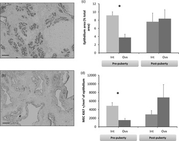
Wallace (Reference Wallace1953) was the first to demonstrate the involvement of ovarian steroids in the development of the ruminant mammary gland at puberty (Wallace, Reference Wallace1953). He showed in the calf that the pre-pubertal removal of the main source of estradiol and progesterone by ovariectomy greatly altered mammogenesis. Normal development was found when heifers received estradiol supplementation. These observations have been confirmed on several occasions, including by Purup et al. (Reference Purup, Sejrsen, Foldager and Akers1993), who found that parenchymal mass and DNA concentration in heifers ovariectomized before puberty were five times lower than those measured in intact heifers (Purup et al., Reference Purup, Sejrsen, Foldager and Akers1993). Control of the development of the mammary parenchyma by ovarian steroids mainly occurs through the modulation of MEC proliferation. The incorporation of thymidine into the nuclei of MEC ducts is greatly increased (46 times) at 96 h after injection of estradiol, but this effect is not observed after injection of progesterone (Woodward et al., Reference Woodward, Beal and Akers1993). More recently, it was shown that cell proliferation in the mammary gland of heifers ovariectomized at the age of 2.5 months is 10 times lower than intact heifers (Berry et al., Reference Berry, Jobst, Ellis, Howard, Capuco and Akers2003a), leading to an 85% to 90% reduction of the development of mammary parenchyma in heifers slaughtered at 9 months of age (Purup et al., Reference Purup, Sejrsen and Akers1995). Ovariectomy in pre-pubescent heifers also leads to changes in the distribution of several components of the extracellular matrix in the mammary gland (Berry et al., Reference Berry, Weber Nielsen, Sejrsen, Pearson, Boyle and Akers2003b). The extracellular matrix is composed primarily of laminin, proteoglycans, fibronectin, tenascin and collagens I and IV. These components are synthesized by different cell types present in the mammary gland. The extracellular matrix plays an essential role in MEC proliferation. In vitro, estradiol can induce the proliferation or extracellular matrix synthesis of MECs when they are co-cultured with stromal cells (Haslam and Woodward, Reference Haslam and Woodward2001). In goats, similar to the heifer, the development of the mammary gland is greatly altered by ovariectomy when performed before puberty. Indeed, ovariectomized goats 1 to 3 months after birth present, at 9 months of age, undeveloped and poorly organized epithelial structures, as well as cell proliferation and tissue remodeling lower than what is measured at the same age in intact goats (Figure 3). The hormonal response to ovariectomy varies depending on the species considered; in the heifer, removing the main source of estradiol and progesterone induces overexpression of the α form of the estradiol receptor (Berry et al., Reference Berry, Jobst, Ellis, Howard, Capuco and Akers2003a), whereas in the goat, this expression is reduced (Dessauge et al., Reference Dessauge, Finot, Wiart, Aubry and Ellis2009; Yart et al., Reference Yart, Finot, Marnet and Dessauge2012a). Thus, although the mechanisms involved in the control of mammary gland development differ in some aspects, the role of ovarian steroids in mammogenesis seems to be confirmed in cattle and goats, and more generally in most mammals. This is not the case for sheep, in which mammogenesis at puberty occurs independently of ovarian secretions (Ellis et al., Reference Ellis, McFadden and Akers1998).
Role of ovarian secretions in the mammary gland development at puberty. Histological sections of mammary gland obtained at the age of nine months in a intact goat (a) and in goat ovariectomized at 1 month of age (b) (scale bar=100μm). Ovariectomy before puberty alters the development of the mammary epithelium (c) and the proliferation of MECs, highlighted by immunohistochemical staining for Ki67 (d), but has no effect on these two parameters after puberty. *P<0.05. Int=intact goats (n=15); Ovx=ovariectomized goats (n=15). (Yart et al., Reference Yart, Finot, Marnet and Dessauge2012a ).

Factors involved in the development of the mammary gland have local but also systemic actions. The interactions between these hormones and their signaling pathways are complex. It is therefore difficult to determine whether their actions on the mammary gland are the result of direct stimulation or other organs. However, it is now known that the stroma and adipose tissue (mammary gland fat pad) not only form an inert matrix but also play an important role in the establishment of epithelial structures. Early studies highlighted the importance of the mammary fat pad via murine mammary epithelial explant transplantation in various organs (Deome et al., Reference Deome, Faulkin, Bern and Blair1959; Hoshino, Reference Hoshino1978). Hoshino (Reference Hoshino1978) observed that mammary epithelium explants grew normally after transplantation in perirenal adipose tissue, but not after transplantation in the peritoneal cavity or in the anterior chamber of the eye (Hoshino, Reference Hoshino1978). The mammary fat pad plays a role in both proliferation and differentiation of MECs. It is an active site for the hormones involved in the development of the mammary gland, especially for ovarian steroids and GH, and participates in the transmission of these hormonal messages. Indeed, Capuco et al. (Reference Capuco, Ellis, Wood, Akers and Garrett2002a) showed that proliferating MECs do not express receptors for ovarian steroids, but these receptors, like GH receptors, are expressed in the stroma (Akers et al., Reference Akers, Beal, McFadden and Capuco1990; Capuco et al., Reference Capuco, Li, Long, Ren, Hruska, Schorr and Furth2002b; Meyer et al., Reference Meyer, Capuco, Boisclair and Van Amburgh2006). The fat pad responds to these hormonal stimuli by synthesizing various growth factors that have mitogenic actions, such as IGFs, fibroblast growth factors or hepatocyte growth factor (Hovey et al., Reference Hovey, McFadden and Akers1999). Thus, administration of estradiol in heifers increases the expression of IGF-I and decreases the expression of IGFBP-3 in the fat pad (Berry et al., Reference Berry, McFadden, Pearson and Akers2001; Meyer et al., Reference Meyer, Capuco, Boisclair and Van Amburgh2006). This increased expression of IGF-I after administration of estradiol is accompanied by a significant increase in MEC proliferation in the mammary gland and an 80% reduction in the expression of estrogen receptor α (ERα) in these cells (Meyer et al., Reference Meyer, Capuco, Boisclair and Van Amburgh2006).
The adult mammary gland
Development of the mammary gland in adults is a fascinating process because during the life of the animal, the mammary gland will undergo many changes in terms of size, structure, composition and activity. This developmental cycle is directly modeled on the reproductive cycle. The developmental cycle, which is initiated by pregnancy, is divided into four phases that partly overlap: mammogenesis (lobulo-alveolar growth), lactogenesis, galactopoiesis and involution.
Although mammogenesis is decisive for subsequent lactations, this phase mainly consists of the establishment of the mammary duct network. The vast majority (60% to 94%, depending on the species) of lobulo-alveolar development occurs during the first pregnancy (Knight and Peaker, Reference Knight and Peaker1982). Lobulo-alveolar structures continue to grow by increasing in size and complexity. During pregnancy, the growth of the mammary gland results in a proportional increase in the parenchyma rather than the fat pad until the cell density is such that the lobes and lobules are separated only by septa composed of connective tissue. This phase of mammogenesis occurs under the action of sex steroids (estrogen and progesterone), which are secreted by the ovaries and by the placental system, combined with pituitary hormones (GH and Prl). Denamur and Martinet (Reference Denamur and Martinet1961) showed that hypophysectomy of pregnant ewes has little impact on the development of the mammary gland. In contrast, administration of placental extracts associated with steroid hormones in virgin ovariectomized and hypophysectomized female rats induces mammary gland development (Ray et al., Reference Ray, Averill, Lyons and Johnson1955).
Estrogen and progesterone secreted during pregnancy have a proliferative effect on MECs (Clarke, Reference Clarke2000). Thus, in rats, the amount of total DNA in mammary tissue (reflecting the number of cells) increases by 200% to 300% during gestation (Knight and Peaker, Reference Knight and Peaker1982). Studies in mice whose estrogen receptor genes have been invalidated have shown that estrogen acts mainly on the development of mammary ducts, while progesterone has a key role in lobulo-alveolar development (Atwood et al., Reference Atwood, Hovey, Glover, Chepko, Ginsburg, Robison and Vonderhaar2000; Aupperlee and Haslam, Reference Aupperlee and Haslam2007). However, estrogen indirectly controls lobulo-alveolar development, insofar as the expression of different forms of PR is under the control of estrogen (Petz et al., Reference Petz, Ziegler, Schultz, Kim, Kemper and Nardulli2004). In addition, during pregnancy, cells expressing PR are very rare (Brisken et al., Reference Brisken, Heineman, Chavarria, Elenbaas, Tan, Dey, McMahon, McMahon and Weinberg2000), and in women, 96% of MECs expressing ERα express PR and are also non-proliferative (Anderson et al., Reference Anderson, Clarke and Howell1998). These results suggest that the proliferative actions of progesterone on MECs occur through a paracrine pathway between epithelial cells.
At the end of gestation, increased mammary size is mainly due to MEC hyperplasia and expansion of the alveoli. MECs undergo differentiation and acquire the physical and biochemical capacities to synthesize the various constituents of milk under the action of the pituitary hormones Prl and GH. The hormonal regulation aspects during lactation will be developed later.
A lactation cycle ends with the involution of the mammary gland. This phase corresponds to the gradual regression of secretory tissue, which returns to a state of development that is slightly more advanced than it was before the beginning of the first pregnancy. Involution begins after young are completely weaned and milking ceases, and it induces a reduction in the secretion of galactopoietic hormones and the accumulation of milk in the udder (Lamote et al., Reference Lamote, Meyer, Massart-Leen and Burvenich2004). In cows as well as goats, the earliest phase of involution is reversible. At a later stage, the phenomenon of programmed cell death (apoptosis) is amplified, and the extracellular matrix is degraded by metalloproteinases, leading to a loss of almost all MECs (Stefanon et al., Reference Stefanon, Colitti, Gabai, Knight and Wilde2002). Schams et al. (Reference Schams, Kohlenberg, Amselgruber, Berisha, Pfaffl and Sinowatz2003) showed a change in the regulation of the expression of ER and PR in the bovine mammary gland during involution (Schams et al., Reference Schams, Kohlenberg, Amselgruber, Berisha, Pfaffl and Sinowatz2003). In adults, the expression of these receptors peaks 2 to 4 weeks after the onset of involution, suggesting that estrogen and progesterone are involved in the regulation of involution. Several studies have examined the effect of estradiol in cows in mid- to late lactation. The administration of estradiol induces a significant decrease in milk production (Mollett et al., Reference Mollett, Erb, Monk and Malven1976; Athie et al., Reference Athie, Bachman, Head, Hayen and Wilcox1996; Delbecchi et al., Reference Delbecchi, Miller, Prud’homme, Petitclerc, Wagner and Lacasse2005). This decline is associated with a decrease in mammary volume (Mollett et al., Reference Mollett, Erb, Monk and Malven1976), an increase in milk stanniocalcin (Delbecchi et al., Reference Delbecchi, Miller, Prud’homme, Petitclerc, Wagner and Lacasse2005) and a change in milk composition (Athie et al., Reference Athie, Bachman, Head, Hayen and Wilcox1996). This phase of involution is necessary before starting a new lactation cycle. Indeed, in the cow, without an intervening involution phase, milk production is much lower than normal during the next lactation (Capuco et al., Reference Capuco, Ellis, Hale, Long, Erdman, Zhao and Paape2003), suggesting that MECs have a limited lifespan and the mammary gland has a strong ability to regenerate. This de novo mammogenesis is essential for proper functioning of the mammary gland. Secretory tissue regeneration with each new pregnancy occurs with the recruitment of pluripotent stem cells that are present in the mammary gland.
The lactation persistency
Evolution of milk production during lactation
During lactation, from parturition to dry-off, the amount of milk produced by the mammary gland changes, following a curve. Immediately after parturition, milk production increases rapidly to reach a peak of production between 6 and 8 weeks of lactation (Knight and Peaker, Reference Knight and Peaker1984). It then follows a phase of decline in milk production, during which the quantity of milk produced by the mammary gland will gradually decrease until milk secretion completely ceases. The speed at which milk production declines after peak lactation is the factor that characterizes lactation persistency. Graphically, the milk production curve has a variable but always negative slope. Several computational models have been proposed to compare the persistency of lactation. Sölkner and Fuchs (Reference Sölkner and Fuchs1987) tested the suitability of three models at different stages of lactation. The persistency was calculated for 305-day lactation cycles, using the first 100 days of lactation as the reference period and two periods of interest: 101 to 200 days of lactation and 201 to 300 days of lactation (Sölkner and Fuchs, Reference Sölkner and Fuchs1987). Following this study, Sölkner and Fuchs (Reference Sölkner and Fuchs1987) found that standard deviations and the ratio of the period of interest for milk production to the reference period of milk production were the most appropriate methods for calculating persistency. They also concluded that this calculation was more relevant if it included the end of lactation. The amount of milk produced by the mammary gland at any given time depends on the number and activity of MECs, as well as the organization of secretory tissue. The evolution of milk production during lactation is actually the result of a joint evolution of these three parameters.
Knight and Peaker (Reference Knight and Peaker1984) have compared the evolution of milk production and the development of mammary tissue during lactation in the goat. To this end, they took several mammary biopsies at different stages of lactation and compared the number and activity of mammary cells between these different stages. They demonstrated that increased milk production in early lactation is primarily due to an increase in the number of cells and the activity of these cells. After the peak of lactation, between the 8th and 23rd weeks of lactation, the decline of milk production is mainly due to a decrease in the number of cells, while at a later stage, this decrease is intensified by a decrease in the secretory activity of the cells. Similar observations were made in the bovine mammary gland after the slaughter of cows at different stages of lactation (Capuco et al., Reference Capuco, Wood, Baldwin, McLeod and Paape2001). In contrast, according to Capuco et al. (Reference Capuco, Wood, Baldwin, McLeod and Paape2001), increased milk production in early lactation is mainly due to increased secretory cell activity and not due to massive MEC proliferation (Capuco et al., Reference Capuco, Wood, Baldwin, McLeod and Paape2001). Capuco et al. (Reference Capuco, Wood, Baldwin, McLeod and Paape2001) also noted that the rate of cell proliferation was relatively stable during lactation (0.3%), and thus changes in MEC numbers were mainly due to modulations in the rate of apoptosis (Capuco et al., Reference Capuco, Wood, Baldwin, McLeod and Paape2001). Stefanon et al. (Reference Stefanon, Colitti, Gabai, Knight and Wilde2002) set forth a proposal concerning the cellular mechanisms involved in the evolution of the milk production during lactation. They described a slight amount of cellular proliferation in early lactation associated with a low rate of apoptosis and tissue remodeling, resulting in an increase in the number of functional MECs and an increase in milk production. In one study on the goat mammary gland, the amount of DNA increased from 3.21 to 4.06 mg/g tissue between the first and third weeks of lactation, reflecting an increase in the number of cells (Knight and Peaker, Reference Knight and Peaker1984). In mid-lactation, cell proliferation decreases and the apoptosis rate increases slightly, leading to decreases in MEC number, cell and mammary size and milk production. Finally, at the time of mammary involution, the apoptosis rate is very high, and the intensity of tissue remodeling increases. Generally, during mammary involution, the apoptosis rate is significantly higher than the rate of cell proliferation, which results in a decrease in MECs, associated with significant tissue remodeling through the actions of matrix metalloproteases (MMPs) and alveolar regression. The rate of cell proliferation is more or less important depending on whether the animal is gestating or lactating. In dairy cattle, lactating cows are commonly inseminated and pregnant during the second half of the lactation. In this case, the mammary gland already begins to regenerate to ensure the next lactation through the recruitment and proliferation of new undifferentiated MECs under the action of pregnancy hormones. It is also interesting to note that it seems that the mechanisms involved in the modulation of the proliferation/apoptosis balance in the goat are different from those observed in cows. Indeed, although the goat mammary gland undergoes a loss of MECs during lactation (Knight and Peaker, Reference Knight and Peaker1984), Linzell (Reference Linzell1973) reported that goats can remain non-pregnant and lactating for 2 to 4 years if they are milked twice a day. Thus, goats may have naturally good persistency, suggesting that loss of apoptotic MECs may be slower than in the bovine mammary gland.
Factors influencing lactation persistency
Lactation persistency is strongly related to breed and parity. Indeed, the lactation curve of Holstein cows with a high dairy potential has a bell shape, while the lactation curve of Jersey cows with smaller dairy potential is much flatter. Jersey cows therefore have better persistency than Holstein cows. Persistency is not related to the level of milk production but the ability of the cow to maintain steady milk production. The principle is the same with respect to parity. In a first lactation, secretory structures in the mammary gland are not yet fully mature, so the lactation potential continues to increase after the peak of lactation, making the lactation curve flatter. During subsequent lactations, the level of production at the peak of lactation increases and the apparent persistency decreases. In cows, lactation persistency is thought to stabilize after the third lactation (Schutz et al., Reference Schutz, Hansen, Steuernagel and Kuck1990). Several physiological and environmental factors can modulate lactation persistency by influencing the proliferation/apoptosis balance and tissue remodeling in the mammary gland. Factors such as feeding level, milking frequency, stage of gestation and the health status of the animal are directly related to farming practices. In the short term, these factors affect the level of milk production, but if the animal is not fit in the longer term, these factors may affect lactation persistency. Other factors, such as photoperiod, are related to the environment but can be integrated into farming practices to modulate lactation persistency. Photoperiod has an effect on milk production (Peters et al., Reference Peters, Chapin, Emery and Tucker1981; Marcek and Swanson, Reference Marcek and Swanson1984; Miller et al., Reference Miller, Stanisiewski, Erdman, Douglass and Dahl1999; Dahl et al., Reference Dahl, Buchanan and Tucker2000). Passage from a short-day photoperiod to a long-day photoperiod (i.e. >16 h of light/day) increases milk production. The exposure of lactating cows to 16 h of light per day can increase the daily MY by 6.7% compared with cows subjected to a natural photoperiod (Peters et al., Reference Peters, Chapin, Emery and Tucker1981). Prl, whose secretion is reduced by melatonin (Auldist et al., Reference Auldist, Turner, McMahon and Prosser2007), plays an intermediary role in the increase of milk production in response to a long-day photoperiod. Dahl et al. (Reference Dahl, Elsasser, Capuco, Erdman and Peters1997) also noted that the increase in milk production induced by long-day photoperiods is associated with an increase in plasma IGF-I, a hormone known for its galactopoietic action (Dahl et al., Reference Dahl, Elsasser, Capuco, Erdman and Peters1997). However, the plasma concentration of IGF-I is not influenced by the administration of melatonin (Auldist et al., Reference Auldist, Turner, McMahon and Prosser2007). In livestock, it is possible to exploit this photoperiod effect by applying a light treatment (Morrissey et al., Reference Morrissey, Cameron and Tilbrook2008) or by programming delivery in winter. In this manner, the beginning of lactation (when Prl secretion is highest) takes place during the declining phase of the photoperiod, and the declining phase of milk production (the phase during which hormone secretion decreases galactopoiesis) takes place at the bottom of the photoperiod, thus limiting the decrease in Prl secretion. Sorensen and Knight (Reference Sorensen and Knight2002) studied the effect of the season of parturition (calving in winter or spring) on lactation persistency and the plasma concentrations of GH, IGF-I and Prl (Sorensen and Knight, Reference Sorensen and Knight2002). Although the authors did not observe any effect of season of calving on the concentration of IGF-I, they did show that cows calved in winter had greater GH than cows calved in spring, and in these cows, Prl was also much more stable during lactation. These differences in the patterns of galactopoietic hormone secretion were associated with greater lactation persistency in cows calved in winter. Lactation persistency is also strongly related to the health status of the animal and particularly the health of the mammary gland. Processes related to bacterial infection of the udder or mastitis, as well as the action of neutrophils in response to infection in the mammary gland, result in an increased rate of cell death in the mammary gland, which affects lactation persistency if the loss of MECs is important. Long et al. (Reference Long, Capuco, Wood, Sonstegard, Tomita, Paape and Zhao2001) noted that, following the induction of mastitis by inoculation of Escherichia coli into the mammary gland of lactating cows, there is an increase in the degradation of extracellular matrix by MMPs and increased expression of proapoptotic marker Bax associated with decreased expression of the anti-apoptotic marker Bcl-2. Mastitis in these cows also increased the number of cells expressing the proliferation marker Ki67. Taken together, these results suggest that a bacterial infection of the udder initially induces a loss of MECs in areas of infected tissue followed by a renewal of these cells by proliferation.
In dairy farms, milk production can be modulated by short-term milking frequency and diet, which have implications for lactation persistency. These two factors quickly affect the amount of milk produced by the mammary gland. Increased milking frequency (three or four milkings per day) induces an increase in milk production, and in the same way, a decrease in milking frequency (one milking per day) induces a decrease in milk production. In dairy cows, these changes affect milk production and lactation persistency after a return to two milkings per day (Hale et al., Reference Hale, Capuco and Erdman2003; Bernier-Dodier et al., Reference Bernier-Dodier, Delbecchi, Wagner, Talbot and Lacasse2010). Dietary restriction also significantly alters milk production. Thus, cows subjected to a restrictive diet (−20% net energy) have a lower daily milk production by 9.8 kg (Norgaard et al., Reference Norgaard, Sorensen, Theil, Sehested and Sejrsen2008) or 13 kg (Dessauge et al., Reference Dessauge, Lollivier, Ponchon, Bruckmaier, Finot, Wiart, Cutullic, Disenhaus, Barbey and Boutinaud2011) compared with cows receiving a basal diet. Milk production cuts that result from a decrease in the frequency of milking (Bernier-Dodier et al., Reference Bernier-Dodier, Delbecchi, Wagner, Talbot and Lacasse2010) or the application of nutrient restriction (Dessauge et al., Reference Dessauge, Lollivier, Ponchon, Bruckmaier, Finot, Wiart, Cutullic, Disenhaus, Barbey and Boutinaud2011) are associated with an increased rate of apoptosis in the weeks following treatment application as well as tissue remodeling by MMPs and decreases in the expression of milk proteins and in the concentration of IGF-I. A decrease in milking frequency also induces an increase in cell proliferation, which is not the case for dietary restriction (Dessauge et al., Reference Dessauge, Lollivier, Ponchon, Bruckmaier, Finot, Wiart, Cutullic, Disenhaus, Barbey and Boutinaud2011). If the stress induced by the treatment is too large, the animal cannot adapt, and the loss will affect MEC persistency. However, the mechanisms involved in the modulation of milk production level by milking frequency appear to be substantially different from those that depend on nutrition. A decrease or increase in milking frequency alters IGF-I but not GH level (Hale et al., Reference Hale, Capuco and Erdman2003), while dietary restriction induces an increase in plasma GH (Elsasser et al., Reference Elsasser, Rumsey and Hammond1989; Dessauge et al., Reference Dessauge, Lollivier, Ponchon, Bruckmaier, Finot, Wiart, Cutullic, Disenhaus, Barbey and Boutinaud2011). In addition, differences in milking frequency for two half-udders on the same cow (i.e. two-quarters milked one time per day and two-quarters milked three times daily) do not increase the concentration of IGF-I in the milk from the quarters milked once per day (Bernier-Dodier et al., Reference Bernier-Dodier, Delbecchi, Wagner, Talbot and Lacasse2010). These results suggest that the influence of milking frequency on lactation persistency mostly involves extramammary factors, including primarily systemic factors such as changes in the secretion of GH.
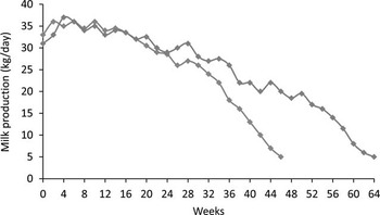
Several studies have examined the effect of pregnancy on lactation persistency. Taken together, these studies show that gestation has a negative effect on lactation persistency (Bachman et al., Reference Bachman, Hayen, Morse and Wilcox1988; Bertilsson et al., Reference Bertilsson, Berglund, Ratnayake, Svennersten Sjaunja and Wiktorsson1997; Sorensen and Knight, Reference Sorensen and Knight2002; Norgaard et al., Reference Norgaard, Sorensen, Theil, Sehested and Sejrsen2008). The point at which pregnancy alters milk production seems to be related to the stage of gestation and not the advancement of lactation because a significant decrease in milk production is observed between 100 and 200 days of gestation (Bachman et al., Reference Bachman, Hayen, Morse and Wilcox1988; Bertilsson et al., Reference Bertilsson, Berglund, Ratnayake, Svennersten Sjaunja and Wiktorsson1997), regardless of the progression of lactation (Bertilsson et al., Reference Bertilsson, Berglund, Ratnayake, Svennersten Sjaunja and Wiktorsson1997). This stage of gestation coincides precisely with the onset of estrogen secretion by the fetoplacental system (Patel et al., Reference Patel, Takenouchi, Takahashi, Hirako, Sasaki and Domeki1999), suggesting that the decline in milk production that is observed is due to the increased levels of circulating estrogens in maternal blood. The administration of estradiol in lactating cows also induces a significant drop in milk production (Mollett et al., Reference Mollett, Erb, Monk and Malven1976; Athie et al., Reference Athie, Bachman, Head, Hayen and Wilcox1996; Delbecchi et al., Reference Delbecchi, Miller, Prud’homme, Petitclerc, Wagner and Lacasse2005). Additionally, it appears that pregnancy and therefore the hormones secreted by the fetoplacental system reduce cell proliferation in the lactating mammary gland but do not affect apoptosis (Norgaard et al., Reference Norgaard, Sorensen, Theil, Sehested and Sejrsen2008). In the majority of dairy systems today, the interval between calving is 12 months, with a lactation period of ∼10 months; cows are inseminated between the second and third months of lactation, with a drying-off period of ∼2 months before calving. Because the length of gestation in cows is 9 months, while the interval between calvings is 12 months, the cow is lactating and pregnant for 7 months (Figure 4). When insemination is performed at a more advanced stage of lactation to have an interval of 18 months between two calvings, lactation persistency is significantly improved (Bertilsson et al., Reference Bertilsson, Berglund, Ratnayake, Svennersten Sjaunja and Wiktorsson1997).
Evolution of milk production in dairy cows with a calving interval of 12 months (n=45, blue curve) or 18 months (n=45, red curve). From Bertilsson et al. (Reference Bertilsson, Berglund, Ratnayake, Svennersten Sjaunja and Wiktorsson1997)

Ovarian steroids and the mammary gland
Ovarian steroids influence the proliferation/apoptosis balance of MECs
Recent studies conducted on non-pregnant lactating cows have highlighted the effects of removing the main source of estradiol and progesterone in lactating cows by ovariectomy at the time of the peak of lactation (Yart et al., Reference Yart, Dessauge, Finot, Barbey, Marnet and Lollivier2012b). These studies, which examined the secretory tissue of the mammary gland at four time points spread on the whole lactation, showed a lower rate of apoptosis in late lactation and an increase in cell proliferation just after the peak of lactation in ovariectomized cows. When working on a model of ovariectomy, it is necessary to keep in mind that this procedure not only removes the main source of estradiol and progesterone but is also capable of inducing a hormonal imbalance that may affect other endocrine axes. Other factors, such as inhibin, activin, bone morphogenic proteins and factors of the TGF-β family, are produced by the ovaries and released into the circulation (Forde et al., Reference Forde, Beltman, Lonergan, Diskin, Roche and Crowe2011). This is also the case for oxytocin, which is a galactopoietic hormone. It is often believed that it is mainly oxytocin from the pituitary that acts on the mammary gland to stimulate milk synthesis and ejection. However, a significant amount of oxytocin is produced continuously in the ovaries in the early luteal phase of the corpus luteum (Flint et al., Reference Flint, Sheldrick, Theodosis and Wooding1986). In sheep, ovarian oxytocin can act on the mammary gland through super-ovulation by inducing a significant increase in milk production (Labussière et al., Reference Labussière, Marnet, Combaud, Beaufils and de la Chevalerie1993). Few studies have examined the effect of progesterone on the mammary gland. Moreover, it appears that the role of estradiol is predominant over that of progesterone in the modulation of milk production level (Mollett et al., Reference Mollett, Erb, Monk and Malven1976) and in the dynamic control of MECs (Zarzynska et al., Reference Zarzynska, Gajewska and Motyl2005; Sobolewska et al., Reference Sobolewska, Gajewska, Zarzynska, Gajkowska and Motyl2009).
The involvement of estradiol in the increasing rate of apoptosis that is measured in intact cows compared with ovariectomized cows has been suggested in several in vitro studies. Studies conducted on MAC-T and BME-UV1 cells have highlighted an increase in the caspase 3 activation (Sobolewska et al., Reference Sobolewska, Gajewska, Zarzynska, Gajkowska and Motyl2009 and Reference Sobolewska, Motyl and Gajewska2011) and increased TGF-β expression, a factor involved in the induction of apoptosis in mammary involution (Zarzynska et al., Reference Zarzynska, Gajewska and Motyl2005), in response to estradiol treatment.
During lactation, the MEC environment changes. The initiation of secretory tissue development occurs under the action of high levels of progesterone and estradiol (Clarke, Reference Clarke2000), and lactation is initiated and maintained by many hormones (Prl, GH, oxytocin, thyroid hormones and glucocorticoids), which stimulate the synthesis of various milk components and act as survival factors for MECs. In early lactation, the blood flow in the mammary gland is important and can provide large amounts of the elements needed for milk synthesis. As lactation progresses, the amount of hormonal secretions and blood flow in the mammary gland decrease (Svennersten-Sjaunja and Olsson, Reference Svennersten-Sjaunja and Olsson2005), which helps to increase the rate of apoptosis and the decline in milk production after the peak of lactation. Recently, Yart et al. (Reference Yart, Dessauge, Finot, Barbey, Marnet and Lollivier2012b) demonstrated that the expression of ERα in the mammary parenchyma increases during lactation and is decreased by ovariectomy in late lactation. This result suggests that the sensitivity of MECs to estradiol increases with advancing lactation and is related to MY. Arguments from the literature support this hypothesis of increased sensitivity to estradiol in connection with a change in the phenotype of MECs. In the heifer, it seems that proliferating cells do not express ERα within the mammary gland (Capuco et al., Reference Capuco, Ellis, Wood, Akers and Garrett2002a). Thus, the acquisition of estradiol receptivity would occur during MEC maturation. In vitro studies have demonstrated that estradiol accelerates the mechanisms of apoptosis and increases the rate of apoptosis in pre-apoptotic MECs. By varying the composition of the culture medium, it is possible to mimic the deprivation of galactopoietic factors (included hormonal and survival factors) and nutrients that gradually takes place in the lactating mammary gland. Zarzynska et al. (Reference Zarzynska, Gajewska and Motyl2005) have observed an increase in the expression of TGF-β and the apoptosis rate due to a restriction of the concentration of fetal calf serum in the culture medium (Zarzynska et al., Reference Zarzynska, Gajewska and Motyl2005). Another in vitro method to mimic senescent MECs in the mammary gland is to place MEC in culture with a medium is rich in Prl and GH that stimulates differentiation and maturation (Huynh et al., Reference Huynh, Robitaille and Turner1991; Zhou et al., Reference Zhou, Akers and Jiang2008). In vivo, these two hormones are essential for the acquired ability to synthesize milk components by MECs. Indeed, even if GH mostly has a proliferative effect on MECs, this hormone is essential for the initiation of lactation (Annen et al., Reference Annen, Fitzgerald, Gentry, McGuire, Capuco, Baumgard and Collier2007). Prl stimulates MEC differentiation and induces the synthesis of various milk components (Akers et al., Reference Akers, Bauman, Capuco, Goodman and Tucker1981a and Reference Akers, Bauman, Goodman, Capuco and Tucker1981b). Placing the cells in such conditions would promote differentiation and then aging.
Ovariectomy alters the interactions of MECs with other cells and the extracellular matrix
In the early 1990s, Woodward (Reference Woodward1991) focused on the effect of ovarian steroids on MEC proliferation during mammogenesis in the heifer. One of his first steps was to inject pharmacological doses of estradiol, progesterone or estradiol combined with progesterone into pre-pubertal heifers. He showed that estradiol stimulates MEC proliferation combined or not with progesterone and that progesterone has no effect on proliferation. The second phase of his work was to investigate the in vitro effects of these two steroids on the proliferation of the MAC-T cell line. Woodward showed no proliferative effect of progesterone or estradiol on MECs. From these studies, it was concluded that ovarian steroids, particularly estradiol, indirectly stimulate the proliferation of MECs in the developing bovine mammary gland. It appears that, in cattle, paracrine communications play a crucial role in the control of mammary development by ovarian steroids. In the heifer, 99% of MECs, which proliferate in response to the administration of estradiol, do not express ERα during mammogenesis (Capuco et al., Reference Capuco, Ellis, Wood, Akers and Garrett2002a). These results confirm the Woodward (Reference Woodward1991) hypothesis that MEC proliferation within the bovine mammary gland in response to estradiol is indirectly initiated by cells expressing ERα via a paracrine signal.
Many studies in the heifer have provided evidence demonstrating the involvement of stroma and, more specifically, adipocytes in estrogen signal transmission during mammogenesis (Capuco et al., Reference Capuco, Ellis, Wood, Akers and Garrett2002a; Meyer et al., Reference Meyer, Capuco, Boisclair and Van Amburgh2006; Connor et al., Reference Connor, Meyer, Li, Van Amburgh, Boisclair and Capuco2007). However, to our knowledge, no study has yet investigated paracrine communication signals linked to estrogen and progesterone in the lactating mammary gland. We have studied the expression of ovarian steroid receptors not only in the parenchyma but also in mammary adipose tissue. Adipose tissue volume is greatly reduced in lactating mammary glands from ovariectomized cows and is mainly located under the skin. The vast majority of mammary volume during lactation is occupied by the secretory parenchyma. This is most likely why adipose tissue has attracted so little interest from different research teams working on mammary gland function. Quantification of the ERα protein in the mammary parenchyma at different stages of lactation has shown that the sensitivity of the secretory tissue to estradiol increases during lactation. Ovariectomy induces a decrease in the expression of ERα in late lactation in the parenchyma and mammary adipose tissue (Yart et al., Reference Yart, Lollivier, Finot, Dupont, Wiart, Boutinaud, Marnet and Dessauge2013b). Recently, we investigate the differential effect of estradiol on bovine MECs mimicking two physiological statuses: active and pre-apoptotic MECs. We demonstrate that estradiol has a major effect on pre-apoptotic MECs and might accelerate MEC apoptosis by caspases activation rather than inducing apoptosis in active MECs. Pre-apoptotic MECs could be compared with senescent cells in the late-lactation mammary gland (Yart et al., Reference Yart, Finot, Lollivier and Dessauge2013a). The results obtained in this study about the evolution of mammary parenchyma sensitivity to ovarian steroids are consistent with those reported by Schams et al. (Reference Schams, Kohlenberg, Amselgruber, Berisha, Pfaffl and Sinowatz2003). Thus, the decrease in ERα level measured at the end of lactation in ovariectomized cows was not related to a downregulation of the expression of this receptor but rather to a decrease in the proportion of cells expressing ERα. Immunohistological staining for ERα performed on sections of mammary parenchyma taken at slaughter supports this hypothesis: the proportion of ERα-positive cells in the mammary parenchyma from ovariectomized cows is five times lower than that measured in control cows.
Ovariectomy not only changes the proliferation/apoptosis balance by reducing apoptosis, but it also reduces the intensity of tissue remodeling. In the lactating mammary gland, tissue remodeling intensity can be measured through the activity of MMPs and other gelatinases released into milk. Degradation of the extracellular matrix by MMPs increases sharply in late lactation and is essential for mammary involution (Stefanon et al., Reference Stefanon, Colitti, Gabai, Knight and Wilde2002). Ovarian steroids, including estradiol, accelerate the process of involution (Athie et al., Reference Athie, Bachman, Head, Hayen and Wilcox1996) and can stimulate the expression and activity of certain gelatinases (Ambili et al., Reference Ambili, Jayasree and Sudhakaran1998). Athie et al. (Reference Athie, Bachman, Head, Hayen and Wilcox1996) used several markers to study the effect of estradiol treatment on mammary involution in Holstein cows. They reported decreases in α-lactalbumin, lactose and mineral concentrations in mammary secretions as well as increases in somatic cell concentrations, lactoferrin and sodium concentrations. Welty et al. (Reference Welty, Smith and Schanbacher1976) have demonstrated an increase in the concentration of lactoferrin in milk from the second day of involution (Welty et al., Reference Welty, Smith and Schanbacher1976), although it appears that this increase does not become significant until the 11th day of involution (Hurley, Reference Hurley1989), and its expression in the mammary parenchyma increases only after 8 days of involution (Singh et al., Reference Singh, Davis, Dobson, Molenaar, Wheeler, Prosser, Farr, Oden, Swanson, Phyn, Hyndman, Wilson, Henderson and Stelwagen2008). Moreover, at the end of lactation, milk composition changes: the number of somatic cells increases and the total protein concentration also increases, resulting from decreases in the levels of casein and α-lactalbumin with increases in lactoferrin and N-acetyl-β-glucosaminidase (Hurley, Reference Hurley1989). The concentration of stanniocalcin-1 (STC-1) is increased in milk during lactation (Miller et al., Reference Miller, Delbecchi, Petitclerc, Wagner, Talbot and Lacasse2006) and in mammary secretions after drying-off (Tremblay et al., Reference Tremblay, Bernier-Dodier, Delbecchi, Wagner, Talbot and Lacasse2009). An increase in STC-1 concentration in milk also accompanied the decrease in MY that followed the administration of estradiol in lactating cows (Delbecchi et al., Reference Delbecchi, Miller, Prud’homme, Petitclerc, Wagner and Lacasse2005). In mammals, this hormone is involved in calcium homeostasis, but it also seems to stimulates apoptosis of MECs in milk. The in vitro treatment of the MAC-T cell line with mammary secretions collected after drying-off, and therefore rich in STC-1, induces an increase in apoptosis (Tremblay et al., Reference Tremblay, Bernier-Dodier, Delbecchi, Wagner, Talbot and Lacasse2009). In our studies conducted on ovariectomized lactating cows (Yart et al., Reference Yart, Dessauge, Finot, Barbey, Marnet and Lollivier2012b), we showed that the ovariectomy reduced serum albumin and lactoferrin concentrations in milk, while the α-lactalbumin concentration remained not significantly affected. Taken together, these results suggest that the removal of the main source of estradiol and progesterone allows the process of mammary involution to slow down.
Ovarian steroids modulate lactation persistency
The removal of ovarian secretions during lactation improves lactation persistency, limiting the drop in MY after the peak of lactation. As we discussed above, although estradiol and progesterone are not the only molecules released by active ovaries, the literature suggests that these two steroids, especially estradiol, negatively influence MY and lactation persistency in dairy cows (Mollett et al., Reference Mollett, Erb, Monk and Malven1976; Athie et al., Reference Athie, Bachman, Head, Hayen and Wilcox1996; Delbecchi et al., Reference Delbecchi, Miller, Prud’homme, Petitclerc, Wagner and Lacasse2005).
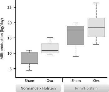
Lactation persistency is dependent on breed and lactation rank. It is assumed that lactation persistency becomes stable after the third lactation (Schutz et al., Reference Schutz, Hansen, Steuernagel and Kuck1990). Various in vivo studies have been conducted on cows of different breeds (Holstein×Normande, Holstein) and different lactation ranks. In our study, the analysis of covariance data for lactation persistency calculated for the periods from 100 to 200 days of lactation (P100-200) and 200 to 300 days of lactation (P200-300) did not reveal any significant effect on lactation rank (P=0.62, 0.58, respectively). However, a strong effect of breed was found (P100-200: P<0.0001; P200-300: P<0.05). Holstein cows have better persistency than Normande×Holstein cows, regardless of treatment and parity. The graphical representation of the distribution of means for individual MY over the last 10 weeks of lactation for ovariectomized cows also illustrates the variability between the two breeds (or cross breeds) studied. It appears, in fact, that individual responses to treatment are different between the two breeds (Figure 5). Median differences between ovariectomized cows and control cows are greater in Normande×Holstein cows than Holstein cows, but the distribution appears larger in Holstein cows.
Individual variability of milk production in response to ovariectomy. The distribution of individual average milk production over the last 10 weeks of lactation in control cows (Sham) and ovariectomized (Ovx) of the two studies (Normande×Holstein blue and red Prim’Holstein study) was represented by a box plot. The data are summarized using five values (bottom to top): the minimum, first quartile, the second quartile (or median), third quartile, and maximum.

In our studies, the difference between MY in ovariectomized cows and control cows was significant only several months after ovariectomy (6th month of lactation in Normande×Holstein cows–4 months after ovariectomy; 10th month of lactation in Holstein cows–8 months after ovariectomy). However, plasmatic ovarian steroids assays during the study showed that both estradiol and progesterone concentrations were significantly reduced in ovariectomized cows. This suggests that the ovaries became necrotic in 2 weeks after the ligation of the ovarian pedicles. It is rare that the application of a treatment results in a direct response 4 to 8 months afterwards. It is therefore reasonable to assume that the removal of ovarian secretions after the decrease in MY at peak lactation does not directly limit but rather slows or partially inhibits the physiological mechanisms involved in the decreased activity of milk synthesis by inverting the proliferation/apoptosis balance or increasing tissue remodeling in the mammary gland. Food transitions (depending on housing conditions: in confinement or pasture) and variations in the natural photoperiod influence MY and lactation persistency (Dahl et al., Reference Dahl, Elsasser, Capuco, Erdman and Peters1997; Dessauge et al., Reference Dessauge, Lollivier, Ponchon, Bruckmaier, Finot, Wiart, Cutullic, Disenhaus, Barbey and Boutinaud2011). Secretion of the galactopoietic hormones Prl and GH is influenced by photoperiod and decreases when the days become shorter (Sorensen and Knight, Reference Sorensen and Knight2002). In the mammary gland, these hormones stimulate the activity of milk synthesis and act as survival factors on MECs (Flint and Knight, Reference Flint and Knight1997; Hovey et al., Reference Hovey, McFadden and Akers1999; Green and Streuli, Reference Green and Streuli2004). Thus, the decrease in GH and Prl secretion due to the decreasing photoperiod in late summer induces a decrease in survival factors, which potentially increases the sensitivity of these cells to ovarian steroids and accelerates the process of apoptosis. Indeed, in vitro studies conducted with the bovine MEC line MAC-T showed that estradiol accelerates the mechanisms of apoptosis by activating caspases and the cleavage of Poly (ADP-Ribose) Polymerase in pre-apoptotic cells. Other studies with the bovine MEC cell line BME-UV1 also showed that estradiol and progesterone stimulate MEC autophagy in vitro, and estradiol also stimulates the activity of caspases, which is characteristic of apoptosis (Sobolewska et al., Reference Sobolewska, Gajewska, Zarzynska, Gajkowska and Motyl2009 and Reference Sobolewska, Motyl and Gajewska2011). Additionally, Accorsi et al. (Reference Accorsi, Pacioni, Pezzi, Forni, Flint and Seren2002) showed the protective effects of Prl, GH and IGF-I on MECs by cultivating explants of late-lactation bovine mammary gland in medium containing estradiol and progesterone and supplemented with Prl, GH and IGF-I, either together or separately.
Conclusion
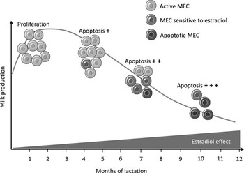
Ovarian secretions appear to be key factors for mammary gland development and function in ruminants. Estradiol and progesterone are essential for pubertal mammogenesis in heifer and goat, by stimulating MEC proliferation and by this way lobuloalveolar expansion. In contrast, ovarian steroids have a negative effect on mammary gland function. During lactation, they influence the evolution of proliferation/apoptosis balance in favor of apoptosis, and stimulate tissue remodeling in the mammary gland, resulting in a decrease in MY. In dairy cows, estradiol receptivity of MEC increases during lactation. The action of estradiol on these cells is different in the beginning, middle and end of lactation. It seems that estradiol acts directly on MEC to induce and accelerate the process of apoptosis. The gradual increases in sensitivity to ovarian steroids after the peak of lactation accelerate the loss of MEC and milk production decrease in non-pregnant cyclical cows. The action modes of estradiol on MEC during lactation, deduced from the various results presented during this review are presented in Figure 6. Some points that could not be addressed remain to be clarified: What is the effect of progesterone on bovine mammary gland in lactation? What are the interactions between estrogen, progesterone, cortisol and prolactin during lactation? The use of complementary models in vivo and in vitro may provide essential elements to answer these questions.
General diagram showing the action of estradiol on ovarian mammary epithelial cells (MECs) during lactation in not pregnant dairy cows. After the peak of lactation, the number of MEC decreases and increases sensitivity to estradiol. Estradiol acts then on MEC to induce and accelerate the process of apoptosis, which increases the loss of MEC and lower milk production. The increasing effect of estradiol during lactation on MEC and milk production is represented by the red triangle.

Acknowledgement
The authors are grateful to American Journal Expert (Durham, NC, USA) for the language editing. This research was supported by the French National Institute of Agricultural Science (INRA), the PHASE department.