


Introduction
The discovery of vitamin D (VD) and the prevention of rickets must be ranked as one of the outstanding clinical achievements of all time. The Nobel Prize in Chemistry (1928) was awarded to German scientist Adolf Otto Reinhold Windaus, who isolated VD and reported the molecular structure of VD. It is now well known that rickets was caused by the lack of this important compound. Now that the role of VD for calcium and phosphate metabolism was understood, rickets is almost eradicated at least in today's world. Thousands of studies were carried out to decipher the role of VD in humans and animals, which led to the finding of VD receptors (VDR) are linked to chromosomes in almost all cells and tissues, thus leading to its important effects on different physiologic processes. VD has recently received enormous attention; the increased scientific papers reported that VD has essential roles in reproduction, protection against infection, prevention of cancer and cardiovascular disease, and treatment of diabetes (Feldman et al., Reference Feldman, Krishnan, Swami, Giovannucci and Feldman2014; Sherman et al., Reference Sherman, Yu, Engle, Ding, Atkins, Tiriac, Collisson, Connor, Van Dyke, Kozlov, Martin, Tseng, Dawson, Donahue, Masamune, Shimosegawa, Apte, Wilson, Ng, Lau, Gunton, Wahl, Hunter, Drebin, O'Dwyer, Liddle, Tuveson, Downes and Evans2014; Wei et al., Reference Wei, Yoshihara, He, Hah, Fan, Pinto, Huddy, Wang, Ross, Estepa, Dai, Ding, Sherman, Fang, Zhao, Liddle, Atkins, Yu, Downes and Evans2018). Despite its name, VD is not really a vitamin, but the precursor to the steroid hormone, 1,25-dihydroxyvitamin D3 (also known as calcitriol and 1,25(OH)2D3), which mediates numerous biochemical reactions in tissues (Holick, Reference Holick2007). In modern swine production, pigs may be VD deficient at varying times of the year, diets may vary (Arnold et al., Reference Arnold, Madson, Acvp, Ensley, Goff, Stevenson, Acvp, Crenshaw, Wang and Horst2015), and VD insufficiency results in metabolic dysfunction. In order to improve the VD status of pig, it is very important to understanding VD metabolism and the physiological effects of VD. The purpose of this review is to summarize up-to-date findings on VD metabolism and the application of VD in pigs, with a comprehensive understanding of the actions of VD, in order to assist researchers and nutritionists investigating potential roles of VD.
Sources, absorption, and metabolism of vitamin D
There are two forms of VD: ergocalciferol (vitamin D2, abbreviated VD2) and cholecalciferol (vitamin D3, abbreviated VD3). All sterols possessing VD activity have the same steroid nucleus. The VD3 is produced by the ultraviolet light action on 7-dehydrocholesterol. VD obtained from the diet is absorbed with other dietary fats in the small intestine (Raimundo et al., Reference Raimundo, Lang, Scopel, Marcondes, Araújo, Faulhaber and Furlanetto2015). The efficient absorption of VD is dependent on the presence of fat in the intestinal lumen, which triggers the release of bile acids and pancreatic lipase. Cutaneous conversion of VD3 from 7-dehydrocholesterol is photochemistry. The formation efficiency of VD3 in the skin is influenced by skin pigmentation and ultraviolet light intensity. Because swine housed in confinement systems often do not have access to adequate sunlight, they are entirely dependent on dietary supplementation to meet their VD requirement. VD3 from intestinal absorption or production in the skin is transported by VD binding protein (DBP), which can also bind VD metabolites, to the liver and is hydroxylated by VD 25-hydroxylase to produce 25(OH)D3 (Fig. 1). The 25(OH)D3 is the major form of VD in circulatory systems and its concentration in blood has served as one of the most reliable biomarkers of VD status. Cytochrome P-450 enzymes (CYPs) are responsible for the conversion of VD3 to 25(OH)D3 (Zhu and Deluca, Reference Zhu and Deluca2012). It has been demonstrated that CYP2R1 was the major VD 25-hydroxylase (Zhu et al., Reference Zhu, Ochalek, Kaufmann, Jones and DeLuca2013).
The metabolic pathway of vitamin D.

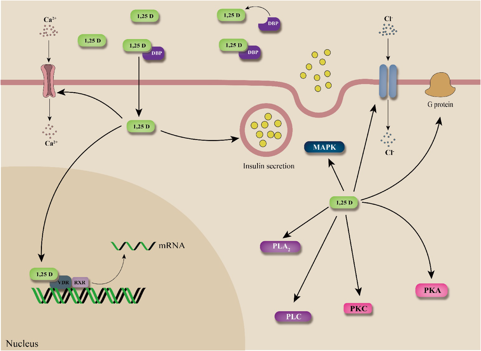
The majority of 25(OH)D3 is bound to DBP to form 25(OH)D3/DBP complexes, which are filtered by kidneys and taken up by proximal tubular cells. In kidneys, the released 25(OH)D3 is hydroxylated by 1α-hydroxylase (CYP27B1) to 1α,25(OH)2D3, abbreviated as 1,25(OH)2D3, which is VD sterol hormone. In addition, 25(OH)D3 can undergo epimerization at the C3 position to 3-epi-25(OH)D3, which accounts for a portion of total 25(OH)D3 in blood circulation. 3-epi-25(OH)D3 was transported to the kidney and converted to 3-epi-1,25(OH)2D3 which is an active metabolite with similar biochemical and biological properties to 1,25(OH)2D3 (Fig. 1; Molnár et al., Reference Molnár, Sigüeiro, Sato, Araujo, Schuster, Antony, Peluso, Muller, Mouriño, Moras and Rochel2011). The genomic mechanism of 1,25(OH)2D3 involves the binding of the 1,25(OH)2D3 activated VDR/retinoic X receptor heterodimeric complex. This complex can move to the nucleus, where it binds genes and modulates the transcription that encodes proteins to regulate the biologic response (Fig. 2; Mizwicki and Norman, Reference Mizwicki and Norman2009).
1,25(OH)2D3 activation of cellular signaling (modified from Mizwicki and Norman, Reference Mizwicki and Norman2009).

In addition, 25(OH)D3 can also be converted to 24,25(OH)2D3 and 1,24,25(OH)3D3 by the CYP24A1 enzyme (Jones et al., Reference Jones, Prosser and Kaufmann2012). When circulating 1,25(OH)2D3 increases, it will be catalyzed into excretory products: 1,24,25(OH)3D3 (calcitriol or calcitroic acid) and 1,25(OH)2D3-23,26-lactone. These inactive metabolites of VD are excreted in the feces via bile. The role of these compounds or inactivation of VD has not been fully evaluated, and key physiological and metabolic roles have not been discovered.
Vitamin D and immune system
It has been found that VDR signaling occurs in several immune cells, including monocytes, macrophages, natural killer cells, dendritic cell, T and B lymphocytes (Lee et al., Reference Lee, Kang, Jeon, Kim, Yoon, Song, Lyu, Piao, Kim, Han, Song, Lee, Song, Kim, Yu and Choi2005; Jeffery et al., Reference Jeffery, Burke, Mura, Zheng, Qureshi, Hewison, Walker, Lammas, Raza and Sansom2009; Baeke et al., Reference Baeke, Gysemans, Korf and Mathieu2010; Chishimba et al., Reference Chishimba, Thickett, Stockley and Wood2010; Chun et al., Reference Chun, Lauridsen, Suon, Zella, Pike, Modlin, Martineau, Wilkinson, Adams and Hewison2010). In vitro, 1,25(OH)2D3 suppressed immune responses by inhibiting T helper/inducer (Th) cell activity, which activated peripheral blood mononuclear cells to suppress immunoglobulin production. The immunosuppressive effect of VD3 is correlated to a decrease in inflammatory cytokine interleukin-2 (IL-2) (Zhao et al., Reference Zhao, Yu, Mao, He, Huang, Zheng, Yu, Han, Liang and Chen2014). In addition, 1,25(OH)2D3 was reported to decrease the expression of interferon-γ (IFN-γ) (Cippitelli and Santoni, Reference Cippitelli and Santoni1998). In Th2 cells, 1,25(OH)2D3 also stimulated the production of IL-4 (Mahon et al., Reference Mahon, Wittke, Weaver and Cantorna2003) and enhanced the ability of macrophages and monocytes to kill bacteria. 1,25(OH)2D3 had also been reported to result in an enhancement of CD4+CD25+ T regulatory cells (a subset of CD4+ T cells important for the inhibition of inflammation). Furthermore, 1,25(OH)2D3 directly inhibits the production of inflammatory Th1-secreted cytokines (Fig. 3), such as IL-2 and IFN-γ, as well as the Th17-derived cytokines IL-17 (induction of matrix destruction) and IL-21 (Jeffery et al., Reference Jeffery, Burke, Mura, Zheng, Qureshi, Hewison, Walker, Lammas, Raza and Sansom2009). T cells secreting IL-9 have also been shown to be 1,25(OH)2D3 targets (IL-9 stimulated chemokine production, enhanced mucus production from airway epithelial cells, and altered barrier function in the intestine). Moreover, 1,25(OH)2D3 diminished Th17 development (Palmer et al., Reference Palmer, Lee, Maynard, Oliver, Bikle, Jetten and Weaver2011). 1,25(OH)2D3 affected the phenotype and behavior of dendritic cells through its early and transcriptionally mediated reprogramming of metabolic pathways and induced oxidative and glycolytic metabolic pathways (Ferreira et al., Reference Ferreira, Vanherwegen, Eelen, Gutiérrez, VanLommel, Marchal, Verlinden, Verstuyf, Nogueira, Georgiadou, Schuit, Eizirik, Gysemans, Carmeliet, Overbergh and Mathieu2015). VD also plays a role in maintaining the mucosal barrier function by upregulating the expression of tight junction and adherent junction proteins and suppressing epithelial cell apoptosis (Del Pinto et al., Reference Del Pinto, Ferri and Cominelli2017). Further, intestinal VDR signaling plays a fundamental role in intestinal homeostasis through its effects on autophagy, as VD potentially influences the composition of the intestinal microbiome (Wu et al., Reference Wu, Zhang, Lu, Xia, Zhou, Petrof, Claud, Chen, Chang, Carmeliet and Sun2015; Waterhouse et al., Reference Waterhouse, Hope, Krause, Morrison, Protani, Zakrzewski and Neale2018). A recent study confirmed the potent effects of VD on cytokine production and in regulating normal innate and adaptive immune functions (O'Brien and Jackson, Reference O'Brien and Jackson2012). In animal models, co-administration of VD with vaccine showed superior immunogenicity and specific antibody response (Sadarangani et al., Reference Sadarangani, Whitaker and Poland2015). VD was also one of the nutritional factors to influence the immune response to vaccination, whether VD deficiency just introduced a state of immune vulnerability was important but not clear (Zimmermann and Curtis, Reference Zimmermann and Curtis2019). Mechanical and systematic studies are needed to understand the impact of VD signaling on the adaptive immune response to vaccines, which could indicate the design of vaccines using VD as an adjuvant.
The immunomodulatory effects of 1,25(OH)2D3.

Based on recent research, supplementation of 25(OH)D3 in pig diets can improve animal health. The effects of supplementing with 1500 IU kg−1 of VD3 or 1500 IU kg−1 of VD3 plus 2000 IU kg−1 of two VD sources (VD3 or 25(OH)D3) on the health of post-weaned pigs were measured (Konowalchuk et al., Reference Konowalchuk, Rieger, Kiemele, Ayres and Barreda2013). Anti-inflammatory responses were positively affected by 25(OH)D3-supplemented diets and resulted in an increase in the total number of leukocytes per milliliter of blood, individual granulocyte, and lymphocyte subpopulations, upregulation of their survival capacity under basal conditions and an increase in phagocytic capacity of discrete leukocyte populations (Konowalchuk et al., Reference Konowalchuk, Rieger, Kiemele, Ayres and Barreda2013). Blood leukocytes are primary contributors to pig anti-inflammatory defenses and are key activators and regulators of inflammation and homeostasis of immune system. Moreover, similar upregulation of phagocytosis was observed among phagocytes isolated from mucosal surfaces of the pig lung. The contributions of 25(OH)D3 to the bone marrow hematopoietic compartment increased the proliferation of leukocyte progenitors and the survivability of hematopoietic cells (Konowalchuk et al., Reference Konowalchuk, Rieger, Kiemele, Ayres and Barreda2013). This has the potential to positively impact long-term production of circulating leukocytes responsible for anti-inflammatory defenses and the regulation of immune homeostasis. In addition, the supplementation of 50 μg kg−1 25(OH)D3 in diets for piglets increased the concentration of immunoglobulin G (Meuter et al., Reference Meuter, Thoby, Lichou and Renouf2016a).
Rotavirus infection is among the most common causes of mortality and subsequent economic loss, particularly at weaning when young pigs are most vulnerable to infection. There is some literature that elucidated VD's protective role against infections by modulating inflammatory responses under this challenge (Liao et al., Reference Liao, Zhang, Ding, Xu and Bai2011; Zhao et al., Reference Zhao, Yu, Mao, He, Huang, Zheng, Yu, Han, Liang and Chen2014, Reference Zhao, Yu, Mao, He, Huang, Zheng, Yu, Han, Liang and Chen2015; Tian et al., Reference Tian, Liang, Chen, Mao, Yu, Zheng, He, Huang and Yu2016). The effects of dietary supplementation with 25(OH)D3 on antibody and cytokines in serum and intestinal contents of weanling pigs with or without rotavirus challenge were investigated (Liao et al., Reference Liao, Zhang, Ding, Xu and Bai2011). 25(OH)D3 supplementation increased the secretion of anti-inflammatory cytokine, IL-4, and reduced the secretion of proinflammatory cytokines, IL-2, IL-6 and IFN-γ. Dietary supplementation with 25(OH)D3 inhibited Th1-type immune response and enhanced Th2-type immune response (Liao et al., Reference Liao, Zhang, Ding, Xu and Bai2011). Moreover, VD3 supplementation could alleviate the negative effects caused by porcine rotavirus challenge (Zhao et al., Reference Zhao, Yu, Mao, He, Huang, Zheng, Yu, Han, Liang and Chen2014), which is based on the early interferon response to rotavirus infection and the effect of VD on host–pathogen interactions. The dietary supplementation of 5000 IU kg−1 VD3, compared to dietary supplementation with 200 IU kg−1 VD3, improved the faecal consistency and decreased the secretion of proinflammatory cytokines, IL-2 and IL-6, in the challenged pigs (Zhao et al., Reference Zhao, Yu, Mao, He, Huang, Zheng, Yu, Han, Liang and Chen2014). Furthermore, VD supplementation activated the RIG-I signaling pathway (Zhao et al., Reference Zhao, Yu, Mao, He, Huang, Zheng, Yu, Han, Liang and Chen2014). In addition, 25(OH)D3 can directly inhibit rotavirus in intestinal epithelial cells in vitro, and the anti-rotavirus effects of 25(OH)D3 are mediated by RIG-I signaling pathways (Zhao et al., Reference Zhao, Yu, Mao, He, Huang, Zheng, Yu, Han, Liang and Chen2015). Additionally, VD3 regulates autophagic maturation and genes expression of porcine antibacterial peptide to attenuate rotavirus infection (Tian et al., Reference Tian, Liang, Chen, Mao, Yu, Zheng, He, Huang and Yu2016). The above-mentioned results support previous observations regarding the positive immunomodulatory role of VD3, 25(OH)D3 as a superior dietary supplementation for weanling pigs in comparison with conventional VD3 during rotavirus infection challenges.
Vitamin D and reproductive health
With specific regard to the potential effects of VD on reproduction (see Fig. 4), it is mediated via VDR signaling. The expression of VDR signaling occurs in mammalian reproductive tissues, including the ovary, uterus, placenta endometrium, placenta, testis, spermatozoa, and the pituitary gland (Shahrokhi et al., Reference Shahrokhi, Ghaffari and Kazerouni2016). Human endometrium research revealed the upregulation of homeobox protein A10 (HOXA10) expression at the time of implantation, which is essential for a normal development, and 1,25(OH)2D3 regulates key target genes (HOXA10) influenced by implantation (Du et al., Reference Du, Daftary, Lalwani and Taylor2005). Female mice (VDR null mutant) showed uterine hypoplasia and impaired folliculogenesis, and male mice (VDR null mutants) exhibited decreased sperm production and decreased sperm motility with histological abnormality of the testis (Kinuta et al., Reference Kinuta, Tanaka, Moriwake, Aya, Kato and Seino2000). Peripubertal VD3 sufficiency is important for an appropriately timed pubertal transition and maintenance of normal female reproductive physiology (Dicken et al., Reference Dicken, Israel, Davis, Sun, Shu, Hardin and Neal-Perry2012). Maternal VD status during pregnancy affects adipose development and metabolic health of offspring (Belenchia et al., Reference Belenchia, Jones, Will, Beversdorf, Vieira-Potter, Rosenfeld and Peterson2018). In vivo and in vitro data supported the notion that VD3 is a key regulator of neuroendocrine and ovarian physiology; VDR signaling has a positive effect on the maintenance of normal male reproduction, female fertility, and embryo implantation.
Overview of vitamin D role in the reproductive system.

There are several lines of evidence suggesting an important role of VD in pig fertility. Birth weight (VD3 = 1.44 ± 0.27 kg versus 25(OH)D3 = 1.55 ± 0.24 kg) can be increased by feeding a diet with 50 μg kg−1 of 25(OH)D3 for sows, the 25(OH)D3 supplementation (50 μg kg−1 diet) is more effective than VD3 (2000 IU kg−1 diet) in increasing sow and fetal VD status (Weber et al., Reference Weber, Witschi, Wenk and Martens2014). The effect of improved VD status using 25(OH)D3 on reproduction in gilts was investigated (Coffey et al., Reference Coffey, Hines, Starkey, Starkey and Chung2012), 25(OH)D3 supplementation resulted in heavier litter weights (VD3 = 6.5 kg versus 25(OH)D3 = 8.2 kg) compared to VD3 supplementation. Although preweaning mortality was not mentioned, feeding 25(OH)D3 increased litter size by 2.5 fetuses compared with VD3 (Coffey et al., Reference Coffey, Hines, Starkey, Starkey and Chung2012). 25(OH)D3-fed sows weaned 1.17 more piglets, and maternal 25(OH)D3 supplementation enhanced bone strength, density, and ash content of piglets compared with VD3 supplementation (Zhou et al., Reference Zhou, Chen, Zhuo, Lv, Lin, Feng, Fang, Che, Li, Xu and Wu2016b). Replacing equal quantities of the dietary VD3 with 25(OH)D3 significantly improved the maternal status of VD and reduced specific post-parturition problems (Meuter et al., Reference Meuter, Thoby, Lichou and Renouf2016b). The effect of supplementation with or without 25(OH)D3 on the performance of gestating sows was studied; maternal supplementation with 25(OH)D3 can positively impact the reproductive performance (Zhang et al., Reference Zhang, Bae, Jeong and Kim2017). Sow farrowing duration was shortened (from 5.38 to 4.71 h), and the average number of mummified fetuses decreased significantly compared with the control group (0.1 versus 0.5) while birth weight was significantly enhanced (from 1.18 to 1.44 kg; Table 1; Zhang et al., Reference Zhang, Bae, Jeong and Kim2017). Newborn pig plasma 25(OH)D3 levels were very low, which may cause VD deficiency. Because plasma 25(OH)D3 level in piglets at birth is closely related to that of the mother, high concentration of VD supplementation for sow diets could be an effective strategy to increase VD status of the neonate, which is transported through mother's placenta and milk to fetuses. Milk VD3 concentration is linearly increased with increasing VD3 supplementation in sow diets (Flohr et al., Reference Flohr, Tokach, Dritz, DeRouchey, Goodband, Nelssen and Bergstrom2014). Piglets consume 25(OH)D3-fortified milk that has contributed to an increase in VD3 status from birth to weaning; improving supplementation levels of VD in diets can alleviate the decreased reproductive performance due to VD deficiency. Live births can be increased by feeding VD for sows (see Table 1), which is due to VD influencing maternal–conceptus interaction further to improve implantation. In addition, those observations indicate that dietary 25(OH)D3 was more effective than commonly used VD3 in rapidly re-establishing the steady status of sows after parturition.
Effects of vitamin D supplementation on reproductive performance of sows

VD, vitamin D3; HD, 25(OH)D3; N, number of pigs in the experiment.
*Statistically significant difference (P < 0.05).
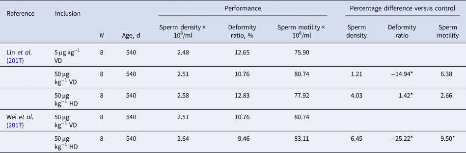
In male reproduction (Table 2), the effect of dietary supplementation of VD3 on boars was investigated. The supplementation of 2000 IU kg−1 VD3 in diets increased sperm motility and effective sperm number, and enhanced testosterone secretion and expression of CYP19, CYP24A1, and VDR mRNAs in sperm (Lin et al., Reference Lin, Lv, Dong, Wu, Tao, Xu, Che, Fang, Bai, Feng, Li and Xu2017). An increased expression of CYP24A1 and VDR is in response to high dietary VD, which suggests that VDR signaling mediating spermatogenesis. Testosterone and estradiol are key hormones associated with male reproduction, which is essential for spermatogenesis and epididymal sperm maturation. The CYP19 gene encodes aromatase, and 1,25(OH)2D3 binds to VD response elements in the promoter of CYP19 and exerts tissue-specific regulation of aromatase. Increased CYP19 expression occurs in response to VD3 treatments, which is associated with an increase in the effective number of sperm and sperm quality (Lin et al., Reference Lin, Lv, Dong, Wu, Tao, Xu, Che, Fang, Bai, Feng, Li and Xu2017). In addition, dietary supplementation of VD3 can increase the enzyme activity, such as aromatase, which promotes testosterone conversion to estradiol for improving sperm quality (Lin et al., Reference Lin, Lv, Dong, Wu, Tao, Xu, Che, Fang, Bai, Feng, Li and Xu2017). 25(OH)D3 is more conducive than VD3 in promoting reproductive performance at the same level of supplementation. Supplementation of 25(OH)D3 (50 μg kg−1 diet) for boars increases VD content in blood, sperm morphology, movement ability, and ultimately improved reproductive performance (Wei et al., Reference Wei, Dong, Lyu, Wu, Xu, Fang, Che, Feng, Li and Lin2017). Although there is limited evidence available in boars about the mechanism of VD3-mediated reproductive performance, the studies currently available show that VD3 has potential benefits of involvement in male reproduction.
Effects of vitamin D supplementation on reproductive performance of boars

VD, vitamin D3; HD, 25(OH)D3; N, number of pigs in the experiment.
*Statistically significant difference (P < 0.05).
Assessment of vitamin D status and pig health
VD supplements have long been recommended for people to treat or prevent osteoporosis. The VD deficiency results from inadequate irradiation of the skin, from insufficient intake from the diet or from impairments in the metabolic activation of the vitamin. In clinical application, serum 25(OH)D is measured to monitor the VD status of the patients, and serum 25(OH)D comprises the sum of 25(OH)D2 and 25(OH)D3. The two most common types of method for determining 25(OH)D level in the blood include antibody-based method, which uses a kit or an automated clinical chemistry platform. Another method used is liquid chromatography (LC) equipped with an automated equipment featuring mass spectrometric (MS) detection for the quantification of 25(OH)D in serum. However, there is still controversy regarding the performance of serum 25(OH)D assays in clinical and research laboratories. Antibody-based methods measure only total 25(OH)D in serum. Various commercial assays differ because of the nature of the antibody used, some claiming an advantage that they do not discriminate between 25(OH)D2 and 25(OH)D3. The consensus among analysts in a clinical setting is that LC-MS/MS assays are the optimal standard for assay performance in the future. A variety of methods are available to determine serum 25(OH)D2 and 25(OH)D3 levels, each with advantages and disadvantages that must be considered in evaluating the data arising from them. The defined threshold for VD deficiency was set at 25(OH)D below 20 ng ml−1 (50 nmol l−1), and VD insufficiency was 25(OH)D of 21–29 ng ml−1 (525–725 nmol l−1), according to the Endocrine Society guideline (Holick et al., Reference Holick, Binkley, Bischoff-Ferrari, Gordon, Hanley, Heaney, Murad and Weaver2011). The fact that piglets are born with the lowest blood concentration of 25(OH)D3 increased their susceptibility to VD deficiency (Arnold et al., Reference Arnold, Madson, Acvp, Ensley, Goff, Stevenson, Acvp, Crenshaw, Wang and Horst2015). Deficiency symptoms of pigs may take 4–6 months to become evident (Lewis and Southern, Reference Lewis and Southern2000) because of the tissue storage capacity. Common symptoms of VD deficiency in pigs include enlarged joints, broken bones, stiffness of the joints, bowed legs, and posterior paralysis. The growth rate is generally adversely affected by VD deficiency. Rickets is a disease of bones and thus only affects young, growing animals. In older animals, VD deficiency causes osteomalacia, in which there is reabsorption of bone already laid down. Osteomalacia due to VD deficiency is not common in pig farms, although a similar condition can occur in pregnant and lactating sows, which require increased amounts of calcium and phosphorus. Rickets and osteomalacia are not specific diseases necessarily caused by VD deficiency; they lead to decreasing concentration of calcium and phosphorus in the organic matrices of cartilage and bone. The signs are similar to imbalance or failure of the utilization of calcium or phosphorus. In swine production, the serum concentration of 25(OH)D3 below 10–15 ng ml−1 is considered as deficient (Lauridsen, Reference Lauridsen2014). To obtain this 25(OH)D3 concentration (10–15 ng ml−1 blood) in gilts or sows, more than 1400 IU of VD3 per kilogram of feed is necessary (Lauridsen et al., Reference Lauridsen, Halekoh, Larsen and Jensen2010). However, it should be underlined that there are no scientific data available to confirm this threshold. A study reported that more than 200 IU of VD3 per kilogram of diets can obtain over 30 ng ml−1 serum concentration of 25(OH)D3 in sows (Flohr et al., Reference Flohr, Woodworth, Bergstrom, Tokach, Dritz, Goodband and DeRouchey2016a). This conclusion is contradictory to the previous study, which may be due to inconsistent supplement of nutrition in the early growth stage and the ability of the tissue to store VD. In addition, the metabolism of 25(OH)D3 is tightly regulated by plasma parathyroid hormone level and serum calcium and phosphorus levels; this can be explained by the contradictory observations in serum 25(OH)D3 response. The kinetics of plasma VD are complex and affected by multiple factors, which include sources (photosynthesis of VD, dietary VD intake), two isomers (VD2 and VD3) with somewhat different properties, variable rate of transfer of VD from chylomicrons to DBP in the plasma, variable rates of conversion to a variety of VD metabolites, and variable release of VD from the body. For piglets, more than 1500 IU of VD3 per kilogram of diets can obtain over 30 ng ml−1 serum concentration of 25(OH)D3, and the serum concentration of 25(OH)D3 was greater when 25(OH)D3 was provided in diets. In addition, supplementation of 50 μg kg−1 25(OH)D3 in piglet diets easily reached a blood level of 25(OH)D at 30 ng ml−1. This conclusion was obtained from previous studies (Witschi et al., Reference Witschi, Liesegang, Gebert, Weber and Wenk2011; Sugiyama et al., Reference Sugiyama, Kusuhara, Chung, Yonekura, Azem and Hayakawa2013; Regassa et al., Reference Regassa, Adhikari, Nyachoti and Kim2015; Flohr et al., Reference Flohr, Woodworth, Bergstrom, Tokach, Dritz, Goodband and DeRouchey2016b; von Rosenberg et al., Reference von Rosenberg, Weber, Erhardt, Höller, Wehr and Rambeck2016; Duffy et al., Reference Duffy, Kelly, Rajauria, Clarke, Gath, Monahan and O'Doherty2018a, Reference Duffy, Kelly, Rajauria, Jakobsen, Clarke, Monahan, Dowling, Hull, Galvin, Cashman, Hayes and O'Doherty2018b). According to previous surveys of serum concentrations of 25(OH)D3 in different growth stages of pigs, it was suggested that an adequate 25(OH)D level for keeping healthy probably exceeds 30 ng ml−1 in serum. Additional research is needed to determine an optimal level of serum 25(OH)D in swine production.
Excessive intake of VD is toxic and can cause soft tissue mineralization, secondary to persistent hypercalcemia and hyperphosphatemia, slow growth, poor feed conversion, and even death (NRC, 2012). Weanling pigs given daily oral doses of 6250 μg of VD3 (250000 IU VD3 equivalent) exhibited poor growth and feed intake, reduced liver and bone weights, and calcification in organs (NRC, 2012). For finishing pigs, feeding super nutritional concentrations (40000 and 80000 IU kg−1 VD3 in diet) of VD3 for 1 or 2 months before slaughter results in serum concentrations above 100 ng ml−1 (Wilborn et al., Reference Wilborn, Kerth, Owsley, Jones and Frobish2004); addition of 40000 IU kg−1 of VD3 in diets improves pork quality whereas 80000 IU kg−1 of VD3 in diets decreases the growth rate of the pigs.
Efficacy and dose requirements of vitamin D
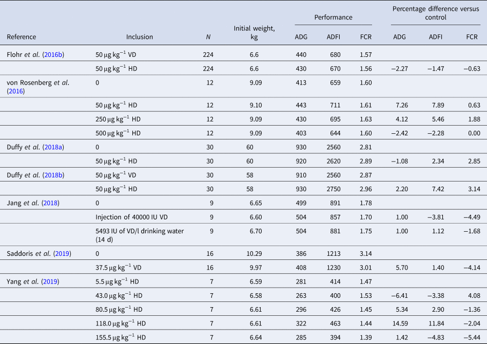
Most terrestrial animals acquire VD by synthesis in skin upon exposure to ultraviolet radiation. The supplementation of VD in animal diets is necessary for absorption, transport, and utilization of calcium and phosphorus (Saddoris et al., Reference Saddoris, Fleet and Radcliffe2019). Pigs do not require additional VD supplementation if sufficient sunlight exposure is provided (Lewis and Southern, Reference Lewis and Southern2000). Under confinement housing, dietary VD fortification is needed. In the National Research Council recommendation of VD3 (NRC, 2012), minimum requirements for VD3 ranged from 220 to 150 IU kg−1 diet for pigs (5–135 kg; NRC, 2012). In addition, the recommendations of VD for gestating and lactating sows in NRC are 800 IU kg−1 diet. The recommendation of VD3 in boar diets is 200 IU kg−1. Published studies have contributed to the recommended higher VD requirement for gestating and lactating sows from 200 IU kg−1 (NRC, 1998) to 800 IU kg−1 (NRC, 2012). The requirement estimate for VD in growing pigs has not changed much over the years (from 1998 to 2012). In practice, the VD3 supplementation is much higher than the NRC recommendation; the level of VD used in commercial swine diets varies widely (Yang et al., Reference Yang, Wang, Li and Ma2020). The design and outcome of published studies on the effects of feeding VD on the growth performance of pigs are displayed in Table 3. The current nutrient recommendation of VD3 is 220 IU kg−1 of the diet for weaned pigs (NRC, 2012). Increasing VD3 concentrations in an oral dose, drinking water, or dry feed increased serum 25(OH)D3 in weaned pigs. Also, feeding weaned pigs 50 μg kg−1 25(OH)D3 potentially improves anti-inflammation. Overall, the evidence does not strongly support improvement in growth performance of weaned pigs, though increasing VD3 status of the weaned pigs improves immune functions. It is suggested the current requirement estimate of 220 IU kg−1 of the diet might not be sufficient for weaned pigs. For finishing pigs, the current requirement estimate of VD3 is 150 IU kg−1 diet (NRC, 2012). Based on published data, increasing the VD3 concentration in the finishing pig diets above the NRC value does not improve growth performance and bone mineralization, but feeding a high concentration of VD to pigs alleviated negative influences from rotavirus infection. If there is no rotavirus challenge, the current NRC recommendation for finishing pigs is sufficient. Compared with NRC recommendations, supplementing 2000 IU kg−1 or more of VD3 shows a benefit for breeding pigs. For gestating and lactating sows, the current NRC estimates for VD3 is 800 IU kg−1 of the diet (NRC, 2012). Some studies reported sows fed 2000 IU kg−1 of VD3 or 25(OH)D3 had more piglets alive than sows fed 800 IU kg−1 of VD3 (see Table 3). Also, pigs from sows fed 2000 IU kg−1 of VD3 had an improvement in neonatal bone strength and mineralization. The majority of studies reported potential benefits of the improvement of maternal VD supplementation strategies; the VD recommendation for sows during gestation and lactation may be required to improve based on current documentation of scientific papers. Moreover, little evidence is available regarding VD in boar reproduction, supplementation of boar diets with 2000 IU kg−1 of VD3 or 25(OH)D3 increased sperm quality (Lin et al., Reference Lin, Lv, Dong, Wu, Tao, Xu, Che, Fang, Bai, Feng, Li and Xu2017). The 25(OH)D3 was more potent in improving boar reproduction than VD3 (Wei et al., Reference Wei, Dong, Lyu, Wu, Xu, Fang, Che, Feng, Li and Lin2017).
Effects of vitamin D supplementation on growth performance of pigs

ADG, average daily gain; ADFI, average daily feed intake; FCR, feed conversion rate; VD, vitamin D3; HD, 25(OH)D3; N, number of pigs in the experiment.
*Statistically significant difference (P < 0.05).
Together, these results of feeding experiments indicated that, when fed equivalent concentrations of VD, 25(OH)D3 is more effective in increasing pig circulating concentrations of 25(OH)D3 compared to VD3. The reason is easy to understand, 25(OH)D3 is absorbed more effectively than VD3 and it bypasses the liver and reaches the blood stream more quickly, and it is further hydroxylated into the active form of VD3. Large-group evaluation experiments are required to determine the impact of using 25(OH)D3 as a VD source on animal production. In addition, with increasing dietary VD3 supplementation, the growth performance of pigs is not increased linearly. High concentration of VD3 (5000 IU kg−1 diet) is beneficial for pig health, especially in rotavirus infection. Also, most studies were focused on dams and piglets with little information about boars. Therefore, VD in boar health and metabolism needs further investigation.
Conclusion
A multitude of studies indicate that VD has an important role in the prenatal development, immune system, and reproduction of pigs. Future studies are needed to also determine the most feasible levels of serum 25(OH)D3 and when supplements or drugs should be introduced under stress challenge. We are only beginning to understand the factors involved in the regulation of the immune and reproductive systems by VD. Although networks regulated by VDR signaling are complex, further studies, including genome, proteome, and metabolic analyses, are required to further elucidate the multiple roles of VD in complex metabolic pathways.
Acknowledgments
This study was financially supported by the China Agricultural University Research Funding (201705510410056) and Ministry of Agriculture and Rural Affairs Funding (21178259). We thank Prof. Lee J. Johnston in the University of Minnesota for reviewing this article and suggestions, and A/Prof. Crystal Levesque in South Dakota State University for revising the manuscript.
Author contributions
The authors' contributions are as follows: Pan Yang wrote and revised the manuscript, Pan Yang and Yongxi Ma contributed to the preparation of figures, Yongxi Ma provided critical comments. All authors read and approved the final manuscript.
Conflict of interest
None.