Introduction
Supracricoid laryngectomy with cricohyoidoepiglottopexy is functional organ preservation surgery suitable for early-to-intermediate and selected advanced laryngeal cancers. In 1959, the Austrians, Majer and Rieder, reported the initial concept of supracricoid laryngectomy; in the 1970s and 1980s, Piquet and Laccourreye, from France, promoted the clinical use of supracricoid laryngectomy.Reference Piquet and Chevalier2, Reference Laccourreye, Laccourreye, Weinstein, Menard and Brasnu3 Today, supracricoid laryngectomy with cricohyoidoepiglottopexy has become one of the major surgical options for larynx preservation worldwide.Reference Sperry, Rassekh, Laccourreye and Weinstein4–Reference Nakayama, Okamoto, Miyamoto, Yokobori, Takeda and Masaki6 In this surgical procedure, approximately three-quarters of the larynx, including the bilateral glottis and paraglottic spaces (except for one or two arytenoids), is removed.Reference Kim, Sun, Park, Cho, Park and Cho7 The major advantage of this procedure is the associated high control rate for the primary cancer.Reference Nakayama, Okamoto, Miyamoto, Yokobori, Takeda and Masaki6, Reference Laccourreye, Laccourreye, Garcia, Gutierrez-Fonseca, Brasnu and Weinstein8
Supracricoid laryngectomy with cricohyoidoepiglottopexy can be utilised for salvage after radiotherapy (RT) failure, but has rarely been performed after partial laryngectomies:Reference Nakayama, Okamoto, Hayakawa, Ishiyama, Kotani and Miyamoto9 there is only one case report of the use of this technique for salvage after failure of vertical partial laryngectomy.Reference Luna-Ortiz, Nuñez-Valencia and Carmona-Luna10 We report a case of salvage supracricoid laryngectomy with cricohyoidoepiglottopexy after failed RT (primary failure) and vertical partial laryngectomy (secondary failure), with successful oncological and functional outcomes. A literature search indicated this to be the first report of the clinical application of this surgical procedure after the failure of two major treatments.
Case report
Primary and secondary treatments
In October 2002, a 58-year-old male company executive was diagnosed with a right glottic squamous cell carcinoma (tumour–node–metastasis (TNM) stage T1aN0M0) at his local hospital. He subsequently had a complete response to RT (62.2 Gy). In April 2006, approximately three and a half years after RT, the first local recurrence (T1aN0M0) developed in the posterior portion of the right vocal fold. He subsequently underwent vertical partial laryngectomy at the same hospital.
According to his attending surgeon, the entire right vocal fold had been invaded by the recurrent cancer. It was therefore removed, along with adjacent false vocal folds and subglottic tissue. The defect of the right hemilarynx was reconstructed and primary closure was performed using a sternohyoid muscular flap covered with a local skin strip. Temporary tracheostomy was performed at the time of vertical partial laryngectomy, and was closed three months later. The vertical partial laryngectomy wound developed a methicillin-resistant Staphylococcus aureus (MRSA) infection, resulting in a temporary fistula that required six months to close with local treatment.
Peri-operative course
In December 2009, approximately three and a half years after vertical partial laryngectomy, a second local recurrence (T2N0M0) developed at the posterior portion of the reconstructed right musculocutaneous flap. Total laryngectomy was recommended by the attending surgeon, but the patient rejected this option because of the associated functional defects. He was then referred to our clinic (approximately 500 km from his local hospital) for possible functional organ preservation treatment.
The 65-year-old patient attended our clinic in January 2010. He had quit smoking seven years previously and maintained a moderate pulmonary status, with blood gases at 73 torr (in room air). He exercised regularly and maintained an excellent physical condition for his age (Figure 1a). Laryngoscopy findings revealed an exophytic recurrence at the post-inferior portion of the reconstructed right musculocutaneous flap. Restricted mobility was identified in the right arytenoid (Figure 1b, c). Contrast computed tomography (CT) revealed enhancement to the right hemilarynx (Figure 1d); the left hemilarynx was intact. Based on these findings, supracricoid laryngectomy with cricohyoidoepiglottopexy was considered a suitable treatment option; however, the possibility of intra-operative conversion to total laryngectomy was discussed when obtaining informed consent.
Clinical evaluation of the patient at the initial visit in January 2010. (a) Scarring at the anterior neck resulting from vertical partial laryngectomy and tracheostomy. (b) A distant view of the glottis (black arrow indicates whitish musculocutaneous flap resulting from vertical partial laryngectomy at the right glottis). (c) A close-up view of the glottis (white arrow denotes a recurrent tumour at the right posterior glottis). (d) Tumour enhancement in the right hemilarynx, as revealed by contrast computed tomography.

Supracricoid laryngectomy with cricohyoidoepiglottopexy was performed in February 2010. For this, a T-shaped skin incision was designed to include the vertical partial laryngectomy and tracheostomy wound scars. The right sternohyoid muscle was missing following vertical partial laryngectomy. The right scarred hemilarynx was carefully resected, along with the left glottis and the remaining portion of thyroid cartilage (Figure 2a). The entire right arytenoid except for the corniculate cartilage was then removed. Nine marginal strips surrounding the resected larynx were submitted for frozen pathological analysis: all margins were tumour negative. Before pexis, the right corniculate cartilage and surrounding pharyngeal mucosa were retracted forward and sutured to the inner edge of the cricoid cartilage to create appropriate airway narrowing of the neoglottis (Figure 2b). After pexis, the right lobe of the thyroid gland was elevated with superior thyroid vessels and relocated so as to overlap and reinforce the pexis gap (Figure 2c) and fill the devoid portion of the strap muscular closure (Figure 2d).
Intra-operative images of supracricoid laryngectomy with cricohyoidoepiglottopexy showing electric cautery directed at the scarred area situated between right hemilarynx (posterior end of the paraglottic space) and pyriform sinus (indicated by finger) (a), right corniculate reconstruction before pexis (b), repositioning of the right lobe of the thyroid gland to overlap and reinforce the pexis gap (c) and use of the right lobe to fill the devoid portion of strap muscular closure (d, white arrow).
Macroscopic observation showed the recurrent cancer to be located at the post-inferior portion of the reconstructed musculocutaneous flap (Figure 3a). Microscopic observation identified moderate- to well-differentiated squamous cell carcinoma at the posterior edge, with multiple foci scattered around the arytenoid cartilage and beneath the musculocutaneous flap (Figure 3b). Abscess formation with extensive fibrosis and a foreign body reaction was noted surrounding the recurrent foci.
Pathological findings. (a) A supracricoid laryngectomy-resected specimen revealing local recurrence in the post-inferior portion of the whitish musculocutaneous flap. (b) H&E staining of a specimen taken from the slice shown by the dotted line in (a) showing exophytic recurrence with multiple foci scattered beneath the musculocutaneous flap (black arrows; low power view).
Post-operative course
The immediate post-supracricoid laryngectomy course was uneventful until the end of the first week, when increased purulent secretion was observed at the T-shaped skin junction and tracheal stoma. An MRSA infection was identified and subsequently treated with an anti-MRSA antibiotic agent. Hyperbaric oxygen therapy was provided for 30 days starting post-surgery day 12. With meticulous local treatment, the infected wound healed completely by post-surgery day 60, and the patient was discharged. After complete healing of the neoglottis (Figure 4), two operations were required to close the tracheal stoma because of local wound infection. In-patient swallowing rehabilitation was provided in September 2010 and the treatment course was completed in October 2010, approximately eight months after supracricoid laryngectomy with cricohyoidoepiglottopexy.
Images showing neoglottis four years post-supracricoid laryngectomy: (a) Respiratory phase; (b) Phonetic phase.
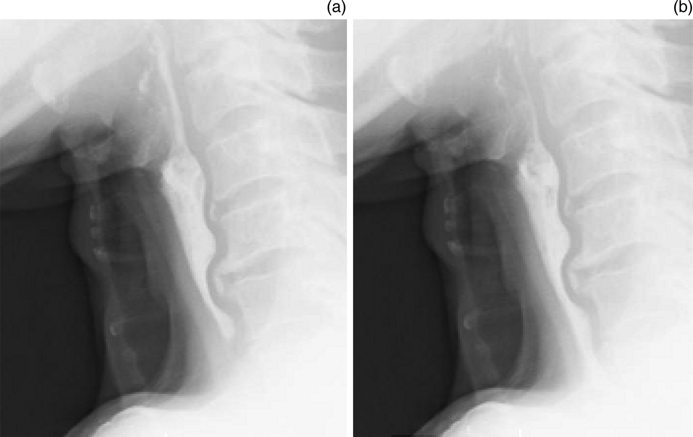
In February 2014, four years after supracricoid laryngectomy with cricohyoidoepiglottopexy, the patient had recovered well and had satisfactory laryngeal function. At 2 years post-supracricoid laryngectomy, he spoke with a hoarse voice and had a grade of hoarseness in rough, breathy, asthenic, and strained scales (‘GRBAS’) score of 22 201 and a maximum phonation time of 12 seconds. At four years post-supracricoid laryngectomy, videofluoroscopic swallowing analysis revealed a favourable swallowing outcome for both jelly and liquid iohexol contrast agent (Figure 5). The patient currently has no dietary restrictions and enjoys dining in public. He has resumed his work as a company executive and enjoys coaching a junior league baseball team.
Images of videofluoroscopic swallowing analysis four years post-supracricoid laryngectomy. No mis-swallowing was observed for jelly (a) or liquid iohexol (b).
Discussion
Supracricoid laryngectomy with cricohyoidoepiglottopexy is by far the most radical open surgical procedure for functional organ preservation in glottic cancer. It is reported to be more beneficial than conventional partial laryngectomy for local control.Reference Zhang, Lu, Chen, Lou, Ge and Song5, Reference Laccourreye, Laccourreye, Garcia, Gutierrez-Fonseca, Brasnu and Weinstein8 It can also be used as salvage surgery following RT failure. In our institute, both oncological and functional outcomes have been satisfactory and did not vary between patients undergoing primary and salvage surgery.Reference Nakayama, Okamoto, Hayakawa, Ishiyama, Kotani and Miyamoto9 Salvage supracricoid laryngectomy with cricohyoidoepiglottopexy is applicable to all recurrent tumour stages and is the most effective for unfavourable T2 and T3a tumours.Reference Nakayama, Okamoto, Hayakawa, Ishiyama, Kotani and Miyamoto9 Despite this positive evidence, the application of salvage supracricoid laryngectomy with cricohyoidoepiglottopexy after RT and vertical partial laryngectomy failure was unprecedented. A final decision to use this surgical procedure was based on clinical evaluations, including targeted CT imaging and a patient interview.
During the supracricoid laryngectomy with cricohyoidoepiglottopexy procedure, the right scarred hemilarynx was carefully resected, along with the left glottis and the remaining portion of the thyroid cartilage. Fortunately, there was no extralaryngeal extension of the cancer. The surgical margins of the resected specimen were negative in all directions owing to the radical nature of supracricoid laryngectomy, including the ability to remove the paraglottic space en bloc.Reference Kim, Sun, Park, Cho, Park and Cho7 The entire ipsilateral arytenoid cartilage can also be removed, but it is crucial to preserve and reconstruct the corniculate cartilage,Reference Loyo, Laccourreye, Weinstein and Helsingr11 which ensures proper airway narrowing of the neoglottis.
In consequence of the previous MRSA infection following vertical partial laryngectomy, we repositioned the right lobe of the thyroid gland to overlap and reinforce the pexis gap. The right lobe also perfectly filled the devoid portion to achieve strap muscular closure. When the ipsilateral thyroid lobe is resected for oncological reasons, the contralateral lobe can be used for the same purpose. In this case, susceptibility to MRSA infection was recognised and anticipated. Therefore, when increased purulent secretion was observed, bacterial cultures and additional anti-MRSA treatment were promptly introduced. Hyperbaric oxygen therapy is also reported to facilitate wound healing.Reference Nakayama, Okamoto, Seino, Miyamoto, Matsuki and Ogawa12 Various surgical complications are reported to be associated with supracricoid laryngectomy with cricohyoidoepiglottopexy.Reference Naudo, Laccourreye, Weinstein, Jouffre, Laccourreye and Brasnu13 Therefore, for successful surgery, it is important to promptly detect and correctly manage all complications.
• Salvage supracricoid laryngectomy with cricohyoidoepiglottopexy after failed radiation therapy and vertical partial laryngectomy had successful oncological and functional outcomes
• This is the first case report of salvage supracricoid laryngectomy with cricohyoidoepiglottopexy performed after the failure of two major treatments
• Appropriate case selection and proficient surgical skills were key to a successful outcome
In addition to a favourable oncological outcome, supracricoid laryngectomy with cricohyoidoepiglottopexy can be used to achieve a functional outcome comparable with that of vertical partial laryngectomy.Reference Laccourreye, Laccourreye, Garcia, Gutierrez-Fonseca, Brasnu and Weinstein8 Special attention must be paid to cases involving extended resection of the arytenoid or cricoid cartilage, which may compromise laryngeal function and require prolonged recovery.Reference Park, Joo, Cho, Kim and Kim14, Reference Laccourreye, Ross, Brasnu, Chabardes, Kelly and Laccourreye15 This patient attained a satisfactory functional outcome, but the recovery process took longer than eight months. However, this long recovery period was expected and had been discussed pre-operatively with both the patient and his family.
Conclusion
Salvage supracricoid laryngectomy with cricohyoidoepiglottopexy was successfully applied after failure of two major treatments (RT and vertical partial laryngectomy). Appropriate case selection and proficient surgical skills were key to a successful outcome for this patient. To make a final decision in such highly challenging cases, the patient benefit should be prioritised over clinical interests. Conversion to total laryngectomy should always be discussed with the patient and viewed as an oncological back-up. Head and neck surgeons should not be afraid to adopt functional preservation open surgical procedures in well-selected and well-motivated patients. A requirement for more challenging surgical procedures and meticulous rehabilitation processes should not exclude appropriate treatments from a surgeon's repertoire.
Acknowledgements
This study was supported by a Grant-in-Aid for Scientific Research (C) award from the Ministry of Education, Culture, Sports, Science and Technology of Japan (24592608: 2012–2015), and a grant from the Kanagawa Health Foundation (2013–2014).