Introduction
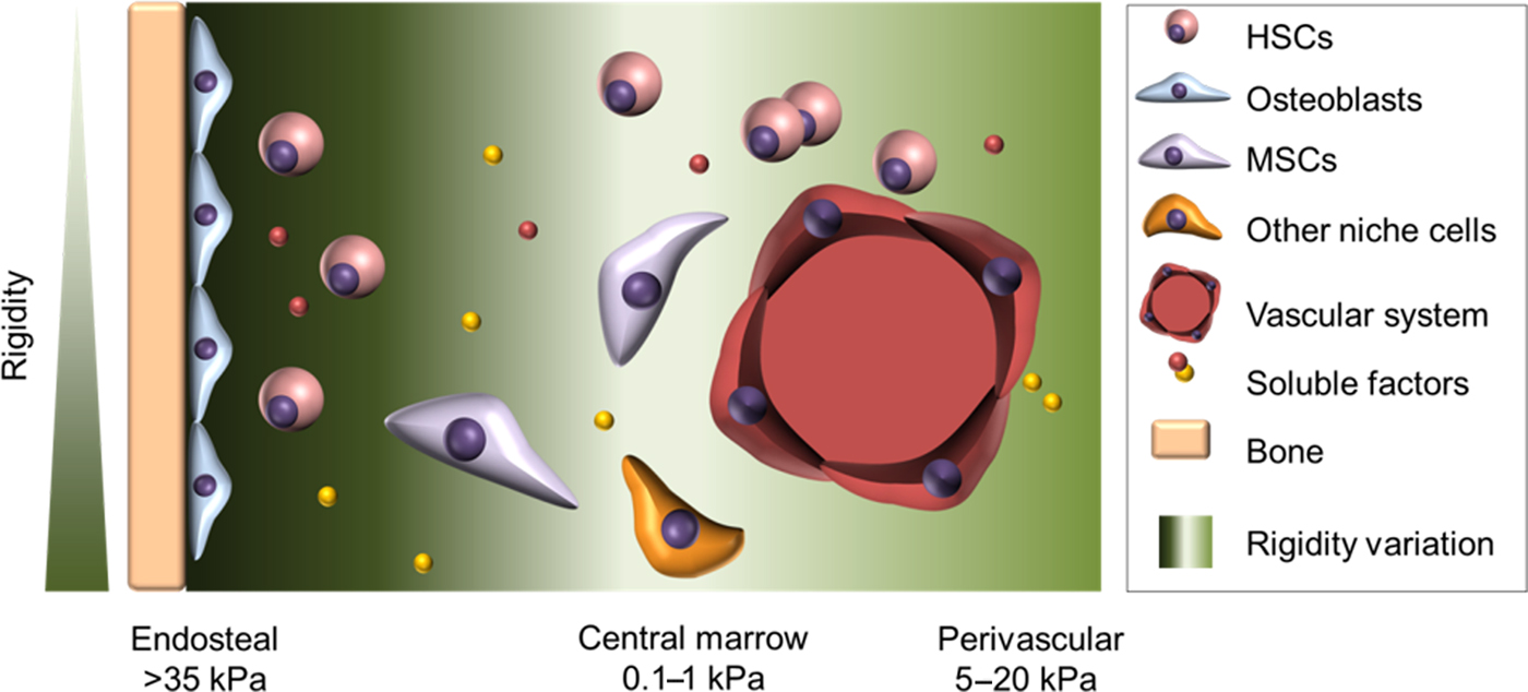
The red bone marrow, located in the cavities of the trabecular regions in long bones, represents a complex blood and bone cell-producing organ. Blood cells develop from immature hematopoietic stem and progenitor cells (HSPCs) and ensure the completion of several essential tasks like immune function, blood clotting or oxygen transport. Whereas most mature blood cells undertake their tasks mainly outside of the bone marrow, the only place where hematopoietic stem cells (HSCs) execute their functions (stem cell maintenance and blood cell production by differentiation) to guarantee lifelong blood production, is in their stem cell niche in the red part of the bone marrow.[Reference Crane, Jeffery and Morrison1] Current studies revealed the existence of such niches at certain sites of the bone marrow. These niches are illustrated in a simplified form in Fig. 1. One of those is described as the endosteal niche, characterized by bone regenerating osteoblasts (OBs) located at the endosteal surface of bone.[Reference Crane, Jeffery and Morrison1] OBs are able to regulate HSC function e.g., by activation of the Notch pathway and production of thrombopoietin (TPO), they are involved in controlling HSCs quiescence.[Reference Yoshihara, Arai, Hosokawa, Hagiwara, Takubo, Nakamura, Gomei, Iwasaki, Matsuoka, Miyamoto, Miyazaki, Takahashi and Suda2, Reference Calvi, Adams, Weibrecht, Weber, Olson, Knight, Martin, Schipani, Divieti, Bringhurst, Milner, Kronenberg and Scadden3] Another site for HSC niches was described close to the vascular structure near the endosteum, specified as arterioles, which have also an impact on HSC quiescence and thus form a unique arteriolar niche.[Reference Kunisaki, Bruns, Scheiermann, Ahmed, Pinho, Zhang, Mizoguchi, Wei, Lucas, Ito, Mar, Bergman and Frenette4] Nevertheless, dividing and non-dividing HSCs are mainly found in the perisinusoidal regions of the bone marrow.[Reference Acar, Kocherlakota, Murphy, Peyer, Oguro, Inra, Jaiyeola, Zhao, Luby-Phelps and Morrison5] Here, HSCs are in close proximity to blood vessel covering endothelial cells, which affect HSC dormancy and self-renewal via cell–cell contacts (e.g., via E-selectin). This region is generally referred to as the endothelial/perisinusoidal niche.[Reference Winkler, Barbier, Nowlan, Jacobsen, Forristal, Patton, Magnani and Levesque6, Reference Kiel, Yilmaz, Iwashita, Yilmaz, Terhorst and Morrison7] At this perivascular site, HSCs are in close contact with enriched mesenchymal stromal cells (MSCs) which are subdivided into Leptin+, C-X-C motif chemokine 12 (CXCL12) abundant reticular (CAR) or Nestin+ cells. These cell populations are an important source of factors required for HSC maintenance.[Reference Crane, Jeffery and Morrison1, Reference Acar, Kocherlakota, Murphy, Peyer, Oguro, Inra, Jaiyeola, Zhao, Luby-Phelps and Morrison5] Apart from these, hematopoietic cells which are found throughout the bone marrow niches, are also able to influence HSC fate.[Reference Crane, Jeffery and Morrison1] Among others, T-cells in the endosteal region regulate stem cell maintenance, whereas an ablation of megakaryocytes, the early precursor of blood clotting thrombocytes, from the bone marrow results in mobilization of HSCs into the peripheral blood system.[Reference Fujisaki, Wu, Carlson, Silberstein, Putheti, Larocca, Gao, Saito, Lo Celso, Tsuyuzaki, Sato, Cote, Sykes, Strom, Scadden and Lin8, Reference Chow, Lucas, Hidalgo, Mendez-Ferrer, Hashimoto, Scheiermann, Battista, Leboeuf, Prophete, van Rooijen, Tanaka, Merad and Frenette9] The named endothelial, perivascular or hematopoietic cells are just a selection out of many niche cells, which are all known to regulate HSC function and are important regulators in the environment of HSCs. The niche cells govern HSCs by providing cell–cell contacts or by the production of soluble factors. The most popular cell–cell contacts in the context of the HSC niche are cell-cell adhesions via N-cadherin, interactions, via vascular cell adhesion molecule 1 (VCAM1) and very late antigen-4 (VLA-4; integrin α4β1) or the Notch ligand–receptor interplay.[Reference Arai, Hosokawa, Toyama, Matsumoto and Suda10, Reference Schroeder and DiPersio11] In addition to these, soluble factors such as fibroblast growth factor 2, WNT ligands, stem cell factor (SCF) or CXCL12 expressed by MSCs as well as bone morphogenic proteins (BMPs) or angiopoietin expressed by other niche cells have an impact on cell cycle, maintenance of HSCs and normal niche function.[Reference Crane, Jeffery and Morrison1] Furthermore, niche cells produce extracellular matrix (ECM) components. The ECM, embedding the cells in their niche, consists of proteins such as collagen IV, glycoproteins such as fibronectin or glycosaminoglycans (GAGs, e.g. hyaluronic acid (HA)) and matrix remodeling enzymes. This protective shelter functions as a growth factor reservoir and determines the balance between stemness and differentiation with the help of cell–matrix interactions.[Reference Klein12–Reference Goncharova, Serobyan, Iizuka, Schraufstatter, de Ridder, Povaliy, Wacker, Itano, Kimata, Orlovskaja, Yamaguchi and Khaldoyanidi15] HSCs can bind to ECM proteins mainly via membrane-bound integrins.[Reference Coulombel, Auffray, Gaugler and Rosemblatt16] On one hand, this interaction can result in a protein sequence-specific cell response; on the other hand, the cells can sense the biophysical properties of the substrate leading to a substrate stiffness-dependent cell response. In the latter case, HSCs rearrange their cytoskeleton after substrate binding and exert traction to the substrate. In a process called mechanotransduction, this mechanical stimulus can be converted into a biochemical signal in the cell, which can influence HSC behavior.[Reference Lee-Thedieck and Spatz17] The stiffness in the bone marrow affecting HSC behavior ranges from 0.1 to 1 kPa in the central marrow and 5–20 kPa in the perivascular region up to >35 kPa in the endosteal part.[Reference Nelson and Roy18]
Schematic drawing of the bone marrow niche in vivo. The simplified representation of the niche in the bone marrow shows the microenvironment of HSCs (light red) consisting of several types of niche cells such as the OBs (blue) located at the endosteum, perivascular endothelial cells (red) or MSCs (purple) and biochemical stimuli due to the expression of soluble factors (red and yellow dots) by niche cells. Furthermore, biophysical parameters (different stiffnesses reviewed in Nelson & Roy[Reference Nelson and Roy18]) throughout the bone marrow which have an impact on cells behavior are illustrated in green.

This summarized overview of bone marrow components exhibits that HSCs are embedded in an intricate environment in which they receive cell-specific, biochemical, and biophysical signals from all three dimensions. This unique environment of the bone marrow represents the only place where HSCs are able to maintain their stem cell properties life-long. So far, in vitro culturing of HSCs is not sufficient to maintain stem cell properties and ends up with differentiation of the stem cells into committed progenitors or mature blood cells. Sufficiently in vitro multiplied HSCs from one donor could be used for cell transplantations into several patients with leukemia and thereby overcome the problem of donor limitations.[Reference Walasek, van Os and de Haan19] Furthermore, malignant diseases concerning the bone marrow can severely alter or even destruct the complex interaction in the HSC niche.[Reference Dombret and Gardin20, Reference Nombela-Arrieta and Isringhausen21] The treatment of such disorders comprises the medication with disease-specialized drugs.[Reference Dombret and Gardin20] For effective targeting of the disease and to improve the therapeutic outcome, new drugs have to be developed. To achieve these goals, more information on how biochemical and biophysical cues regulate HSC fate and how these signal transductions are changed in a diseased bone marrow, are required.
In order to cope with these scientific tasks, bone marrow model systems are needed which (i) allow analysis of human hematopoiesis in steady state and disease, (ii) can be used as drug-testing systems and (iii) facilitate the in vitro expansion of HSCs. For this purpose, animal models are often used, accepting the major drawback of insufficient transferability to human beings.[Reference Knight22] Furthermore, the ethical aspects and in this context the implementation of the 3Rs principle to replace, reduce and refine animal experiments urges alternatives.[Reference Törnqvist, Annas, Granath, Jalkesten, Cotgreave and Öberg23] Therefore, in vitro models have steadily been developed during the last years. The usage of conventional 2D cell culture devices is becoming less common due to the increasing knowledge of the in vivo conditions revealing several drawbacks of the highly artificial 2D environment. The complex interplay of cells with their surrounding in a 3D architecture affects the morphology of stem cells as well as their differentiation into mature cells disclosing the importance of a 3D environment for HSCs.[Reference Lee-Thedieck and Spatz17, Reference Choi, Mahadik and Harley24] In the following chapters, we review state-of-the-art 3D cell culture models mimicking the bone marrow in vivo in health and disease as well as methods for their construction with special emphasis on 3D bioprinting. Furthermore, their implementation for in vitro cell analysis and future aspects in context to refine bone marrow models are discussed.
Evolution of bone marrow models
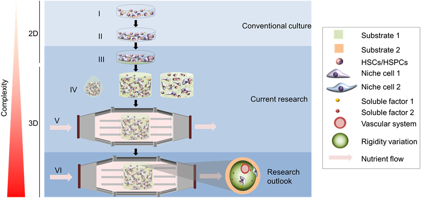
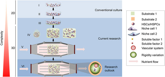
As already mentioned, improvements of the HSC expansion for clinical needs and of bone marrow models for fundamental studies and drug-testing are indispensable in future research. To reach this goal, HSC cultures which mimic relevant characteristics of their natural healthy niche in the bone marrow, need to be developed. The evolution of such bone marrow models, which comprised more and more aspects of a natural niche and increased in complexity in the last decades, is described in the following and summarized in Fig. 2. Conventionally, suspension culture systems are used to expand umbilical cord blood HSPCs in research or in clinical studies before transplantations. By adding cytokines to the culture medium, which is drafted in Fig. 2(I), HSC expansion could be improved significantly. Commonly used cytokines for HSC expansion are for example TPO, FMS-like tyrosine kinase 3 ligand (Flt3L), SCF, granulocyte colony-stimulating factor (GCSF), interleukin-6 (IL-6) and interleukin-3 (IL-3). Furthermore, several developmental regulators such as the Notch ligand Delta-1 or small molecules such as the copper chelator tetraethylenepentamine (TEPA), nicotinamide and StemReginin-1 were used in addition to the cytokine cocktails, which further improved the total cell and CD34+ HSPC expansion.[Reference Walasek, van Os and de Haan19] Likewise, several cell types that occur naturally in the bone marrow were described to assist HSC proliferation and maintenance by secreting HSC-supporting factors in cell cultures, which is delineated in Fig. 2(II). This includes, e.g., OBs, naturally present in the endosteal niche, endothelial cells, in vivo an important cell type of the vascular niche, and others that were mentioned before in the introduction. The most commonly used supporting cells in HSC cultures are MSCs because they express higher levels of HSC-supporting factors than other stromal cells,[Reference Mendez-Ferrer, Michurina, Ferraro, Mazloom, Macarthur, Lira, Scadden, Ma'ayan, Enikolopov and Frenette25] and they were already used to expand HSCs for transplantations in clinical studies. In clinical studies, neutrophil and platelet recovery were slightly improved in patients with previous in vitro HSPC expansion before transplantations compared with direct transplantations without HSPC expansion. However, no significant improvement in the survival of the patients was reported, as recently reviewed.[Reference Kiernan, Damien, Monaghan, Shorr, McIntyre, Fergusson, Tinmouth and Allan26] Conventional protocols supported the proliferation of HSCs but they did not foster the self-renewal of HSCs to a large extent, which led to a rapid decline of stemness in such in vitro cultures.[Reference Pineault and Abu-Khader27] With the purpose to improve the expansion of HSCs while maintaining stemness, researchers looked more closely at the natural stem cell niche and started trials to mimic it in its entirety, including chemical conditions, cell and ECM compositions as well as biophysical properties.
Schematic drawing of the development of 3D models. Evolution of conventional HSPC cultures into sophisticated 3D biomimetic bone marrow niches. In conventional culture systems, HSPCs are cultivated and expanded on 2D polystyrene devices in combination with cytokines and small molecules (i) or in addition to feeder cells (ii). By functionalizing the substrate and varying its physical parameters such as stiffness, ECM properties were mimicked in research applications (iii). Allowing cells to grow in a 3D environment, e.g., as spheroids, embedded in a polymer matrix or in the cavities of pores of polymer scaffolds (iv), reformed culturing processes of HSPCs. Using bioreactors or microfluidic devices further improved nutrient supply and added more parameters of the niche to the culture system (v). In future research, more attempts should be made to mimic the niche in more detail, combining various substrates (shown in light green and light orange), stiffnesses (displayed as brightness of the substrate-colors), cells (depicted by the blue and the purple cell), soluble factors (presented by yellow and red dots) and a vascular system (shown as a red circle) in one model (vi).

In this attempt, researchers tried to imitate the ECM of the bone marrow in many different ways. In standard cell culture, conventional tissue culture plates, which are made from polystyrene and are thus stiff (E = 3000 MPa[Reference Brandrup, Immergut and Grulke28]), were used to culture HSCs in suspension. By coating the culture devices with natural components of the bone marrow ECM, such as fibronectin or collagen, as shown in green in Fig. 2(III), HSCs were enabled to attach to this matrix. The attachment of HSCs does not only affect the binding itself but also influences proliferation and stemness of the cell e.g., by mechanotransduction, which is why it is important to allow attachment of the HSCs also in in vitro cultures. Instead of proteins, also smaller bioactive sequences, such as the minimal integrin recognition motif RGD or the fibronectin fragment CS-1, were applied to allow the attachment of the cell to synthetic matrices. Placing these proteins and peptides in a controlled manner by nanopatterning added another biophysical parameter to culture systems since nanoscale topography and nanopatterning also have effects on cell behavior. For example, scientists found out that a distance less than 45 nm between cyclic RGD peptides is required for proper HSC attachment and that efficient cell-peptide adhesion may foster proliferation and differentiation of HSCs.[Reference Muth, Steinl, Klein and Lee-Thedieck29] Another physical parameter, which influences stem cell behavior, is the stiffness of the substrate. Cells can track the stiffness of a substrate by mechanosensing and thereupon change their behavior. For example, when MSCs were grown on a stiff substrate, they differentiated and expressed markers of osteogenic lineages whereas on soft surfaces they expressed neurogenic lineage markers.[Reference Engler, Sen, Sweeney and Discher30] For HSPCs it was found that they adhered better, were more motile and kept a higher level of stemness when they were cultured on stiffer hydrogels.[Reference Lee-Thedieck and Spatz31] They proliferated the most if they were cultured on surfaces with intermediate stiffness (approximately 12.2 kPa).[Reference Kumar, Hsiao, Ling, Dulinska-Molak, Chen, Chang, Chang, Chen, Chen, Hsu and Higuchi32]
In an in vitro 2D culture it is almost impossible to reciprocate complex biochemical and biophysical aspects as they can be generated in 3D in vitro cultures. For instance, the 2D environment does not mimic the 3D bone marrow in vivo in architecture, leading to an irrelevant phenotype of cells.[Reference Zhang and Lodish33] In contrast to a 3D culture, cells in 2D grow in an unnatural dorsal-ventral polarization affecting their contact surface for cell–cell or cell–matrix interactions, which in turn influences cell morphology. For example, MSCs flattened when cultured on 2D surfaces, which further affected the tension in the MSC cytoskeleton and the expression of pluripotency genes.[Reference Zhou, Chen, Li and Wu34] On top of that, cells in 2D culture are covered by large volumes of medium. Secreted molecules are diluted immediately in the medium and no signal gradients can be created which impairs autocrine and paracrine signaling.[Reference Rodling, Schwedhelm, Kraus, Bieback, Hansmann and Lee-Thedieck35] Due to these drawbacks, the idea came up to also consider dimensionality in cell culturing.[Reference Lee-Thedieck and Spatz17] Many different 3D cell culture systems with different complexity were developed and found to support HSC expansion and maintenance to a higher level than 2D systems. In the following part, some of them will be presented.
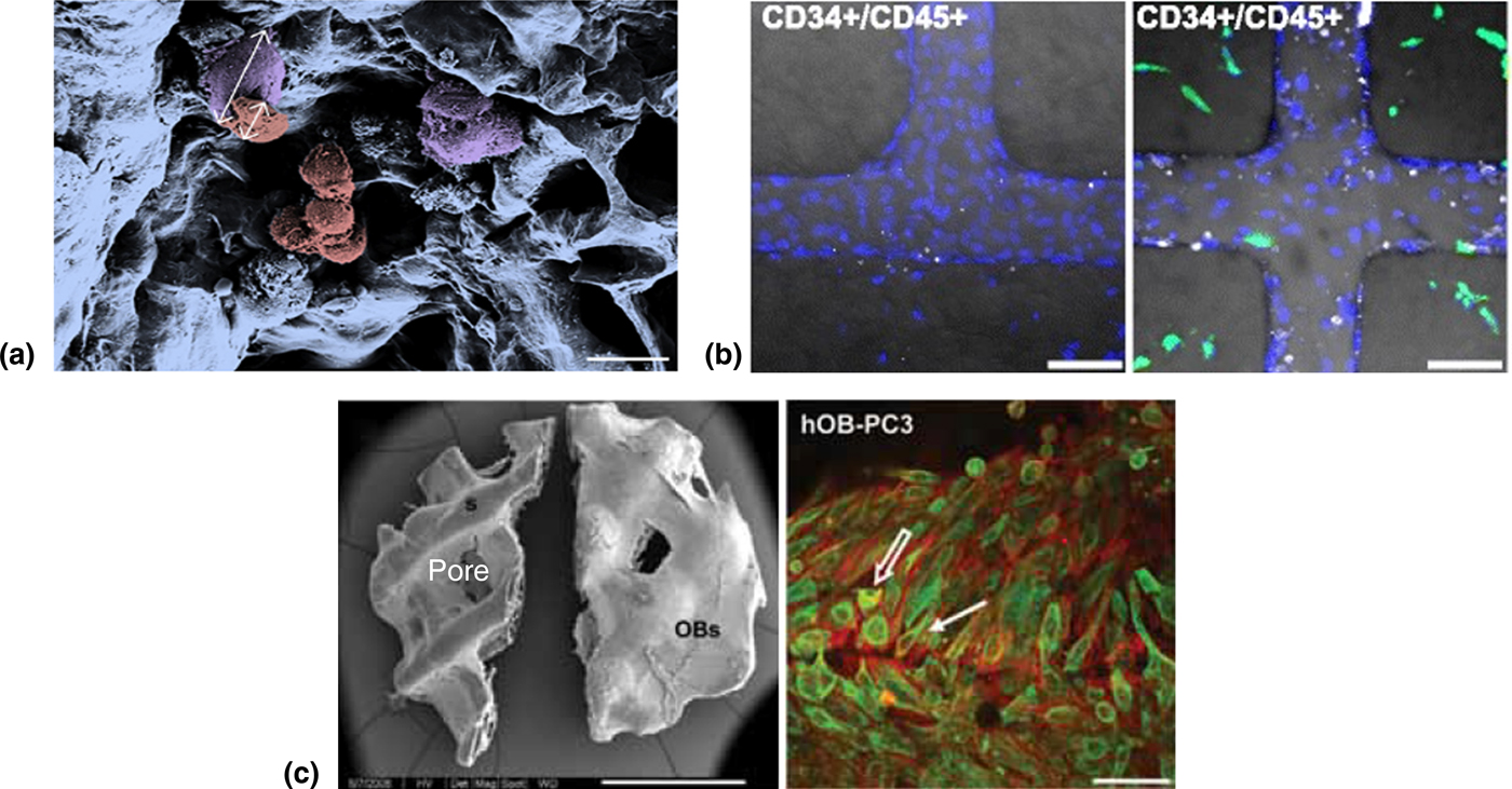
The simplest 3D in vitro cultures are spheroids. To generate spheroids, cells can be cultured in hanging drops or on non-adhesive surfaces to force them to form aggregates. In Figure 2(IV) a hanging drop culture is sketched, comprising an aggregate of different cell types within a drop of medium. An example is the high-throughput coculture system with MSCs and HSCs presented by Cook and colleagues.[Reference Cook, Futrega, Osiecki, Kabiri, Rice, Atkinson, Brooke and Doran36] They used surface modified microwells to support the formation of microaggregates, which then developed spatial structures. It could be observed that, in comparison with 2D cultures, MSCs in the aggregates expressed higher levels of hematopoietic niche factors and the HSC expansion increased. Such spheroids have the advantages that the cells can form proper cell–cell contacts and tissue-like patterns, they keep their natural shape, signaling molecules are not diluted into the medium (except at the spheroid–medium interface) and thus cells can communicate properly.[Reference Costa, de Melo-Diogo, Moreira, Carvalho and Correia37] Currently, a widespread method to culture HSCs in 3D is the incorporation of cells into a polymer solution that is cross-linked to form a gel (depicted in green in the second part of Fig. 2(IV)) in the following. The cells are encapsulated in the forming hydrogel, which resembles the ECM in vivo. Natural or synthetic polymers, which are able to swell in water, were used for this purpose. Materials such as fibronectin, collagen, laminin, and GAGs occur in the bone marrow in vivo and HSCs attach easily to them, which is why they were often used in 3D cell cultures.[Reference Lee-Thedieck and Spatz31] Other natural materials used for scaffolds were chitosan, alginate, Matrigel™ (gelatinous protein mixture), agarose, and bacterial nanocellulose.[Reference Salamanna, Contartese, Maglio and Fini38, Reference Sitarski, Fairfield, Falank and Reagan39] By functionalizing synthetic matrices with small peptides, such as RGD, the culture conditions can be controlled precisely, while native ECM molecules, such as HA, not only affect adhesion but also influence a lot of other signaling pathways.[Reference Necas, Bartosikova, Brauner and Kolar40] Conventional synthetic polymers used for hydrogels are, e.g., poly (lactic acid) (PLA), poly (ε-caprolactone) (PCL), poly (lactic-co-glycolic acid) (PLGA) and poly (ethylene glycol) (PEG). These materials were used alone or in conjunction with each other. In some models, inorganic components of the bone such as hydroxyapatite (HAP) and tricalcium phosphate (TCP) were also incorporated.[Reference Salamanna, Contartese, Maglio and Fini38, Reference Sitarski, Fairfield, Falank and Reagan39] Proper attachment of cells and cell compatibility are major requirements of these gels and the possibility to modulate the gel properties, such as stiffness, is a further advantage to improve them as culture systems. For the further usage of HSCs, e.g., in transplantations, it is important that the cells can be efficiently recovered from the gels without harming them. This requirement could be met by degradable gels. Gels with natural polymers can, for example, be degraded by enzymes whereas in synthetic materials special cross-linkers (e.g., polymers with disulfide bonds), which are cleaved by special stimuli such as light (photocleavable linkers, e.g., Fairbanks et al.[Reference Fairbanks, Singh, Bowman and Anseth41]) or reducing agents (e.g., Zhang et al.[Reference Zhang, Skardal and Prestwich42]) can be introduced.[Reference Kharkar, Kiick and Kloxin43] Such stimuli-responsive gels gain more and more importance nowadays. An interesting application is the usage of biodegradable hydrogels. Cytokines, small molecules or other factors that influence cells are incorporated into the gel and undergo a sustained release during the process of degradation induced by the cells themselves (e.g., Cheng et al.[Reference Cheng, Su, Cao, Luo, Li, Pu and He44]). Also cell migration, cell–cell contacts and stemness of the cells was improved, if cells were able to remodel their matrix.[Reference Madl, LeSavage, Dewi, Dinh, Stowers, Khariton, Lampe, Nguyen, Chaudhuri, Enejder and Heilshorn45] The advantages of encapsulation are that cells have full contact with the matrix and that signaling molecules are not diluted quickly. However, the cells are often not in direct contact. Since the natural bone marrow does not consist of a homogeneous matrix but is perforated by pores, meshworks made of fibers and porous sponge-like scaffolds, such as the sketch in the third part of Fig. 2(IV), were developed to mimic this architecture. The cells were added after scaffold preparation. Thus, in these approaches, the cells were not directly surrounded by the gel, but they were gathered in the pores of the gel. This permitted the formation of close cell–cell contacts and a 3D architecture for cell-matrix contacts.[Reference Lee-Thedieck and Spatz17] The pores could be generated by different methods, such as salt leaching, freeze-drying or 3D printing and by changing parameters of the manufacturing protocols, pore sizes and shapes could be varied. To mimic the in vivo conditions and to provide an adequate architecture for the 3D organization of the cells, while allowing for enough place for nutrient exchange, pore sizes between 70 and 300 µm were chosen.[Reference Henderson, Ladewig, Haylock, McLean and O'Connor46, Reference Raic, Rodling, Kalbacher and Lee-Thedieck47] A full interconnectivity of the pores was achieved, so that the medium and the cells could completely penetrate the scaffold. Examples for successful applications of porous 3D hydrogels for the expansion of HSPCs in vitro are the scaffolds of Ferreira et al.[Reference Ferreira, Jahnen-Dechent, Labude, Bovi, Hieronymus, Zenke, Schneider and Neuss48] and Raic et al.[Reference Raic, Rodling, Kalbacher and Lee-Thedieck47]. Ferreira et al. tested several natural polymers as scaffolds in combination with either cytokines or MSCs as support and found fibrin in combination with MSCs to be the most promising system with a 3 × 10⁷-fold expansion of CD34+ cells compared with conventional 2D systems.[Reference Ferreira, Jahnen-Dechent, Labude, Bovi, Hieronymus, Zenke, Schneider and Neuss48] Figure 3(a) shows a scanning electron micrograph of HSCs and MSCs attaching to a synthetic poly (ethylene glycol) diacrylate (PEGDA)-RGD scaffold. Raic et al. showed that HSPC expansion was upregulated in this scaffold in comparison to the expansion in 2D tissue culture plates if HSPCs were cocultured with MSCs.[Reference Raic, Rodling, Kalbacher and Lee-Thedieck47]
In vitro models of the bone marrow in health and disease. (a) Example of a bone marrow niche model in healthy conditions: Pseudo colored scanning electron micrograph of a porous PEGDA-RGD hydrogel scaffold (blue) from Raic et al. The 3D culture system was seeded with MSCs (purple) from the bone marrow and HSPCs (red). Arrows indicate the size of the cells (MSC ≈ 28 µm, HSPC ≈ 12 µm). Scale bar = 20 µm. Adapted/reproduced with permission from Elsevier, ref. Reference Raic, Rodling, Kalbacher and Lee-Thedieck47 (b) In vitro model of a leukemic niche: Microvessel system lined by endothelial cells mimicking the vascular niche developed by Kotha and colleagues. The LCs (blue) were perfused in the microvessel system which was embedded in collagen type I (left). Addition of fibroblasts (HS5-cells; green) into the matrix around the vessels led to increased adherence and migration of LCs (right). Scale bars = 100 µm. Adpated/reproduced with permission from BioMed Central, ref. Reference Kotha, Hayes, Phong, Redd, Bomsztyk, Ramakrishnan, Torok-Storb and Zheng49 (c) A model simulating prostate cancer bone metastasis: On the left is a scanning electron microscopy image of the scaffold prepared from mPCL-TCP and then seeded with human OBs. Within 3 weeks, OBs formed a sheet on the scaffold. Scale bar = 2 mm. On the right is the immunofluorescent staining of OBs with phalloidin (red) and PC-3 cells with anti-pan CK (green) after 6 days of coculture. PC-3 cells are characterized by a spherical (white open arrow) and fibroblast appearance (white closed arrow). Scale bar = 75 µm. Adapted/reproduced with permission from Taylor and Francis Ltd. ref. Reference Sieh, Lubik, Clements, Nelson and Hutmacher50.

To culture large amounts of cells in an active state over a long period of time, bioreactors were used. Here, we use the term bioreactor to describe an engineered system that allows automated control over medium supply, waste removal, and agitation. Bioreactors were combined with 2D or 3D applications. Rödling et al. developed a bioreactor containing a PEGDA-RGD scaffold, which resembles the drawing in Fig. 2(V). It could mimic the niche in an active or in a steady-state condition that either supported HSPC differentiation or HSPC maintenance.[Reference Rodling, Schwedhelm, Kraus, Bieback, Hansmann and Lee-Thedieck35] Microfluidic devices can also accomplish a continuous nutrient and oxygen supply and, furthermore, gradients (e.g., oxygen, calcium ions, cytokines, and small molecules) can be generated.[Reference Bello, Park and Lee51] Such systems operate with extremely small amounts of fluids (10−6 to 10−15 mL) using laminar flow microchannels (1–1000 µm). To mimic blood vessels that are pervading the tissue in vivo, microfluidic devices were used and in this way, another important parameter of the bone marrow—the mimicry of vasculature and perfusion—could be included into models. Kotha et al. developed a sophisticated model that mimics very well the vascular niche and the process of HSC homing in a microfluidic device, which comprises various cell types, a flow system, and a soft matrix.[Reference Kotha, Hayes, Phong, Redd, Bomsztyk, Ramakrishnan, Torok-Storb and Zheng49] They constructed an endothelialized vascular network in a collagen matrix with MSCs and other bone marrow stromal cells. After perfusing the network with medium containing monocytes (CD14+/CD45+) or HSPCs (CD34+/CD45+), they could observe these cells adhering to or migrating into the soft matrix. Very complex microfluidic systems are organs-on-a-chip. An interesting example is the bone marrow-on-a-chip from Torisawa and colleagues.[Reference Torisawa, Spina, Mammoto, Mammoto, Weaver, Tat, Collins and Ingber52] They implanted a collagen gel with demineralized bone powder and BMP subcutaneously into a mouse where it developed into a bone with an intact bone marrow. Afterward, the bone was taken out and put into a microfluidic device where it could be cultured for at least 1 week. This system proved to be a proper in vitro model for irradiation toxicity tests. However, it was of murine origin and did thus not represent the human bone marrow. Reinisch et al. developed humanized bone marrow-ossicles by implanting human MSCs into mice and let them form bones, which were then chimeric, of murine and human origin.[Reference Reinisch, Hernandez, Schallmoser and Majeti53] Such microfluidic systems are also one of many possible approaches to mimic the bone marrow in malignant diseases by imitating the invasion of cancer cells from the bloodstream into the bone marrow in vitro. In the following two chapters, such approaches for models of leukemia and bone metastasis will be presented and discussed.
Leukemic niche models
In hematological disorders such as leukemia, the bone marrow is overflooded by malignant hematopoietic cells leading to a disturbed or insufficient hematopoiesis.[Reference Riether, Schurch and Ochsenbein54] Transformation of hematopoietic cells into malignant cells can occur due to genetic events, hereditary or environmental conditions and affect the myeloid or lymphoid cell lineage.[Reference Riether, Schurch and Ochsenbein54, Reference Stieglitz and Loh55] A small proportion of these cells regain stem cell properties, such as an unlimited self-renewal potential and the ability to initiate leukemia as well as a reduced differentiation potential.[Reference Riether, Schurch and Ochsenbein54, Reference Lapidot, Sirard, Vormoor, Murdoch, Hoang, Caceres-Cortes, Minden, Paterson, Caligiuri and Dick56] The malignant leukemic cells (LCs) occupy the bone marrow niche for their own survival and an uncontrolled proliferation contributing to disease progression.[Reference Brenner, Nepstad and Bruserud57, Reference Schepers, Pietras, Reynaud, Flach, Binnewies, Garg, Wagers, Hsiao and Passegue58] The highly organized HSC niche is rearranged by LCs and a leukemic niche is formed, accompanied by a loss of important signals and niche cells, impairing normal homeostasis.[Reference Schepers, Pietras, Reynaud, Flach, Binnewies, Garg, Wagers, Hsiao and Passegue58] In this protective environment, LCs are not assailable for some therapeutic procedures aiming at the destruction of malignant cells.[Reference Jacamo, Chen, Wang, Ma, Zhang, Spaeth, Wang, Battula, Mak, Schallmoser, Ruvolo, Schober, Shpall, Nguyen, Strunk, Bueso-Ramos, Konoplev, Davis, Konopleva and Andreeff59] Due to their acquired stem cell properties and the interception of anti-apoptotic signals, they can survive in the marrow despite harsh conditions during treatment and can lead to a reappearance of the disease.[Reference Riether, Schurch and Ochsenbein54, Reference Jacamo, Chen, Wang, Ma, Zhang, Spaeth, Wang, Battula, Mak, Schallmoser, Ruvolo, Schober, Shpall, Nguyen, Strunk, Bueso-Ramos, Konoplev, Davis, Konopleva and Andreeff59, Reference Saito, Uchida, Tanaka, Suzuki, Tomizawa-Murasawa, Sone, Najima, Takagi, Aoki, Wake, Taniguchi, Shultz and Ishikawa60] Development of new therapeutic strategies to address this problem needs in vitro tools overcoming the limitations of animal models, in most cases mouse-based model systems.[Reference Cook and Pardee61] Apart from the obvious discrepancies between human and mice, such as bodyweight, lifespan or fertility, the differences in the assembly of the peritumoral milieu limit the often used xenotransplantation models which allow the investigation of human cells in an in vivo system.[Reference Cook and Pardee61–Reference Demetrius63] Therefore, in vitro models gain more and more importance in investigating the origin and the progression of hematopoietic malignancies and the associated signaling pathways to develop new treatment strategies. For this purpose, current studies are focused on the establishment of in vitro models which allow analysis of cell–cell and cell–matrix interactions as well as the interplay between LCs and soluble factors in a 3D environment resembling the in vivo conditions. In the following part, in vitro models are presented that allow investigations of these regulatory cell interactions and contribute new hints about the function of the human leukemic bone marrow. Similar to HSCs, LCs are also found in the vascular or endosteal part of the bone marrow and can interact with cell-populations of these microenvironments.[Reference Ishikawa, Yoshida, Saito, Hijikata, Kitamura, Tanaka, Nakamura, Tanaka, Tomiyama, Saito, Fukata, Miyamoto, Lyons, Ohshima, Uchida, Taniguchi, Ohara, Akashi, Harada and Shultz64, Reference Cogle, Goldman, Madlambayan, Leon, Masri, Clark, Asbaghi, Tyner, Dunlap, Fan, Kovacsovics, Liu, Meacham, Hamlin, Hromas, Scott and Fleming65] Bray et al. constructed a 3D PEG-heparin hydrogel with human umbilical vein endothelial cells (HUVECs) amongst others as environmental stimuli to investigate the impact of the vascular niche in acute myeloid leukemia (AML). They found that cells from the vascular network in a 3D scaffold support the resistance of LCs against chemotherapeutics.[Reference Bray, Binner, Korner, von Bonin, Bornhauser and Werner66] Furthermore, Kotha and colleagues also developed an artificial vascular microenvironment which allowed studying 3D multicellular interactions and cell trafficking between distinct fibroblasts and LCs. In the in vitro vascular marrow, they showed that LCs respond to distinct fibroblasts which were embedded in a collagen type I matrix around the vessels with increased adhesion and migration within the vessel network which is shown in Fig. 3(b).[Reference Kotha, Hayes, Phong, Redd, Bomsztyk, Ramakrishnan, Torok-Storb and Zheng49] In an engineered microfluidic 3D collagen matrix, presenting an endosteal in vitro platform, Bruce and colleagues showed direct cell–cell interactions in 3D between MSCs and in vivo endosteally located OBs with LCs from acute lymphoblastic leukemia.[Reference Bruce, Evans, Mezan, Shi, Moses, Martin, Gibson and Yang67] Using in vitro transwell filter devices revealed an influence of MSCs, which represent the main population in bone marrow stroma, on survival and viability of LCs isolated from patients with chronic lymphatic leukemia. In addition, they found that a coculture of MSCs with leukemic B cells promote the cytokine production of MSCs, concluding that LCs are able to rearrange their cytokine milieu.[Reference Trimarco, Ave, Facco, Chiodin, Frezzato, Martini, Gattazzo, Lessi, Giorgi, Visentin, Castelli, Severin, Zambello, Piazza, Semenzato and Trentin68] These extracellular communication molecules are important for cell homing, migration and growth. Changing their composition by increasing the expression of homing or anti-apoptotic factors in the bone marrow can support LCs survival.[Reference Jacamo, Chen, Wang, Ma, Zhang, Spaeth, Wang, Battula, Mak, Schallmoser, Ruvolo, Schober, Shpall, Nguyen, Strunk, Bueso-Ramos, Konoplev, Davis, Konopleva and Andreeff59, Reference Tavor, Petit, Porozov, Avigdor, Dar, Leider-Trejo, Shemtov, Deutsch, Naparstek, Nagler and Lapidot69] Therefore, studying cell behavior in response to soluble molecules is of great importance to find new treatment strategies that are based on abrogating the connection between LCs and their supportive environment. With the objective of finding molecular mechanisms to reach this goal, migration studies in a 3D filter system revealed the involvement of the Raf/MEK/ERK1/2 pathway in the cell response of leukemic T cells after binding of the CXCL12 homing factor.[Reference Mills, Goh, Kudatsih, Ncube, Gurung, Maxwell and Mueller70] CXCL12 is known to mediate chemoresistance after binding to its receptor C-X-C chemokine receptor type 4 (CXCR4) on the cell surface of LCs in different leukemia types.[Reference Cho, Kim and Konopleva71, Reference Liou, Delgado-Martin, Teachey and Hermiston72] Due to its great importance, several 3D cell culture platforms were developed to analyze the impact of the CXCL12/CXCR4 axis on LCs migration. Promising in vitro studies concerning disruption of these interactions showed a decreased drug resistance of chronic myeloid leukemia cells after pretreatment with a CXCR4 inhibitor.[Reference Weisberg, Azab, Manley, Kung, Christie, Bronson, Ghobrial and Griffin73] The combination of a CXCL12 inhibitor and a tyrosine kinase inhibitor reduced cell migration to a greater extent than either drug alone.[Reference Weisberg, Sattler, Azab, Eulberg, Kruschinski, Manley, Stone and Griffin74] In the endosteal region of the bone marrow, OBs express osteopontin which can bind to its receptor on LCs leading to protection against apoptosis. In a 3D polystyrene scaffold coated with OBs and LCs, the addition of an inhibitor for the osteopontin receptor together with a tyrosine kinase inhibitor disrupted the adhesion between the LCs and the protective microenvironment and increased their sensitivity to drugs. Therefore, blocking the LC-binding to osteopontin could be an efficient therapy for patients with AML.[Reference Shen, Zeng, Wang, Ma, Zhang and Kong75] Cells encapsulated in a 3D surrounding not only communicate with other niche cells and their expressed cytokines but also their interactions with the ECM allocate cell behavior. Porous scaffolds fabricated from polymeric materials coated with the ECM protein collagen were proven to be suitable for the culture of LC lines in contrast to uncoated controls, highlighting the importance of cell-ECM interactions in cell growth.[Reference Blanco, Mantalaris, Bismarck and Panoskaltsis76] Furthermore, migration experiments in a 3D transwell assembly showed an improved migration of a leukemic myeloid cell line during culture with collagen IV.[Reference Favreau, Vary, Brooks and Sathyanarayana77] Investigations of matrix mechanics and its role on LCs revealed that matrix stiffness of 3D alginate hydrogels regulates proliferation of AML cell lines through autocrine signaling. In addition, matrix stiffness, as well as the integrin ligand (RGD) functionalized scaffolds modulated resistance of AML cells against protein kinase inhibiting drugs.[Reference Shin and Mooney78] 3D alginate hydrogels, mimicking the soft marrow, supported cell growth to a higher extent than the 2D control, led to more diverse myeloid progenitor cells and had an impact on cell cycle.[Reference Vu, Lim and Lim79] The discussed studies indicate that the bone marrow microenvironment including cell–cell, and cell–matrix contacts as well as soluble factors play a critical role in leukemia progression and survival and protects the malignant cells against chemotherapeutic treatment. Results from a previous study, showing that the drug sensitivity of LCs depended on the dimensionality of the environment, further stress the need for new 3D in vitro cell culture platforms.[Reference Bruce, Evans, Mezan, Shi, Moses, Martin, Gibson and Yang67] Rödling et al. developed a reactor system for perfusion of 3D scaffolds mimicking the bone marrow in vivo. They could demonstrate the relevance of perfusion during drug treatment as results differed with and without perfusion.[Reference Rodling, Schwedhelm, Kraus, Bieback, Hansmann and Lee-Thedieck35] Therefore, using 3D systems can not only provide new information about the regulation of LCs in an in vivo like environment but is also a promising tool as a drug-testing platform to find new therapeutic strategies for patients with leukemia.
Bone metastasis models
Hematological malignancies such as leukemia, lymphoma, and multiple myeloma are not the only cancers affecting the bone. Cancer may arise in any organ of the human body. However, it is the metastases, rather than the primary tumor, that result in majority of the cancer-related deaths.[Reference Gupta and Massagué80] Metastasis is the multistage progression of cancer cells from the primary tumor to other organs, which, despite extensive research, remains the most enigmatic phenomenon in cancer pathophysiology.[Reference Gupta and Massagué80, Reference Fidler81] In fact, bone occupies the third position among the metastatic sites, briefly trailing behind lungs and liver.[Reference Macedo, Ladeira, Pinho, Saraiva, Bonito, Pinto and Goncalves82]
Metastasis is a complex series of events beginning with the detachment of cancer cells from the primary tumor and entry into the blood and lymph vessels (intravasation). These circulating tumor cells translocate to secondary organs, fighting immune resistance and tolerating mechanical forces in the process. At this point, they exit from the bloodstream (extravasation), settle in and adapt to the new microenvironment. Now, the disseminated tumor cells may either enter dormancy as single cells or as small clusters (micrometastases), during which they develop therapeutic resistances and long-term survival strategies. The disseminated tumor cells deploy survival signals which give rise to drug-resistant clones and finally proceed to form macrometastases.[Reference Fidler81, Reference Massagué and Obenauf83, Reference Peitzsch, Tyutyunnykova, Pantel and Dubrovska84] Events behind metastasis, such as the invasion of circulating tumor cells, growth and survival of disseminated tumor cells, evasion of host-tissue defenses, and amplification of survival signals are not only dependent on the innate characteristics of the primary tumor but also on the secondary tissue microenvironment. Different types of cancer show organ-specific metastasis, simultaneously or sequentially. Prostate cancer predominantly spreads to the bone, breast cancer to bone, liver, brain, and lungs and colorectal cancer begins with liver metastasis and moves on to lungs and brain.[Reference Obenauf and Massagué85, Reference Chambers, Groom and MacDonald86]
About 70% of breast and prostate cancers lead to skeletal metastases, followed by thyroid, lungs, kidney and bladder carcinomas.[Reference Salamanna, Contartese, Maglio and Fini38, Reference Macedo, Ladeira, Pinho, Saraiva, Bonito, Pinto and Goncalves82] Multiple myeloma can also be considered as a bone metastatic disease as it not only originates in the bone but also spreads throughout the bone marrow.[Reference Macedo, Ladeira, Pinho, Saraiva, Bonito, Pinto and Goncalves82, Reference Roodman and Silbermann87, Reference Sitarski, Fairfield, Falank and Reagan88] Bone metastasis has an extremely poor prognosis and is mainly characterized by severe chronic pain, impaired mobility, hypercalcemia, spinal cord compression, pathological fractures, and bone marrow aplasia. The appearance of these symptoms significantly decreases the quality of life of the patient and, in most cases, results in death.[Reference Salamanna, Contartese, Maglio and Fini38, Reference Macedo, Ladeira, Pinho, Saraiva, Bonito, Pinto and Goncalves82, Reference Roodman and Silbermann87]
So, what is the reason behind such organ-specific metastasis? In 1889, Dr. Stephan Paget, an English surgeon, put forward his “seed and soil” hypothesis, shedding some light on this phenomenon. He proposed that specific organs provide a comfortable home or the “soil” to certain tumor cells, the “seed”. Progression of metastatic colonization occurs only when “the seed” has found its hospitable “soil”.[Reference Fidler81] The bone marrow is a highly vascularized tissue possessing a rich milieu of cytokines, chemokines and growth factors. It also harbors an arsenal of different cell types such as immune cells, MSCs, HSPCs, bone-forming OBs and bone-resorbing osteoclasts, which express specific molecules that facilitate tumor invasion.[Reference Butcher, Alliston and Weaver89] Kang and colleagues showed that osteolytic factor IL-11, connective tissue growth factor, membrane receptor/adhesive proteins CXCR4 and osteopontin as well as the metalloproteinase1 (MMP1) are important parameters that lead breast cancer cells to metastasize to bone.[Reference Kang, Siegel, Shu, Drobnjak, Kakonen, Cordón-Cardo, Guise and Massagué90]
Stem cell niches present in the bone marrow are of particular interest to the cancer cells. Shiozawa et al. presented that prostate cancer cells and HSCs actively compete with each other for the occupation of endosteal niches. Bone marrow stromal cells and OBs express adhesion molecules annexin A2 (AXA2) and CXCL12 and facilitate the CXCR4 and the AXA2-expressing cancer cells and HSCs to bind and home to the bone marrow. When the cancer cells succeed in hijacking the niche, they force the HSCs to differentiate terminally.[Reference Shiozawa, Pedersen, Havens, Jung, Mishra, Joseph, Kim, Patel, Ying, Ziegler, Pienta, Song, Wang, Loberg, Krebsbach, Pienta and Taichman91]
Such settings provide the perfect hotbed for the cancer cells to invade, thrive and expand. The bone always undergoes active remodeling which requires a strict balance between OBs and osteoclasts. However, during metastatic colonization, this equilibrium is disrupted and leads to an abnormal increase in either cell type.[Reference Massagué and Obenauf83] Osteolytic lesions are usually common in breast cancer and multiple myeloma whereas prostate cancers are more prone to osteoblastic metastasis.[Reference Macedo, Ladeira, Pinho, Saraiva, Bonito, Pinto and Goncalves82, Reference Roodman and Silbermann87] This is why it is important to investigate cancer metastasis in a bone marrow microenvironment.
In general, tumor microenvironment (TME) is the amalgamation of all cellular and extracellular components surrounding cancer cells with which they maintain an effective crosstalk. Cancer-associated fibroblasts, transformed epithelial cells, tumor-infiltrating MSCs, immune cells, adipocytes, and endothelial cells of the blood and lymphatic systems are some of the cellular constituents of the TME. Cadherins are the most important molecules aiding interactions among the cellular components whereas cell–matrix interactions are usually established by integrins present on the cells. In the TME, various growth factor- and cytokine-mediated signalings take place, in an autocrine or a paracrine manner.[Reference Insua-Rodríguez and Oskarsson92–Reference Xu, Farach-Carson and Jia94] Such interactions with the tumor cells influence various biologic characteristics such as migration, proliferation, quiescence, immune, and therapeutic resistance. The presence of a physicochemical gradient in an avascular tumor mass not only limits the access of oxygen, nutrients, and soluble factors but also alters the diffusion profile of drugs into the tumor.[Reference Insua-Rodríguez and Oskarsson92, Reference Nath and Devi93] Apart from biochemical events, changes in cellular rheology is also a hallmark of cancer metastasis. As the formation of macrometastasis progresses, cells are exposed to a myriad of physical forces including hydrostatic pressure, shear stress, compression, and tension forces.[Reference Butcher, Alliston and Weaver89] In an in vitro 2D culture it is almost impossible to reciprocate a multitude of biochemical and biophysical aspects. Therein lies the importance of in vitro 3D models, which provide researchers with a wider horizon. Cancer cells can be cultured alone or in conjunction with other cell types in a spatially-relevant manner, supporting cell–cell and cell–matrix interactions.[Reference Salamanna, Contartese, Maglio and Fini38, Reference Butcher, Alliston and Weaver89, Reference Nath and Devi93] A lion's share of such studies has incorporated breast and prostate cancer cells. Apart from them, myeloma, lung, renal, colorectal and ovarian cancer cells have also been investigated in mono or multi-cultures with TME components.[Reference Salamanna, Contartese, Maglio and Fini38]
Naturally-derived materials, which are commonly utilized in hydrogel fabrication, influence cancer cell adhesion, and their metastatic potential. HA is a preferred substance for bone marrow scaffold formations due to its mechanical properties.[Reference Sitarski, Fairfield, Falank and Reagan88] Pan et al. performed a comparative study between 2D and 3D culture systems using the renal carcinoma cell line 786-O in thiol-modified HA and PEG-based scaffolds. Compared to the 2D cultures, there was a strong resemblance between the gene expression patterns of the cells in 3D culture to those observed in vivo. Adhesion molecules, osteolytic and angiogenic factors, associated with bone metastasis, were strongly expressed in 3D rather than 2D.[Reference Pan, Fong, Martinez, Harrington, Lin, Farach-Carson and Satcher95] Silk has proven to be a promising material for bone metastatic models owing to its biocompatibility, strength, toughness, porosity, thermal, and chemical stability.[Reference Sitarski, Fairfield, Falank and Reagan88] Kwon et al. demonstrated the “osteomimicry” or bone cell-like behavior of metastatic prostate cancer cell line PC-3 when cultured on silk scaffolds. They displayed migration, invasion, survival, and proliferation similar to their in vivo environment.[Reference Kwon, Kim, Rice, Subramanian, Park, Georgakoudi and Kaplan96] Similar observations were made by Cox and colleagues, by conducting experiments with breast cancer cells in collagen-GAG scaffolds.[Reference Cox, Jenkinson, Pohl, O'Brien and Morgan97] Fitzgerald et al. prepared scaffolds using nanoHAP and collagen and cocultured prostate cancer cells (PC-3 and LNCaP) with OBs. MMP9, a classic marker for prostate cancer invasiveness, was enhanced in comparison with the 2D counterparts.[Reference Fitzgerald, Guo, Raftery, Castaño, Curtin, Gooding, Darcy, O'Brien and O'Driscoll98] Synthetic polymer-based scaffolds are also reliable tools for the mechanistic understanding of bone metastasis. In 2010, Sieh et al. fabricated tissue-engineered bone constructs using medical grade PCL and TCP and then seeding them with human OBs isolated from bone tissue. In Figure 3(c) we can see that after an incubation period of 3 weeks in osteogenic medium, a mineralized sheet of OBs was formed and the scaffolds were encapsulated in it. Prostate cancer cells (PC-3 and LNCaP) were seeded on these constructs and an augmentation of MMP2 and 9 was noted in the cocultures. Prostate-specific antigen, which is also a hallmark of prostate cancer progression, was significantly upregulated in the cocultures. Interestingly, all of these markers were absent in the monocultures without OBs.[Reference Sieh, Lubik, Clements, Nelson and Hutmacher50]
Cancer cell dormancy is also an important issue that needs addressing. Marlow and colleagues devised coculture systems involving different metastatic breast cancer cell lines (MDA-MB-231, MDA-MB-453, SUM159, MCF7, BT474, ZR75-1, T47D) with bone marrow-derived MSCs in a 3D collagen biomatrix. The cellular compositions mimicked supportive and inhibitory niches. They proved that the cellular components of the TME play a pivotal role both in inducing dormancy and in re-entering the cell cycle.[Reference Marlow, Dontu and del Mar Vivanco99]
Since bioreactors and microfluidic systems provide an opportunity to implement parameters like vascularization, several research groups have used them to establish the bone marrow microenvironment and recreate naturally occurring forces in the TME.[Reference Salamanna, Contartese, Maglio and Fini38, Reference Xu, Farach-Carson and Jia94] Dhurjati and coworkers have used a bioreactor to recapitulate cancer–stromal cell interactions in a bone marrow microenvironment by coculturing osteoblastic tissue with the highly invasive breast cancer cell line MDA-MB-231. A 3D osteoblastic tissue was grown from isolated pre-OBs for up to 5 months in a specialized bioreactor during which active bone formation was observed in the form of OBs maturation to osteocytes. In the coculture, the cancer cells penetrated the mature osteoblastic tissue (lower cell-ECM ratio and mainly osteocyte phenotype) by ECM degradation. Downregulation of collagen and osteocalcin synthesis and upregulation of pro-inflammatory IL-6 cytokine proved disruption of osteoblastic bone formation.[Reference Dhurjati, Krishnan, Shuman, Mastro and Vogler100] In further studies by Krishnan et al. osteoclasts were incorporated in the coculture, effectively recreating the active bone remodeling process. Degradation of collagen-rich matrix confirmed active osteoclast resorption and the addition of the breast cancer cells further aggravated the process. They underwent chemotaxis towards the active remodeling site and formed colonies comprising osteoclasts, cancer cells, and pre-osteoclasts. This tri-culture model could effectively mimic the “vicious cycle” of bone metastasis and can be considered an interesting tool to evaluate the efficiency of therapeutic drugs in skeletal metastasis.[Reference Krishnan, Vogler, Sosnoski and Mastro101]
By exercising meticulous spatial and temporal control over the gradients of soluble factors and cell–cell contacts, microfluidic systems can be used to replicate the physiological signals in a TME and hence be used to observe tumor invasion, progression, and neovascularization.[Reference Xu, Farach-Carson and Jia94] A 3D microfluidic model developed by Bersini et al. recreated a vascularized bone microenvironment with a triple culture using MDA-MB-231 cells, osteogenic-differentiated human bone marrow-derived MSCs and HUVECs. It was revealed that extravasation was significantly higher in MSC-conditioned medium and within 5 days the extravasated cells proliferated to form micrometastases ranging between 4 and 60 cells. Bone-secreted C-X-C motif chemokine 5 and breast cancer cell C-X-C motif chemokine receptor 2 played important roles in the extravasation process.[Reference Bersini, Jeon, Dubini, Arrigoni, Chung, Charest, Moretti and Kamm102]
In the recent past, scores of works have been published which integrate combinations of cancer and supportive cell types in a variety of 3D in vitro bone marrow models. Indeed, these models have unfolded several avenues in the complex molecular mechanisms involved in the bone metastasis and helped in making headway in therapeutic studies. They have also averted the time and resource-consuming in vivo models and unsuccessful clinical trials. However, an efficient, reliable and cost-effective tool is yet to be commercially available.
3D printing of bone and bone marrow tissues
3D printing emerged as an innovative solution to address many technical challenges that scientists have been facing in various fields of science, engineering, and medicine.[Reference Campbell, Williams, Ivanova and Garrett103] This technique is promising in the field of biofabrication as it allows precise control over placement of different cell types and biomaterials.[Reference Patra and Young104] Therefore, 3D printing has attracted scientists who are working in the field of tissue engineering as it could facilitate the preparation of novel 3D scaffolds which could recapitulate the hierarchical complexity of different types of biologic tissues, which is not achievable by following the conventional methods of 3D scaffold preparation. Additionally, 3D printing potentially allows not only to construct hierarchical 3D hybrid scaffolds with specific physical and biologic characteristics using heterogeneous materials and biologic cells but also to personalize the final morphological shape based on a scanned image obtained using computed tomography (CT) of a patient which would revolutionize treatment processes.[Reference Arrigoni, Gilardi, Bersini, Candrian and Moretti105]
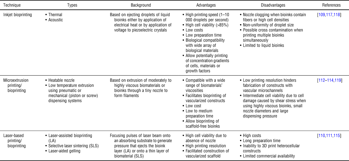
Producing such types of sophisticated scaffolds and models is, in particular, imperative in the field of bone tissue engineering in order to: (i) fabricate vascularized artificial grafts, which can easily integrate in the human body, for the treatment of jaw bone defects as well as zygomatic bone fractures,[Reference Temple, Hutton, Hung, Huri, Cook, Kondragunta, Jia and Grayson106, Reference Chia and Wu107] (ii) build in vitro models to develop and test new treatment strategies for bone disease, (iii) get a deeper understanding of the emergence and evolution of various types of hematological and bone-related diseases and (iv) be used as a platform for expanding HSCs for transplantation. The term “3D bioprinting” is usually used to describe the process of patterning and assembling living and non-living materials to produce bioengineered structures.[Reference Groll, Boland, Blunk, Burdick, Cho, Dalton, Derby, Forgacs, Li and Mironov108] In the following, we use the term 3D printing to describe the printing of non-living materials and 3D bioprinting if living components such as cells are used. Almost all known 3D printing techniques have been used to prepare 3D bone scaffolds. including inkjet bioprinting,[Reference Gao, Schilling, Yonezawa, Wang, Dai and Cui109] laser-assisted bioprinting,[Reference Catros, Fricain, Guillotin, Pippenger, Bareille, Remy, Lebraud, Desbat, Amédée and Guillemot110] laser-aided gelling,[Reference Chang, Lin, Liu, Chen, Lin, Ho and Liao111] 3D printing with a heatable nozzle,[Reference Wüst, Godla, Müller and Hofmann112] fused deposition modeling (FDM),[Reference Hwang, Choi, Kim, Chung, Jin, Shim, Yun, Jeong and Huh113] extrusion,[Reference Alluri, Jakus, Bougioukli, Pannell, Sugiyama, Tang, Shah and Lieberman114] stereolithography (SLA),[Reference Zhang, Lian, Li, Wang, Hao, Bian, He and Jin115] and laser sintering.[Reference Zhu, Xu, Ma, Zheng, Li, Ma, Wu and Weng116] Table I shows a comparison between different printing/bioprinting techniques that have been used in this field.
Comparison of 3D printing/bioprinting techniques.

Broad spectra of natural and/or synthetic biomaterials in form of either bioceramics, composites, i.e., a mixture of bioceramics and polymers, or polymers have been used as bioinks for 3D printing of bone tissue.[Reference Gungor-Ozkerim, Inci, Zhang, Khademhosseini and Dokmeci120] This wide diversity of biomaterials and composites that have been used in the 3D printing of bone scaffolds or grafts allowed scientists to tune the final biomechanical and biophysical properties of the constructed scaffolds or grafts beside their biodegradability, biocompatibility, and osteogenic ability that allow differentiation of MSCs to OBs.
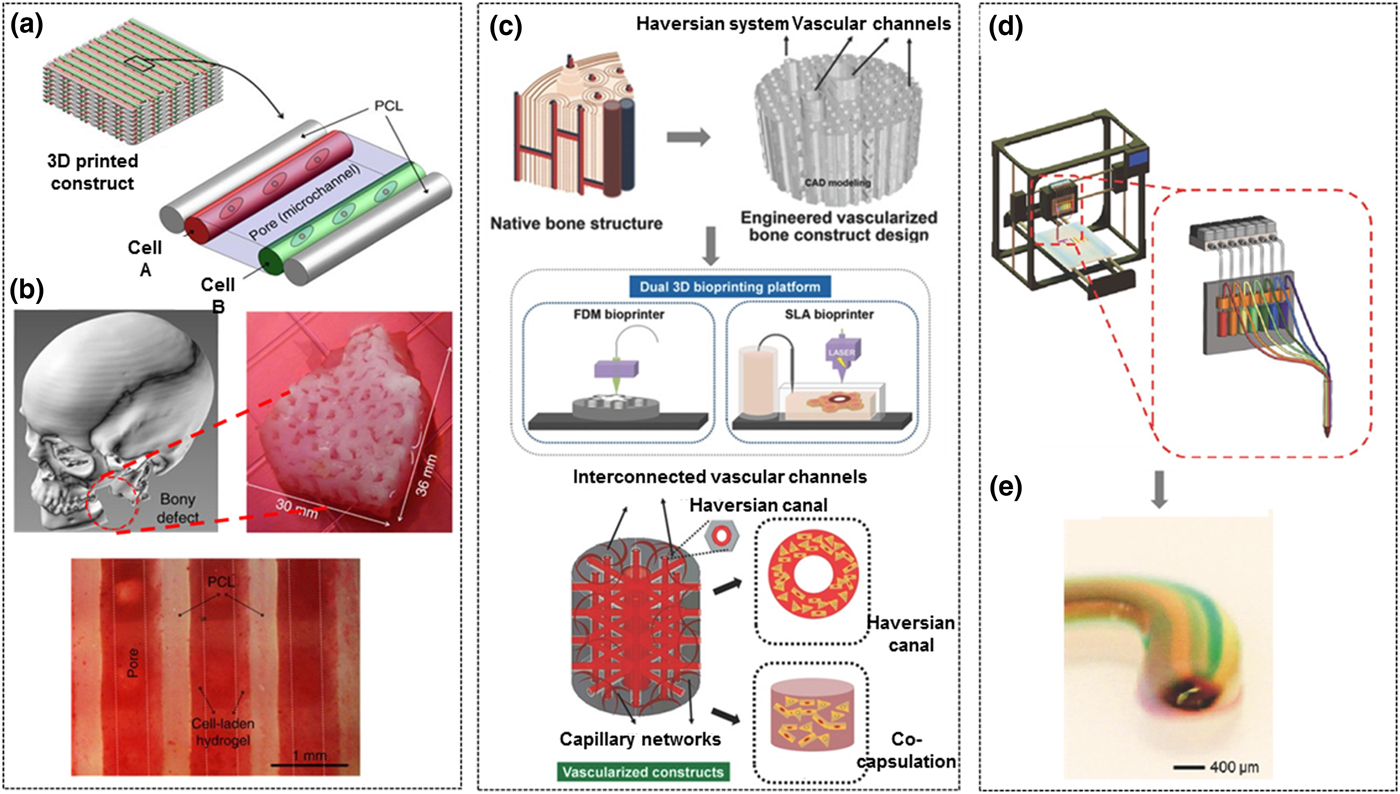
Nevertheless, in vast majority of these scientific efforts, a simple approach to mimic the bone tissue has been used which was based on 3D bioprinting of several layers with well-defined thickness. Each of these layers was composed of parallel lines that were vertically aligned to the lines of the next and previous layers to yield interconnected porous scaffolds with pore diameters that were mainly determined through the spaces between the printed parallel lines of each layer. Presence of interconnected pores is critical for cell survival as it allows the diffusion of nutrients as well as oxygen into the printed scaffolds and could allow the vascularization process.[Reference Cui, Zhu, Nowicki, Zhou, Khademhosseini and Zhang121] These simple models, however, could not fulfill some essential requirements that are needed to develop reliable bone or bone marrow models as well as bone grafts that could mimic the hierarchical, biophysical and biologic complexity of native bone tissue. This is mainly attributed to some technical hurdles of current 3D bioprinting techniques, as they do not allow simple, simultaneous and regional deposition of several different biomaterials, composites, growth factors, stem-, progenitor- and differentiated cells with high resolution and within an acceptable printing speed.[Reference Hospodiuk, Dey, Sosnoski and Ozbolat122, Reference Jungst, Smolan, Schacht, Scheibel and Groll123] Some elegant strategies, however, have been made recently in this direction and a few of them are highlighted here. Atala and his research team developed a multi-dispensing printing module, named “integrated tissue-organ printer” (ITOP), which allowed simultaneous printing of various cells and polymers through a sophisticated printing nozzle with high printing resolution.[Reference Kang, Lee, Ko, Kengla, Yoo and Atala124] The ITOP system was equipped with multiple cartridges to facilitate the printing of multiple cell-laden composite hydrogels beside the supporting polymer (PCL) and sacrificial pluronic F-127 hydrogel. This allowed the formation of microchannels in the scaffold structure which could facilitate the diffusion of nutrients and oxygen into the printed tissue as it is shown in Fig. 4(a). A human-scale mandible bone construct, seeded with human amniotic fluid-derived stem cells, could be fabricated, using ITOP technique, with a geometric structure designed based on the CT scan of a human mandible defect after traumatic injury which can be seen in Fig. 4(b). In fact, such combination of ITOP technique and CT scan of bone tissue could open the door towards reliable models of bone marrow. Cui et al. presented an integrated approach to fabricate a hierarchically-vascularized bone construct with a biphasic structure by utilization of a dual 3D printing/bioprinting system composed of FDM and SLA printing techniques.[Reference Cui, Zhu, Nowicki, Zhou, Khademhosseini and Zhang121] The fabricated construct had a geometric structure and biologic function mimicking the osteon or Haversian system of natural bone as it is depicted in Fig. 4(c). The stiff regions of the constructed scaffold were fabricated using biodegradable PLA fibers printed with an FDM printer, which resulted in cylindrically shaped constructs containing a series of interconnected vertical and horizontal vascular channels. This was followed by coating the surface of the construct with dopamine as a prelude to immobilize BMP2, to facilitate the osteogenic differentiation, via mussel-inspired chemistry. Afterward, human MSCs were seeded into the scaffold. Next, the elastic vascular structure was bioprinted using SLA printer, which could fill the interconnected channels with gelatin methacrylate (GelMA) hydrogels containing human MSCs and HUVECs. Vascular endothelial growth factor peptide was attached to the GelMA during the printing process via “thiol-ene” click reaction. Interestingly, the constructed scaffold showed disparate mechanical properties based on region, the compressive modulus of about 0.38 GPa in the stiff parts of the construct whereas in the vascular region the elastic modulus was in the range of 10 to 30 kPa, which mimics native bone tissue. This strategy of pattering two adjacent regions with different mechanical and biologic properties and coating with different growth factors could support one day the current efforts to fabricate a reliable bone marrow model since, as it was mentioned in the introduction, a native bone marrow owns several niches. Another advanced technological approach that might be used also in the future to support the scientific efforts in this field was recently reported by Khademhosseini and his research team who developed an extrusion 3D printer, which is shown in Figs. 4(d) and 4(e) that was able to achieve fast and continuous extrusion of several materials, i.e., bioinks, with different properties through one nozzle.[Reference Liu, Zhang, Heinrich, De Ferrari, Jang, Bakht, Alvarez, Yang, Li, Trujillo-de Santiago, Miri, Zhu, Khoshakhlagh, Prakash, Cheng, Guan, Zhong, Ju, Zhu, Jin, Shin, Dokmeci and Khademhosseini125]
Schematic drawings of some advanced 3D printing techniques for biofabrication. (a) Illustration of the basic 3D architecture which could be achieved by using the ITOP technique to print multiple-cell hydrogels and supporting PCL polymers. (b) The upper left represents a mandible bone reconstruction designed based on a human CT image of a mandible bony defect, the upper right image shows a photograph of the 3D printed construct after culturing it in osteogenic medium for 28 days, and the lower image depicts osteogenic differentiation of stem cells in the printed construct confirmed by Alizarin Red S staining. Adapted/reproduced with permission from Springer Nature, ref. Reference Kang, Lee, Ko, Kengla, Yoo and Atala124 (c) Schematic illustration of designing and 3D printing of a biomimetic vascularized bone scaffold, the upper image shows the computer modeling of vascularized bone, the middle images depicts the FDM/SLA 3D bioprinting platform, while the lower image represents the microstructural design of vascularized construct. Adapted/reproduced with permission from John Wiley and Sons, ref. Reference Cui, Zhu, Nowicki, Zhou, Khademhosseini and Zhang121 (d) Schematics illustrating the design of 3D printer with seven-channel printhead which is in turn connected to seven cartridges that are individually regulated by programmable pneumatic valves. (e) Side view of one end of a printed microfiber showing its internal 3D structure consisting of seven individually segmented bioinks. Adapted/reproduced with permission from John Wiley and Sons, ref. Reference Liu, Zhang, Heinrich, De Ferrari, Jang, Bakht, Alvarez, Yang, Li, Trujillo-de Santiago, Miri, Zhu, Khoshakhlagh, Prakash, Cheng, Guan, Zhong, Ju, Zhu, Jin, Shin, Dokmeci and Khademhosseini125.

Despite the great potential of 3D printing in the field of biofabrication, this technique has not been used yet in the development of a reliable model of bone marrow tissue that recapitulates the exact anatomy and physiology of the different niches in bone marrow, but rather to construct simple models of porous scaffolds with well-controlled pore size and pattern. Zhou et al., for example, used a tabletop SLA 3D printer to fabricate several 3D highly porous and interconnected scaffolds with different geometric patterns.[Reference Zhou, Castro, Zhu, Cui, Aliabouzar, Sarkar and Zhang126] PEGDA containing RGD and/or nanocrystalline HAP were used as bioinks to fabricate these scaffolds. The authors investigated the effects of pore sizes, geometric patterning, and presence of low intensity pulsed ultrasound on the adhesion, the growth and osteogenic differentiation of MSCs. Braham et al. fabricated an advanced model of 3D bone marrow in order to study the expansion and interaction of primary myeloma cells.[Reference Braham, Ahlfeld, Akkineni, Minnema, Dhert, Öner, Robin, Lode, Gelinsky and Alblas127] The model was composed of two separate but interacting niches, i.e., the endosteal niche, prepared by using a bioprintable paste of calcium phosphate cement with seeded osteogenic multipotent MSCs and the perivascular niche, which was prepared using Matrigel™ containing endothelial progenitor cells and MSCs. This was the first time that both—the endosteal and perivascular components of HSC niches—were included in one mold, of which one part, the endosteal one, was 3D printed.
However, as it was mentioned above, even by 3D printing, obtaining an ideal bone marrow model is not straightforward, but rather faces several critical technical challenges regarding the currently available 3D printing techniques as well as the utilized bioinks. Regarding the technical aspect, developments should be made in order to increase the speed of printing and to facilitate precise and simultaneous bioprinting of several types of bioinks in a way that they would not mix with each other during or after the printing process and does not cause damage to the printed cells. In order to achieve this goal, the development of 3D bioprinters which are able to use two or more printing techniques within one printer, and thereby facilitating printing of several biomaterials of different nature using one 3D printer, is advantageous. Furthermore, new functional bioinks should be developed to allow high-speed printing using biocompatible materials with an acceptable rate of degradation and immobilization of different types of growth factors.
Outlook
Utilization of 3D printing to construct a simple bone scaffold has shown some successes in the area of bone graft fabrication, but so far this was not the case for the models aimed to be used as a platform for expanding stem cells, or as in vitro models to test new treatment strategies or to study the different factors affecting the occurrence of bone malignant diseases. Therefore, developing a reliable model that can precisely mimic the different niches, appears to be mandatory [Fig. 2 (b, VI)]. In order to reach this goal, several interrelated parameters which should be taken into consideration are: (i) chemical and biochemical conditions, e.g., gradient concentration of calcium ions and sustained release of growth factors, (ii) cellular composition, (iii) binding sites for cell attachment, (iv) nanotopography and –patterning, (v) stiffness of the different niches, (vi) 3D architecture, (vii) tailorable degradation rates, (viii) suitable supply of nutrient and oxygen, and (ix) vascularization of the system. Reaching this aim will enable scientists, for instance, to construct reliable leukemic models which are able to support the diffusion of soluble factors, resembling the in vivo gradient. It should allow also cell migration along the gradient and provide an architecture with enough space to analyze cell–cell contacts in in vitro cocultures. Such a constructed model facilitates the investigation of the anchorage of LCs in the leukemic bone morrow in consequence of other cells or soluble factors in their environment. A large number of studies has been dedicated to the biochemical pathways associated with metastatic progression, but the associated biophysical cues have been highly underrepresented. These parameters are equally important in tumor pathophysiology and investigating them could open doors for new therapeutic strategies.
Besides enabling fundamental research in the context of human healthy and diseased bone marrow, bone marrow models undeniably serve an array of applications, such as in vitro HSC expansion for therapeutic treatment of hematological malignancies and as tools for drug-testing. Preparing such models using 3D culture techniques, has opened up new vistas to replicate the complexities present in an in vivo microenvironment. As discussed earlier, these systems exert a significant advantage not only over conventional 2D culture systems but also on in vivo models. They facilitate co-localization of different cell types, equip perfusion of biochemical factors and provide a mechanically stable ECM in 3D, all the while circumventing the investment of resource and time necessary for in vivo studies. They have also imparted valuable insights into the multifaceted nature of cancer biology and aided studies on leukemic niches and tumor dissemination to the bone. Over the past years, numerous bone marrow models have been researched and several strategies have been developed. However, there still remains an urgent need for cost-effective and time-saving applications. Nevertheless, in the case of research, systems incorporating as many characteristics as possible should be devised. The principal idea which should be kept in mind while developing such models is “as complex as necessary but as simple as possible”. 3D printing is a technology that stands out in this regard. It has the potential to mimic the healthy and diseased bone marrow, in varying degrees of complexities, tailored to the prerequisites of each individual study. Having said that, this technique also faces several challenges and further progress is necessary for successful practical applications.
Acknowledgments
We thank the publishers Elsevier, Taylor and Francis Ltd., BioMed Central, Springer Nature, John Wiley and Sons for the permission to reprint published data shown in Figs. 3(a)[Reference Raic, Rodling, Kalbacher and Lee-Thedieck47], 3(b)[Reference Kotha, Hayes, Phong, Redd, Bomsztyk, Ramakrishnan, Torok-Storb and Zheng49], 3(c)[Reference Sieh, Lubik, Clements, Nelson and Hutmacher50], Figs. 4(a), 4(b)[Reference Kang, Lee, Ko, Kengla, Yoo and Atala124], 4(c)[Reference Cui, Zhu, Nowicki, Zhou, Khademhosseini and Zhang121], 4(d), 4(e)[Reference Liu, Zhang, Heinrich, De Ferrari, Jang, Bakht, Alvarez, Yang, Li, Trujillo-de Santiago, Miri, Zhu, Khoshakhlagh, Prakash, Cheng, Guan, Zhong, Ju, Zhu, Jin, Shin, Dokmeci and Khademhosseini125]. This project has received funding from the European Research Council (ERC) under the European Union’s Horizon 2020 research and innovation programme (grant agreement No 757490). C.L.-T. and A.R. acknowledge funding from the BMBF NanoMatFutur programme (FKZ 13N12968) and the programme BioInterfaces in Technology and Medicine (BIFTM) by the Helmholtz Association.