




No CrossRef data available.
Right ventricular function in congenital cardiac disease: noninvasive quantitative parameters for clinical follow-up
Published online by Cambridge University Press: 24 May 2005
Abstract
Right ventricular function is of great importance in patients with both acute and chronic ventricular overload. The early detection of right ventricular dysfunction may have an impact on therapeutic decision making, helping to prevent or further delay functional deterioration of the right ventricle.
In patients with right ventricular overload due to congenital cardiac diseases, dobutamine stress testing combined with magnetic resonance imaging, electrocardiographic changes, and monitoring of concentrations of plasma brain natriuretic peptide are very suitable parameters for the early detection of ventricular dysfunction, and should therefore be used in the follow-up of these patients.
It is apparent that no single measurement of anatomy or function can ever adequately describe the form or performance of the right ventricle. Rather, we should be looking more towards an integrated approach of different parameters for right ventricular function. The quantitative parameters described in this study can serve this purpose. The strong correlation found between these non-invasive and independent parameters encourages their clinical implementation.
Information
- Type
- Review
- Information
- Copyright
- © 2003 Cambridge University Press
Adult patients with congenital cardiac malformations represent a continuously growing population, owing to improvements in medical care, advances in surgical techniques, and meticulous postoperative care. Current surgical standards make mortality the exception in many diseases, and results are judged more in terms of morbidity, residual defects, freedom from reoperation, and long-term functional outcome. Closer follow-up of these patients becomes mandatory, and non-invasive modalities are increasingly being recommended and used.
In a substantial number of patients with congenital defects, the right ventricle is in a functionally or anatomically abnormal position, thereby sustaining a state of chronic pressure or volume overload. Diagnoses include transposition, congenitally corrected transposition, tetralogy of Fallot, and certain subsets of double outlet right ventricle.
Except for qualitative data, such as clinical status, functional classification, and echocardiography, there are few other possibilities to diagnose deterioration of right ventricular function. Although these established diagnostic modalities may allow for appropriate adjustment of medical therapy, or decision making with regards to surgical timing in symptomatic patients or in patients with obvious myocardial impairment, this is not the case in more subtle situations. Newer non-invasive quantitative diagnostic tools would be of particular value in asymptomatic or minimally symptomatic patients with borderline right ventricular function, for whom there are currently several therapeutic dilemmas. Management of these patients would be improved by establishing accurate quantitative determinants of right ventricular function, and by relating them to the already existing qualitative ones. The combined information could thus be implemented in daily clinical practice.
In this review, we discuss the various states of right ventricular functional overload in certain congenital cardiac malformations, and present noninvasive quantitative data obtained in our institution for the assessment of right ventricular function that has proven to be applicable to clinical follow-up in these patients.
Congenital cardiac malformations with right ventricular overload
Tetralogy of Fallot
Residual lesions after repair of tetralogy may include suboptimal relief of the obstructed right ventricular outflow tract, or residual pulmonary artery stenosis, leading to a state of chronic pressure overload (Fig. 1). After complete repair, particularly when a patch has been placed across the ventriculo-pulmonary junction, various degrees of pulmonary valvar insufficiency may create volume overload, and eventual dilation, of the right ventricle.

Figure 1. A spin echo resonance image showing pulmonary valvar stenosis with a wide pulmonary trunk in a patient subsequent to surgical correction of tetralogy of Fallot. RV: right ventricle; LV: left ventricle.
Double outlet right ventricle with a subaortic ventricular septal defect and pulmonary stenosis schematically follows this physiology, and is corrected surgically in a similar fashion to tetralogy of Fallot. The long-term fate of the right ventricle may, therefore, be diagnostically considered and followed in the same manner.
Transposition
There remains a large cohort of young adults with concordant atrioventricular and discordant ventriculo-arterial connections (“transposition”) who have undergone an atrial switch of the Senning or Mustard type in childhood.1 After the atrial switch, the morphologically right ventricle functions as the systemic ventricle, leading to long-term problems of chronic right ventricular pressure overload, and to tricuspid valvar regurgitation with right ventricular volume overload (Fig. 2). Refractory atrial arrhythmias due to multiple incisions and suture lines in the atriums may also induce or exacerbate right ventricular failure. Obstructions or leaks across the atrial baffle may respectively lead to systemic or pulmonary venous obstruction, and left-to-right shunts. The morphology of the morphologically right ventricle in patients after a Senning or Mustard procedure is analogous to that in patients with unoperated congenitally corrected transposition, the combination of discordant connections at both atrioventricular and ventriculo-arterial junctions, where the ventricle is round, and the ventricular septum is convex towards the morphologically left ventricle.2

Figure 2. A gradient echo resonance image showing the “4 chamber” view after surgical correction of transposition using the Mustard procedures. The arrows indicate the baffles. Abbreviations as for Figure 1.
Congenitally corrected transposition
Although the natural history of congenitally corrected transposition may allow asymptomatic survival into adulthood, the majority of unoperated patients face long-term consequences of failure and dilation of the morphologically right ventricle, tricuspid valvar insufficiency, and complete heart block3–6 (Fig. 3). The etiology of right ventricular failure is multifactorial, and probably leads to right ventricular dysfunction that worsens in relation to the development of tricuspid valvar insufficiency and/or complete heart block.7 This remains true after physiologic, also called “classical” repair, where the morphologically right ventricle continues to support the systemic circulation.8

Figure 3. A gradient echo resonance image in “4 chamber” plane from a patient with congenitally corrected transposition. Abbreviations as for Figure 1, TP: tricuspid valvar prosthesis.
Quantitative diagnostic methods for noninvasive assessment of right ventricular function
The electrocardiogram
A 12-lead electrocardiogram may give considerable insight into the degree of pressure and/or volume overload, and allows a simple and inexpensive first glimpse at right ventricular function after repair of various congenital cardiac malformations.
A retrospective study was undertaken in our institution of a cohort of 48 patients with chronic right ventricular pressure overload subsequent to surgical correction of various congenital lesions.9 The main criterion for inclusion was chronic right ventricular pressure overload, with a right ventricular systolic pressure greater than 35 millimetres of mercury as assessed by echocardiography, after surgical repair of the congenital cardiac disease, without important additional hemodynamic lesions, and in asymptomatic or minimally symptomatic patients in the first or second classes of the functional categorization of the New York Heart Association. The patients were classified into three groups according to their diagnosis: patients with congenitally corrected transposition; patients who had undergone an atrial switch procedure for transposition, and patients with a subpulmonary pressure overloaded right ventricle. The last group was divided into two subgroups: those with tetralogy of Fallot, and those with right ventricular pressure overload due to other congenital cardiac diseases. Our study showed a gradual prolongation in the duration and dispersion of the QRS complexes in patients with chronic right ventricular pressure overload, regardless of the nature of the congenital cardiac disease. In the studied population, right ventricular end diastolic volume, right ventricular mass, and brain natriuretic peptide were increased compared to the known reference values for healthy volunteers. A significant correlation was found between the duration of the QRS complex and right ventricular end diastolic volume in patients with a subpulmonary right ventricle submitted to chronic pressure overload. We also demonstrated a significant correlation between the duration of the QRS complex and right ventricular mass in patients with tetralogy of Fallot.
Several studies have described the importance of the duration and dispersion of the QRS complex as a predictor of malignant tachycardias and sudden death in patients with congenital cardiac disease.10, 11 Gatzoulis et al. found that, in patients with tetralogy, a QRS wave wider than 180 milliseconds was a strong predictor for malignant ventricular tachycardias. They also introduced dispersion of the QRS complex as a marker for inhomogeneity of ventricular depolarization.10 These studies, nonetheless, were limited to specific types of cardiac lesions.10, 11 Our study showed changes in electrocardiogram markers over time in patients with chronic right ventricular pressure overload, regardless of the nature of the congenital cardiac malformations.
Magnetic resonance imaging
Magnetic resonance imaging represents the diagnostic noninvasive technique of choice for the evaluation of right ventricular function in congenital cardiac disease, by its potential to obtain both anatomic detail and quantification of flow. It is useful in detecting intracardiac narrowings, and obstruction of homograft conduits, following cardiac surgery in patients with complex malformations.12–15 Its accuracy, and superior reproducibility, are setting a new gold standard for the quantification of right ventricular ejection fraction, cardiac output, myocardial mass, and wall thickness.12, 16 Technical advantages of magnetic resonance imaging in comparison with other techniques are the excellent spatial resolution, the characterisation of myocardial tissue, multi-plane versatility, and the potential for three-dimensional reconstruction.17 Cardiac magnetic resonace imaging allows an accurate noninvasive functional assessment of the right ventricle, but further development of this diagnostic method needs to be done in a multidisciplinary fashion amongst cardiologists, radiologists, physicists, software scientists, and imaging machine producers.
Results of dobutamine testing assessed by resonance imaging in patients with tetralogy, congenitally corrected transposition, and transposition
We explored the effect of dobutamine stress and its possible clinical implications in different groups of asymptomatic patients with chronic right ventricular overload due to congenital cardiac disease.18–20 We studied asymptomatic and minimally symptomatic patients in the first and second classes of the functional classification of the New York Heart Association with chronic right ventricular pressure overload. In all, we assembled 29 patients with a systemic morphologically right ventricle, 16 after an atrial switch for transposition, and 13 patients after physiologic repair of congenitally corrected transposition, 22 patients with a chronically pressure-overloaded subpulmonary right ventricle, and 11 age and sex-matched healthy volunteers. Magnetic resonance imaging was applied both at baseline, and during dobutamine stress, to determine right ventricular volumes and ejection fraction.
Compared to the left ventricle of healthy controls, all patients had larger right ventricular volumes. We demonstrated heterogeneity in response to dobutamine stress, between the different groups of patients. We found decreased right ventricular end diastolic volumes, suggesting impaired filling, and decreased stroke volume in patients after an atrial switch for transposition. In recent studies using invasive techniques, the group of Redington21, 22 also observed reduced diastolic filling and right ventricular stroke volume in response to dobutamine stress testing in patients with surgically corrected transposition. Based on their results, they speculated that the capacitance and conduit function of the abnormal, and often calcified, intraatrial pathways, may be responsible for the failure of augmentation of stroke volume during exercise.22
During dobutamine stress, our patients who had undergone repair of tetralogy of Fallot showed a remarkable decrease in right ventricular stroke volume, accompanied by both failure to augment right ventricular ejection fraction, and impaired right ventricular filling. In a recent study using magnetic resonance imaging, including 15 asymptomatic patients with tetralogy of Fallot, Roest et al.23 described no significant change in ejection fraction during physical exercise. In discordance with our results, these authors described a minimal increase in right ventricular end diastolic volume, and in stroke volume, during exercise. Two reasons may explain the differences between these two studies. In our study, we used magnetic resonance imaging with pharmacological stress, while Roest et al.23 used physical stress. Second, our patients were markedly older, namely 27 years of age, compared to 17.5 years, and therefore probably having less compliant right ventricles, due to longer term overload and hypertrophy. Similar to our results, Gatzoulis et al.24 used radionuclide angiography in 95 patients with tetralogy of Fallot. They noticed failure of the right ventricle to increase its ejection fraction during physical exercise, interpreting their findings as indicative of early right ventricular dysfunction. The right ventricle in tetralogy of Fallot is subject from birth to hypoxia and pressure overload. This results in structural and functional changes, such as hypertrophy, interstitial fibrosis, cellular atrophy, and myofibrillar disorganization. These may all represent possible causes for an inappropriate response to stress in asymptomatic patients with tetralogy of Fallot.21
In our small, but consecutive, group of patients with congenitally corrected transposition, we found an appropriate response to dobutamine stress. Markers of right ventricular function of our unoperated patients with congenitally corrected transposition were closer to controls than our patients who had undergone physiological repair. Dobutamine stress assessed by resonance imaging may help to define the subgroups of patients with favorable anatomy who might proceed in asymptomatic fashion into adulthood without the need for surgery.
Echocardiography
Echocardiography is the most frequently used technique for imaging the right ventricle. Doppler echocardiography can provide morphologic and hemodynamic information through segmental analysis of most congenital cardiac defects.25 Also, echocardiography provides indirect documentation of pulmonary arterial hypertension, and can estimate its severity by the presence of right ventricular dilation and/or hypertrophy, the presence of tricuspid or pulmonic valvar regurgitation, along with Doppler estimation of right ventricular systolic pressure.26
In clinical practice, cardiologists and echocardiographic technicians are faced with a wide variety of right ventricular lesions caused by congenital cardiac disease. It is likely that some abnormalities may be recognized, but all the characteristics of a certain congenital lesion may be underappreciated.26 For this reason, and considering that echocardiography is still the first diagnostic choice in patients with congenital cardiac malformations, it is necessary that both the interpreting cardiologist and the echocardiographic technician have special competencies in the evaluation of congenital cardiac disease.
Plasma neurohormones
Recent publications have shown that concentrations of atrial natriuretic peptide and brain natriuretic peptide are elevated in the plasma of patients with asymptomatic systolic dysfunction, and that these parameters are highly accurate for the detection of cardiac failure.27, 28 Brain natriuretic peptide is a marker for ventricular dysfunction,29 and is secreted in both atriums and ventricles, especially in failing ventricles.30 Brain natriuretic peptide has potent diuretic and systemic vasorelaxant properties.31
In normal subjects, atrial natriuretic peptide is synthesized and secreted almost exclusively in the atrium, whereas patients with congestive cardiac failure have increased production of atrial natriuretic peptide in both atriums and ventricles, in response to increased atrial stretch.29 Although previous studies have suggested that neurohumoral markers play a role not only in left ventricular dysfunction, but also in right ventricular dysfunction,31–33 their clinical applicability in the detection of right ventricular dysfunction is not yet established. Speculation as to their usefulness in noninvasive detection of right ventricular failure is all but too tempting, especially in patients with congenital cardiac malformations.
We measured the levels of these peptides in 21 asymptomatic or minimally symptomatic patients with chronic right ventricular pressure overload due to a congenital cardiac malformation, and in seven healthy volunteers.34 Right ventricular ejection fraction was determined using magnetic resonance imaging. Right ventricular ejection fraction of the volunteers was significantly higher than right ventricular ejection fraction of the patients. Between patients and volunteers, there was a significant difference in the concentrations of the peptides in the plasma. Right ventricular ejection fraction was inversely correlated with the levels of both peptides (Fig. 4). In a recent paper, Bolger et al.33 found a highly significant stepwise increase of neurohumoral activation according to New York Heart Association functional class and systemic ventricular function. The authors interpreted these results as a state of chronic cardiac failure in adults with congenital cardiac disease, relating to the severity of symptoms and ventricular dysfunction, and not necessarily to the underlying anatomic substrate. Our study was focused on asymptomatic or minimally symptomatic patients with chronic right ventricular pressure overload, but basically both studies showed similar results. The levels of the peptides were elevated in the patients as compared to healthy controls, and a strong correlation was found between right ventricular function and neurohumoral activation.

Figure 4. Correlation between levels of brain natriuretic peptide (BNP) in the plasma and right ventricular ejection fraction (RVEF – Fig. 4a), and between levels of atrial natriuretic peptide (ANP) in the plasma and ejection fraction (Fig. 4b). The solid lines represent the linear regression and dotted lines are 95% confidence intervals of the regression line. (Reproduced with permission from Heart).
The results of recent studies involving right ventricular neurohumoral activation are encouraging clinical implementation of the levels of neurohormones in the plasma, brain natriuretic peptide in particular, as quantitative markers for right ventricular function.34–37 It is very likely that, in the near future, this peptide might serve as a marker to assess the efficacy of treatment in patients with right ventricular dysfunction, such as that encountered after acute pulmonary embolism, or after various repairs of congenital cardiac malformations that place the right ventricle under strain.
Discussion
The fate of the morphologically right ventricle in congenital cardiac disease is an increasing source of concern and debate, both with regards to modalities of follow-up and issues of management. The long-term prognosis of these patients is open-ended, and is mainly dependent on right ventricular function, the eventual occurrence of right ventricular failure,7 and disorders of rhythm.38, 39 Because of the growing population of adults with congenital cardiac disease, irrespective of whether they have undergone surgery, new modalities of noninvasive follow-up are required which are accurate, reproducible, and allow for earlier detection of pressure or volume overload of the right ventricle, prior to the occurrence of failure or irreversible myocardial damage. Early detection of right ventricular failure may permit better medical management and a better insight as to the optimal timing for preventive surgery.40
Patients with atrial switch procedures for transposition, or for double outlet right ventricle with subpulmonary ventricular septal defect, generally show reduced exercise tolerance, which has been attributed to factors such as an abnormally low response of heart rate, filling abnormalities of the right ventricle, and systolic dysfunction. Intrinsic myocardial damage of the right ventricle in the systemic position, obstructions to the systemic or pulmonary pathways, and leaks across and rigidity of the baffles in the heart, may all contribute to this picture.
Failure to increase stroke volume during dobutamine stress was confirmed as the primary hemodynamic abnormality in patients with tetralogy of Fallot. Failure to increase right ventricular ejection fraction, and the significant decrease in right ventricular end diastolic volume during dobutamine stimulation, suggested impaired systolic and diastolic function in these patients.
In patients with congenitally corrected transposition, the natural history of right ventricular pressure and/or volume overload allows survival to adulthood. The possibility of right ventricular failure, nonetheless, and the almost inevitable tricuspid valvar insufficiency, mandate accurate and reproducible diagnostic imaging to improve management of the patient and fine-tuning of surgical correction. Magnetic resonance imaging undoubtedly provides the best imaging modality for preoperative assessment, and gives a comprehensive visualization of intracardiac pathways, allowing for determination of the optimal surgical strategies.
Magnetic resonance imaging has become an established diagnostic modality for follow-up, as well as for preoperative assessment of patients with congenital cardiac disease requiring first time surgical intervention, or reoperations. Broadening indications for the use of plasma neurohormones, such as brain and atrial natriuretic peptides, may reveal them to be promising future quantitative markers for the early detection of right ventricular dysfunction, in asymptomatic patients with congenital cardiac malformations.
In conclusion, although substantial knowledge has been gained about right ventricular function in various congenital cardiac malformations, both in the native state and after surgery, there is much left to do. Imaging and noninvasive quantitative parameters already play a role in the diagnosis of right ventricular function. These diagnostic modalities will gradually take an even more important part in clinical practice. Implementation of reliable and reproducible parameters for right ventricular function will enable a more aggressive follow-up tailored to the needs of the individual patient.
Acknowledgement
Igor I. Tulevski is supported by the Netherlands Heart Foundation and the Interuniversity Cardiology Institute of the Netherlands.
References
Paul MH, Wessel HU. Exercise studies in patients with transposition of the great arteries after atrial repair operations (Mustard/Senning): a review. Pediatr Cardiol 1999; 20: 49–55.Google Scholar
Lorenz CH, Walker ES, Graham TP Jr, Powers TA. Right ventricular performance and mass by use of cine MRI late after atrial repair of transposition of the great arteries. Circulation 1995; 92: II233–II239.Google Scholar
Warnes CA. Congenitally corrected transposition: the uncorrected misnomer. J Am Coll Cardiol 1996; 27: 1244–1245.Google Scholar
Voskuil M, Hazekamp MG, Kroft LJ, et al. Postsurgical course of patients with congenitally corrected transposition of the great arteries. Am J Cardiol 1999; 83: 558–562.Google Scholar
Graham TP Jr, Parrish MD, Boucek RJ Jr, et al. Assessment of ventricular size and function in congenitally corrected transposition of the great arteries. Am J Cardiol 1983; 51: 244–251.Google Scholar
Connelly MS, Liu PP, Williams WG, Webb GD, Robertson P, McLaughlin PR. Congenitally corrected transposition of the great arteries in the adult: functional status and complications. J Am Coll Cardiol 1996; 27: 1238–1243.Google Scholar
Perloff C. Congenital Heart Diseases in Adults, 2nd edn. W.B. Saunders, Philadelphia, 1998.
Vouhe P. Congenitally corrected transposition: results of “classical” surgery. In: Redington AN, Brawn WJ, Deanfield JE, Anderson RH (eds). The Right Heart in Congenital Heart Disease. Greenwich Medical Media Ltd, London, 1998, pp 231–236.
Neffke JGJ, Tulevski II, Van der Wall EE, et al. ECG determinants in adult patients with chronic right ventricular pressure overload due to congenital heart disease: Relation with plasma neurohormones and MRI parameters. Heart 2002; 88: 266–270.Google Scholar
Gatzoulis MA, Till JA, Redington AN. Depolarization–repolarization inhomogeneity after repair of tetralogy of Fallot. The substrate for malignant ventricular tachycardia? Circulation 1997; 21: 401–404.Google Scholar
Gatzoulis MA, Walters J, McLaughlin PR, Merchant N, Webb GD, Liu P. Late arrhythmia in adults with the mustard procedure for transposition of great arteries: a surrogate marker for right ventricular dysfunction? Heart 2000; 84: 409–415.Google Scholar
The clinical role of magnetic resonance in cardiovascular disease.Task Force of the European Society of Cardiology, in collaboration with the Association of European Paediatric Cardiologists. Eur Heart J 1998; 19: 19–39.
Helbing WA, Rebergen SA, Maliepaard C, et al. Quantification of right ventricular function with magnetic resonance imaging in children with normal hearts and with congenital heart disease. Am Heart J 1995; 130: 828–837.Google Scholar
Rebergen SA, Niezen RA, Helbing WA, van der Wall EE, de Roos A. Cine gradient-echo MR imaging and MR velocity mapping in the evaluation of congenital heart disease. Radiographics 1996; 16: 467–481.Google Scholar
van der Wall EE. Do magnetic resonance techniques contribute to the management of clinical problems? Eur Heart J 1996; 17: 666–673.Google Scholar
Sechtem U, Pflugfelder PW, Gould RG, Cassidy MM, Higgins CB. Measurement of right and left ventricular volumes in healthy individuals with cine MR imaging. Radiology 1987; 163: 697–702.Google Scholar
Van der Wall EE, Vliegen HW, de Roos A, Bruschke AV. Magnetic resonance imaging in coronary artery disease. Circulation 1995; 92: 2723–2739.Google Scholar
Tulevski II, Lee PL, Groenink M, et al. Dobutamine-induced increase of right ventricular contractility without increased stroke volume in adolescent patients with transposition of the great arteries: evaluation with magnetic resonance imaging. Int J Card Imaging 2000; 16: 471–478.Google Scholar
Tulevski II, Van der Wall EE, Groenink M, et al. Usefulness of magnetic resonance imaging dobutamine stress in asymptomatic and minimally symptomatic patients with decreased cardiac reserve from congenital heart disease (complete and corrected transposition of the great arteries and subpulmonic obstruction). Am J Cardiol 2002; 89: 1077–1081.Google Scholar
Dodge-Khatami A, Tulevski II, Bennink GBWE, et al. Comparable systemic ventricular function in healthy adults and patients with unoperated congenitally corrected transposition using MRI dobutamine stress testing. Ann Thor Surg 2002; 73: 1759–1764.Google Scholar
Redington AN. Right ventricular function. In: Redington AN, Brawn WJ, Deanfield JE, Anderson RH (eds). The Right Heart in Congenital Heart Disease. Greenwich Medical Media Ltd, London, 1998, pp 17–24.
Derrick GP, Narang I, White PA, et al. Failure of stroke volume augmentation during exercise and dobutamine stress is unrelated to load-independent indexes of right ventricular performance after the mustard operation. Circulation 2000; 102: 154–159.Google Scholar
Roest AA, Helbing WA, Kunz P, et al. Exercise MR imaging in the assessment of pulmonary regurgitation and biventricular function in patients after tetralogy of Fallot repair. Radiology 2002; 223: 204–211.Google Scholar
Gatzoulis MA, Elliott JT, Guru V, et al. Right and left ventricular systolic function late after repair of tetralogy of Fallot. Am J Cardiol 2000; 86: 1352–1357.Google Scholar
Houston A, Hillis S, Lilley S, Richens T, Swan L. Echocardiography in adult congenital heart disease. Heart 1998; 80: S12–S26.Google Scholar
Cheitlin MD, Alpert JS, Armstrong WF, et al. ACC/AHA guidelines for the clinical application of echocardiography. A report of the American College of Cardiology/American Heart Association task force on practice guidelines. Circulation 1997; 95: 1686–1744.Google Scholar
McDonagh TA, Robb SD, Murdoch DR, et al. Biochemical detection of left-ventricular systolic dysfunction. Lancet 1998; 351: 9–13.Google Scholar
Bettencourt P, Ferreira A, Sousa T, et al. Brain natriuretic peptide as a marker of cardiac involvement in hypertension. Int J Cardiol 1999; 69: 169–177.Google Scholar
Yasue H, Yoshimura M, Sumida H, et al. Localization and mechanism of secretion of B-type natriuretic peptide in comparison with those of A-type natriuretic peptide in normal subjects and patients with heart failure. Circulation 1994; 90: 195–203.Google Scholar
Kambayashi Y, Nakao K, Kimura H, et al. Biological characterization of human brain natriuretic peptide (BNP) and rat BNP: species-specific actions of BNP. Biochem Biophys Res Commun 1990; 173: 599–605.Google Scholar
Nagaya N, Nishikimi T, Okano Y, et al. Plasma brain natriuretic peptide levels increase in proportion to the extent of right ventricular dysfunction in pulmonary hypertension. J Am Coll Cardiol 1998; 31: 202–208.Google Scholar
Matsuo K, Nishikimi T, Yutani C, et al. Diagnostic value of plasma levels of brain natriuretic peptide in arrhythmogenic right ventricular dysplasia. Circulation 1998; 98: 2433–2440.Google Scholar
Bolger AP, Sharma R, Li W, et al. Neurohormonal activation and the chronic heart failure syndrome in adults with congenital heart disease. Circulation 2002; 106: 92–99.Google Scholar
Tulevski II, Groenink M, van der Wall EE, et al. Increased brain and atrial natriuretic peptides in patients with chronic right ventricular pressure overload: Correlation between plasma neurohormones and right ventricular dysfunction. Heart 2001; 86: 27–30.Google Scholar
Morrison LK, Harrison A, Krishnaswamy P, Kazanegra R, Clopton P, Maisel A. Utility of a rapid B – natriuretic peptide assay in differentiating congestive heart failure from lung disease in patients presenting with dyspnea. J Am Coll Cardiol 2002; 16: 202–209.Google Scholar
Tulevski II, Van Veldhuisen DJ, Mulder BJM. Utility of a B-natriuretic peptide as a marker for right ventricular dysfunction in acute pulmonary embolism. J Am Coll Cardiol 2002; 89: 2080.Google Scholar
Tulevski II, Hirsch A, Sanson BJ, et al. Increased brain natriuretic peptide as a marker for right ventricular dysfunction in acute pulmonary embolism. Thromb Haemost 2001; 86: 1193–1196.Google Scholar
Fontaine JM, Kamal BM, Sokil AB, Wolf NM. Ventricular tachycardia: a life-threatening arrhythmia in a patient with congenitally corrected transposition of the great arteries. J Cardiovasc Electrophysiol 1998; 9: 517–522.Google Scholar
Dimas AP, Moodie DS, Sterba R, Gill CC. Long-term function of the morphologic right ventricle in adult patients with corrected transposition of the great arteries. Am Heart J 1989; 118: 526–530.Google Scholar
Vliegen HW, Van Straten A, De Roos A, et al. Magnetic resonance imaging to assess the hemodynamic effects of pulmonary valve replacement in adults late after repair of tetralogy of Fallot. Circulation 2002; 106: 1703–1707.Google Scholar

A spin echo resonance image showing pulmonary valvar stenosis with a wide pulmonary trunk in a patient subsequent to surgical correction of tetralogy of Fallot. RV: right ventricle; LV: left ventricle.

A gradient echo resonance image showing the “4 chamber” view after surgical correction of transposition using the Mustard procedures. The arrows indicate the baffles. Abbreviations as for Figure 1.

A gradient echo resonance image in “4 chamber” plane from a patient with congenitally corrected transposition. Abbreviations as for Figure 1, TP: tricuspid valvar prosthesis.
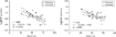
Correlation between levels of brain natriuretic peptide (BNP) in the plasma and right ventricular ejection fraction (RVEF – Fig. 4a), and between levels of atrial natriuretic peptide (ANP) in the plasma and ejection fraction (Fig. 4b). The solid lines represent the linear regression and dotted lines are 95% confidence intervals of the regression line. (Reproduced with permission from Heart).
 You have
Access
- 35
- Cited by