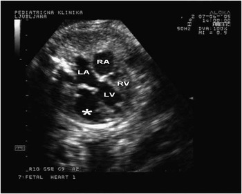
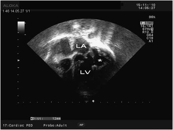
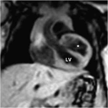
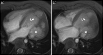
A 5-year-old boy was diagnosed with congenital left ventricular outpouching at 33 weeks of gestation when his mother was referred for foetal echocardiography because of suspected congenital cardiac anomaly (Fig 1). Outpouching of the left ventricle was an isolated finding, connected widely to the ventricle at the region of the lateral wall just beneath the mitral valve. In the four-chamber view of the foetal heart, the surface area of the outpouching equalled the surface area of the left ventricle. There was mild-to-moderate mitral regurgitation, with no signs of congestive cardiac failure. The child was born at term; the delivery and postnatal course were uneventful. The prenatal diagnosis was verified by postnatal echocardiography (Fig 2). In addition, the M-mode echocardiography showed synchronous contractions of the outpouching with the rest of the left ventricle, thus enabling us to define the outpouching as a diverticulum rather than an aneurysm. Later, additional information regarding size, location, and contractility of the diverticulum was gathered using cardiac magnetic resonance imaging (Figs 3 and 4). Owing to the favourable outcome that has been reported for patients with non-apical congenital ventricular diverticulum,Reference Marijon, Ou and Fermont1 a conservative management with acetylsalicylic acid at anti-aggregation doses and enalapril was opted for rather than surgery. In addition to a single episode of convulsions during the febrile illness at the age of two and a history of mildly delayed neurological milestones in the first year of life, which normalised later on, the patient's clinical course so far was completely uneventful.
Apical four-chamber view – 33 weeks of gestation: the size of the left ventricular outpouching (*) is equal to the size of the main left ventricular chamber. LV = left ventricular chamber; LA = left atrium; RA = right atrium; RV = right ventricle.

Apical view – recent echocardiogram: the main left ventricular chamber and left atrium are of normal size. Congenital left ventricular diverticulum (*) is widely connected to the main left ventricular chamber. LV = left ventricular chamber; LA = left atrium.

Cardiovascular magnetic resonance: coronal localiser already shows a double-chamber-like congenital left ventricular diverticulum. LV = left ventricular chamber; * = congenital left ventricular diverticulum.

Cardiovascular magnetic resonance: axial cine – magnetic resonance imaging (free breathing gradient – echo) in diastole (A) and systole (B). The wall of the diverticulum has the same signal intensity as the rest of the myocardium and contracts synchronously with the main left ventricular chamber. Communication between both chambers is just below the mitral valve. LV = left ventricular chamber; * = congenital left ventricular diverticulum.