INTRODUCTION
Cyclophilins (CyPs) catalyse the cis/trans-isomerization of peptidyl-prolyl bonds in peptides or proteins (PPIase activity) (Takahashi, Hayano and Suzuki, 1989; Fischer et al. 1989). Genomes of some prokaryotes, reviewed by (Galat, 2003) and all sequenced-to-date eukaryotes encode from one to several different paralogues of cyclophilins. Their nominal masses may vary from about 17 kDa (monodomain CyP) to several hundred kDa where one cyclophilin-like domain (CLD) is a part of larger proteins that may comprise tetratricopeptide repeats (TPRs), RNA-binding domains (RBD), ranGTP-binding sites, leucine-zippers, zinc-fingers, Ca2+ and calmodulin binding domains, WD40 repeats and other domains. CyPs have been localized to different cellular compartments, namely the cytoplasm (Handschumacher et al. 1984), the membranes and mitochondria (Schneider et al. 1994), the endoplasmic reticulum (Bergsma et al. 1991), the secretory pathway (Colley et al. 1991), the spliceosome (Tiegelkamp et al. 1998), and the nucleus and its membranes (Anderson et al. 1993; Yokoyama et al. 1995; Dolinski et al. 1997). Three other families of proteins possess similar activity, namely FK506-binding proteins (FKBPs), parvulins (prokaryotes) and Pin1 proteins (eukaryotes). PPIases are involved in protein folding and disaggregation, and they interact with many proteins that in turn control diverse cellular processes (reviewed by Galat, 2003). CyPs are Cyclosporin A (CsA) binding proteins (Handschumacher et al. 1984), and the CyP-CsA complex binds to the serine/threonine phosphatase calcineurin (Liu et al. 1991) that interferes with the expression of interleukin-2 in T cells and leads to immunosuppression (Ho et al. 1996). For more than 2 decades CsA has been used in humans as an immunosuppressive drug after organ transplantation. Besides the immunosuppressive activity, CsA has proved to be a parasiticidal drug on some protozoa and helminths (reviewed by Page, Kumar and Carlow, 1995). Its parasiticidal property has also been demonstrated in vitro for Trypanosoma cruzi (McCabe, Remington and Araujo, 1985) that is the aetiological agent of Chagas' disease.
Since the current chemotherapy for this American trypanosomiasis is inadequate, mainly for being unspecific and with toxic side-effects (Urbina and Docampo, 2003), it was interesting to search for new potential chemotherapeutic targets in the parasite's proteome, namely the entire family of the CsA-binding proteins. We have previously shown that some non-immunosuppressive CsA derivatives exhibited a potent anti-T. cruzi activity in vitro without toxic effects on mammalian cells, and interfered with the PPIase activity of the 19 kDa cyclophilin TcCyP19 (Búa et al. 2004). As this protein proved to be an interesting new drug target, we have studied several isoforms in this gene family in the parasite's genome, with a special interest in the CyPs that were actively expressed.
In this paper we present an analysis of the molecular organization of the T. cruzi CyP gene family, their classification into different CyP groups and an overview of diverse cyclophilins encoded in different parasites such as Plasmodium falciparum and the invertebrates Anopheles gambiae and Drosophila melanogaster. We also show the assessment of CyP mRNA transcription in amastigotes and trypomastigotes and the identification of the expressed CyPs as CsA-binding proteins in the parasite. This CsA binding could have implications in the trypanocidal mechanism of action of the non-immunosuppressive CsA analogues previously tested.
MATERIALS AND METHODS
DNA and protein databases
Databases of the Trypanosoma cruzi sequencing genome project (TSK-TSC, www.tigr.org and GeneDB, www.genedb.org) were used. Analysis of DNA and protein homologies were performed at the NCBI Blast server (Altschul et al. 1990). Cyclophilins from the Anopheles gambiae genome are on: http://www.ensembl.org/Anopheles_gambiae; the C. elegans genome http://www.wormbase.org/db/seq/ (Harris et al. 2003); http://www.sanger.ac.uk/Projects/P_falciparum. (Gardner et al. 2002); the Zebra fish genome http://www.sanger.ac.uk/Projects/D_rerio/; the N. crassa genome http://mips.gsf.de/proj/neurospora (Galagan et al. 2003). The cyclophilin protein database (CyP.dab.2003) was updated with the entries obtained from the NCBI (the National Center of Biotechnology Information, http://ncbi.nlm.nih.gov) (Wheeler et al. 2004) and PIR (Protein Information Resources; http://pir.georgetown.edu) (Wu et al. 2003). Hidden Markov model searches were made using the PIR server (Wu et al. 2003). The Data-SQ program (Galat, 2004) was used to select the cyclophilins for multiple sequence alignment (MSA).
Analysis of multiple sequence alignments of cyclophilins
MSAs were made with the ClustalW-60 program (Thompson, Higgins and Gibson, 1994) using Blossum 30 amino acid (AA) exchange matrix, gap penalty 10 and gap extension penalty 0·1. The MSAs were analysed with the Pola-SQ library of programs (Galat, 2004). Total hydrophobicity indexes (HIs) were established using a 9-AA residue moving frame and the hydrophobicity scale (Kyte and Doolittle, 1982). The set of AA residues used for the calculation of the conservation level of the functional consensus remained the same as that used (Galat, 2004) the consensus has been established from diverse crystallographic and mutational studies (Ke, 1992; Zydowsky et al. 1992). The Pola-SQ algorithm searched the large sets of cyclophilins from different genomes and grouped their orthologues and paralogues that retain the highest correlation of their hydrophobicity and bulkiness profiles, sequence similarity (ID) scores, amino acid composition (AAC), pIs and HIs (Galat, 2004). The clustering process allowed us to establish plausible correlations between the T. cruzi cyclophilins paralogues with their orthologues in other organisms. The Form10 program (Galat, 1999) was used for structuring the MSAs; the CyP secondary structure assigned on the top of the MSA was taken from the X-ray structure of human CyP-18a (Ke, 1992).
cDNA clones
cDNA clones were derived from a T. cruzi CL Brener cDNA library previously constructed (Urmenyi et al. 1999) and results of expressed sequence tags (ESTs) (Verdun et al. 1998; Porcel et al. 2000) were analysed. ESTs with cyclophilin homologies were selected, requested and kindly given by Lena Aslund, Department of Genetics and Pathology, Rudbeck Laboratory, Uppsala, Sweden; and Daniel Sánchez, IIB-INTECH, UNSAM-CONICET, Buenos Aires, Argentina. The following 7 cDNA clones, with their GenBank Accession numbers in parentheses, were further characterized in this study. CyP19 gene (AF191832) encoded by cDNA clones TENU0559 (AI021872), TENU1241 (AI046155), TENU1547 (AI053382), TENU1715 (AI053286), TENU3939 (AI110383), TENU0315 (AI005683) and TENU4889 (AW330143). CyP20 gene (AF286042) encoded by cDNA clone TENU2950 (AI0069902). CyP22 gene (AY349021) encoded by cDNA clone TENU2993 (AI073322). CyP25 gene (AF286342) encoded by cDNA clones TENU2805 (AI069760) and TENS0472, (AA882895). CyP28 gene (AJ010066) encoded by cDNA clones TENS1311 (AA952373) and TENS1862 (AA952678). CyP34 gene (AY349020) encoded by cDNA clones TENU1732 (AI053302) and TENU0421 (AI007401). CyP40 gene (AY349022) encoded by cDNA clone TENU2977 (AI073306).
DNA sequencing
cDNA clone plasmids were purified from Escherichia coli using Wizard miniprep kits (Promega, USA). Both DNA strands were sequenced with the Big Dye fluorescent terminator kit (Perkin Elmer). Reactions were loaded on an automated Sequencer ABI 377 (Perkin Elmer).
Pulsed field gel electrophoresis (PFGE) analysis
Chromosomes from T. cruzi CL Brener clone were separated by PFGE using different run conditions and transferred to nylon filters (kindly given by Mario Galindo, Instituto de Ciencias Biomédicas, Facultad de Medicina, Universidad de Chile). cDNAs were PCR-amplified and DNA products were purified with PCR Concert columns (Gibco, BRL, USA). Probes were labelled with [α-32P] dCTP by random priming (Prime a gene, Promega, USA) and DNA hybridizations were performed as described (Búa et al. 2001).
Parasites
Epimastigotes
T. cruzi CL Brener clone parasites were cultured in biphasic medium containing 1·5% nutrient agar with the addition of 0·2% rabbit defibrinated blood and Brain Heart Infusion medium (Difco Michigan, USA). Epimastigotes were collected at 7 days after culture, at 28 °C.
Trypomastigotes
Brazil strain T. cruzi culture derived trypomastigotes were grown in monolayers of Vero cells in RPMI supplemented with 5% horse serum as previously described (Piras, Piras and Henriquez, 1982). Emergent trypomastigotes were harvested daily and examined by light microscopy to determine the percentages of amastigotes and trypomastigotes. Only preparations containing >95% trypomastigotes were used in the subsequent studies.
Amastigotes
Parasites were axenically induced to undergo trypomastigote to amastigote transformation as described previously (Tomlinson et al. 1995). Briefly, trypomastigote samples were centrifuged at 3000 g for 15 min at room temperature, brought to a final cell density of 5×10⁶/ml in DMEM with 0·4% BSA at pH 5·0, and incubated at 37 °C for 2 h. The percentages of trypomastigotes and amastigotes were determined by microscopic examination before and after induction.
RNA isolation and RT-PCR
Parasites were harvested by centrifugation at 3000 g and RNA was isolated by standard methods (Chomczynski and Sacchi, 1987). Aliquots of RNA to be used for RT-PCR analysis were DNAse I treated, purified, and first-strand cDNA synthesis was performed. These procedures were described by Minning et al. (2003). For each RNA sample, duplicate control cDNA reactions were prepared in which the reverse transcriptase was omitted. RT-PCR was performed on triplicate RNA samples from trypomastigotes and amastigotes, in which PCR for the detection of cyclophilin mRNA transcription in trypomastigotes was performed using first-strand cDNA templates, T. cruzi spliced-leader forward primer (5′-aacgctattattgatacagtttctgtactatattg-3′) and sequence-specific reverse primers (described in Table 3). Reactions were prepared containing 25 pmoles forward and reverse primers, 1×Jump Start Taq buffer (Sigma, St Louis, MO, USA), 0·2 mM dNTP, 1·25 U Jump Start Taq (Sigma), and 2 μl diluted (1[ratio ]10) cDNA in a final volume of 50 μl. The cycling parameters were as follows: 1 cycle of 94 °C for 2 min; 35 cycles of 94 °C 15 sec, 55 °C 15 sec, 72 °C 1 min; and 1 cycle of 72 °C for 7 min. Reaction products were analysed by 1·5% agarose gel electrophoresis.
Preparation of parasite extracts
T. cruzi CL Brener clone epimastigotes were disrupted in a 50 mM phosphate buffer, pH 8·0, containing 20 μM PMSF, 20 μM TLCK, 20 μM TPSK and 20 μM o-phenanthroline and then subjected to 10 pulses of sonication of 15 sec each. Cellular debris was removed by centrifugation at 15000 g for 30 min. All steps were performed on ice.
Purification of CsA-binding proteins
An affinity matrix was prepared as described previously (Roberts, Sternberg and Chappell, 1995), using 25 ml of Affi-Gel 10 resin (Bio-Rad, Hercules, USA) and 25 mg of 8-Alanyl-CsA, a CsA derivative (Novartis AG, Basel, Switzerland). Parasite epimastigote extracts were passed through a 1 ml affinity column, washed with 50 mM phosphate buffer, pH 8·0, and eluted with 50 mM phosphate buffer, pH 3·0. The eluate was immediately neutralized with 0·4 M phosphate buffer, pH 9·0, containing 0·035% Triton X-100. Purified proteins were concentrated and analysed by SDS-PAGE (Laemmli, 1970) on 13% acrylamide gels run in a Mini Protean II System (Bio-Rad). Silver staining was done according to a previously published protocol (Shevchenko et al. 1996).
Mass spectrometry analysis
Silver-stained proteins bands were selected for identification using mass spectrometry. Each band was manually excised from the gel and destained with a mixture of 30 mM potassium ferricyanide and 100 mM sodium thiosulphate 50% (v/v), for 20 min at room temperature. After extensive washing with 50 mM ammonium bicarbonate (pH 8·3), gel pieces were rinsed twice with acetonitrile and then dried under vacuum. Proteolytic in-gel digestion was performed with sequencing grade trypsin (Promega, USA) at 35 °C. Peptides were extracted from gels with 60% acetonitrile in 0·2% trifluoroacetic acid, concentrated by vacuum centrifugation and desalted using POROS 10 R2 (Applied Biosystems, Foster City, USA) home-made microcolumns. Peptides were eluted with 3 μl of matrix solution (saturated solution of α-cyano-4-hydroxycinnamic acid in 60% acetonitrile, 0·2% trifluoroacetic acid) directly into the mass spectrometer sample plate. MALDI-TOF MS measurements were carried out in a Voyager DE-PRO system (Applied Biosystems) equipped with a N2 laser source (337 nm). Mass spectra were acquired in reflector mode and were internally calibrated with autolytic fragments of trypsin. Proteins were identified after database searching with monoisotopic peptide masses using MASCOT search engine and including the following searching parameters: taxonomy, Eukaryota; monoisotopic mass tolerance, 0·05 Daltons; partial methionine oxidation; and one missed tryptic cleavage allowed. When possible, confirmation of partial peptide sequences by Post Source Decay (PSD) MS was performed.
RESULTS
Cyclophilins encoded in the Trypanosoma cruzi genome
Searches of the T. cruzi draft sequences of the Genome Project, using the protein sequence of the archetypal cyclophilin TcCyP19 and the Blast program (Altschul et al. 1990) at the available databases, in July 2005, revealed that the T. cruzi genome encodes 27 ORF of cyclophilins but only 15 of them are encoded by unique sequences. The remaining 12 ORF are either fragments or repeated sequences of those 15 unique TcCyP cyclophilins paralogues. Table 1 summarizes the T. cruzi cyclophilin sequence parameters and chromosome localization. The nominal masses of the paralogues vary from 19 to 110 kDa and each of them contains one CLD (Fig. 1) that is a typical feature of cyclophilins encoded in other genomes (Adams et al. 2000; Venter et al. 2001; Lander et al. 2001; Harris et al. 2003).

Fig. 1. Schematic representation of the Trypanosoma cruzi cyclophilins. White rectangles represent the cyclophilin-like domain. Whole rectangles represent the open reading frame of the gene. The black square SL represents the forward Splice Leader sequence present in all Trypanosomatid mRNA. The lines before and after the ORF are the 5′ and 3′ untranslated sequences. The AAAA represents the poly-adenine tail.

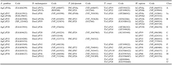
TcCyP19 is a classical cytosolic cyclophilin (83–89% identity to other Trypanosomatids) but it shares high sequence similarity with hCyP-18a (ID=72%). The archetypal cyp-A is encoded in the genomes of many parasites (Klinkert et al. 1996; Ma et al. 1996). TcCyP22 displays a hydrophilic CLD and it was clustered within the group of cytosolic cyclophilins. TcCyP20 showed the highest sequence similarity (64%) with CeCyP16 (P52017) encoded by the cyp10 gene in the Caenorhabditis elegans genome. TcCyP20 also has its orthologues in many other genomes, e.g. 18 kDa cyclophilin from Anopheles gambiae (AgCyP18b), a 18 kDa CyP from Drosophila melanogaster (DmCyP18) or 2 human cyclophilins encoded by the PPIL1 (Ozaki et al. 1996) and PPIL3 genes (Zhou et al. 2001) (see Table 2). Furthermore, in TcCyP20, the tryptophan residue at position 115, essential for CsA binding (Ke, 1992) that is present in many small cyclophilins (Galat, 1999) was substituted by a serine residue, as is the case for AgCyP18b and the large RBD-containing cyclophilin DmCyP75. TcCyP21 and TcCyP24 have the ER-targeting signal sequence and clustered to class B. In the TcCyP25 and TcCyP40 proteins, the Trp residue essential for the CsA binding has been substituted by a histidine residue. The 258 AA long ORF coding for TcCyP28 is terminated by a coiled-coil region that is rich in basic residues and contains several predicted nuclear localization signals (NLS). Its sequence comparison to the NCBI sequence databases showed highest similarity (68%) to a L. major cyclophilin (AL121851.1) that has a similar C-terminus that is a unique sequence feature of the cyclophilin family of proteins. TcCyP28 clustered with the cytoplasmic class A cyclophilins that indicates its probable origin, namely it might have been created by gene fusion of the archetypal T. cruzi CyP-A and a unique gene that contained NLS; TcCyP28 may have a specific function in the nucleus. HMM searches localized in TcCyP40 one N-terminal CLD and three C-terminal TPR domains whereas in all other paralogues only the CLD domain was detected. No other known domain except these 2 was localized by the HMM in the series of the paralogues.
Table 2. Clustered orthologues and paralogues of cyclophilins from five different genomes (The codes that are written in italics have low confidence to belong to the class indicated.)

Global analyses of the Multiple Sequence Alignment comprising 15 paralogues of cyclophilins from T. cruzi and those encoded in other genomes
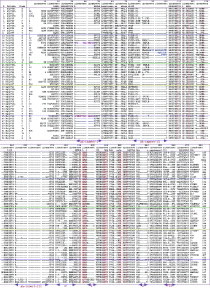
Many sequencing projects of higher organisms and various prokaryotes and lower eukaryotes including the parasite T. cruzi have been recently completed (El-Sayed et al. 2005). Those advances have prompted us to make a comparative study of the cyclophilins encoded in the T. cruzi genome and those encoded in diverse genomes. A total of 495 cyclophilins and cyclophilin-like domains (CLDs), coming both from the NCBI and PIR resources (see Materials and Methods section) as of July 2005, were chosen by the Data-SQ program and were aligned with the Clustal W60 program (Thompson et al. 1994). A subset of the cyclophilins encoded in the genomes of the vertebrates H. sapiens (h), the invertebrates Anopheles gambiae (Ag), Drosophila melanogaster (Dm) and C. elegans (Ce), the parasites T. cruzi (Tc) and Plasmodium falciparum (Pf) and the yeast Neurospora crassa (Nc) were aligned and shown in Fig. 2.

Fig. 2. Alignment of selected cyclophilins from T. cruzi and 4 different genomes. Sequences 1–11 (human cyclophilins, blue line); 12–23 (T. cruzi, green line); 24–32 (P. falciparum, orange line); 33–44 are from A. gambiae (violet line); 45–54 are from N. crassa. The numbers correspond to the beginning and ending AA of the aligned sequence whereas the sign ‘*’ means the C-terminal AA. The secondary structure was taken from (Ke, 1992); the AA residues crucial for PPIase activity were marked in red. The drawing was made with the Form10 program and arranged with ClarisDraw (Microsoft). Some of the database Accession numbers of the aligned proteins are given in Table 2; the NC cyclophilins, NcCyP24, (CSNCM); NcCyP22, (NF00904102); NcCyP30, (NF01487278); NcCyP19, (NF01484255); NcCyP40, (NF00648058); NcCyP18, (NF01527836); NcCyP70, (NF01486260); NcCyP16, (NF01488902); NcCyP90, (NF01484914); NcCyP60, (NF01483993). Note: “the version shown was manually corrected in several places to obtain a better sequence overlap”
Pola – SQ analyses of the multiple sequence alignments (MSAs) revealed that the cyclophilins encoded in the T. cruzi genome can be divided into 2 groups. First, those that have similar sequence organizations as the cyclophilins encoded in the other genomes, such as the P. falciparum, P. yoeli or H. sapiens genomes (TcCyP19, TcCyP20, TcCyP21, TcCyP22, TcCyP24, TcCyP35, TcCyP40, TcCyP42 and TcCyP110); second, those that have unique sequences attached to their CLDs, namely TcCyP25, TcCyP28, TcCyP29, TcCyP30 and TcCyP34. For example, only 5 clearly discerned classes could be assigned to the T. cruzi cyclophilins, namely, TcCyP19 is an orthologue of the archetypal cytosolic cyclophilins (class A, historical name CyP-A) whereas relatively hydrophilic TcCyP22 also clustered with the cytosolic cyclophilins. TcCyP21 and TcCyP24 have sequence features typical for class B cyclophilins that are localized in the ER, the mitochondrial membranes and the secretory pathways (Bergsma et al. 1991; Colley et al. 1991). TcCyP24 has an AA extension of the loop at the N-terminus, which is a typical feature of some cyclophilins from yeast that are known to be associated with the mitochondrial membrane. In historical terms, the small cyclophilins were named as CyP-B (ER-associated cyclophilin); CyP-C, membrane associated cyclophilins; and CyP-D, its mitochondrial form (see Bergsma et al. 1991). TcCyP40 clustered with the heat-shock cyclophilins (Kieffer et al. 1993) that are encoded in the genomes of diverse eukaryotes starting from different fungal organisms and ending on mammals. The nominal masses of the heat-shock cyclophilins vary within a certain range. The sequences of the ‘heat shock cyclophilins’ at their C-terminus possess 3 well-conserved TPR motifs (class F). The sequence organizations of TcCyP42 and TcCyP110 may suggest that they are the orthologues of the Eglin C binding human cyclophilin (hCyP58; classes G) (Wang et al. 1996) and the large cyclophilin that has WW domains (class J), respectively, although both of them do not possess the full set of the sequence attributes that constitute the typical features of classes G and J. The class J cyclophilins are encoded in the genomes of some yeasts (N. crassa), parasites (e.g. P. falciparum, P. yoeli), in the invertebrates, (e.g. A. gambiae, D. melanogaster) and in all higher animals (see Table 2). Moreover, its CLD as well as the protein itself have high hydrophobicity index (57·9% for its CLD and 48·3% for the entire protein, respectively), which might suggest that the protein is associated with the membranes. In fact it clustered with some hydrophobic CLDs that are typical for the membrane-associated cyclophilins (class B). Recent data suggest that this group of cyclophilins is involved in the G1 cell cycle checkpoint (Edgar et al. 2005). Class G cyclophilins are encoded in the genomes of some filarial parasites such as Dirofilaria immitis (Page and Winter, 1998; Hong, Ma and Carlow, 1998) and diverse invertebrates and vertebrates. Its orthologue in C. elegans is involved the cell-cycle checkpoint that bifurcate the cells between the states of mitosis and meiosis (Belfiore et al. 2004). TcCyP25 and TcCyP34 CLDs also have high total hydrophobicity indices.
At least 7 paralogues of the T. cruzi cyclophilins have unique sequence attributes and organizations and were found to possess some distant sequence correlations with classes C, D, E, H and I (Galat, 2004). U-snRNP-associated cyclophilins (class C) that are encoded in the genomes of many lower eukaryotes and in all sequenced to date higher eukaryotic genomes (Teigelkamp et al. 1998) may be represented by TcCyP30 that has some weak correlation with class C although its CLD is not as hydrophobic as it is in the typical members of the class. U-snRNP-associated cyclophilins are encoded in the genomes of some parasites, namely a 20 kDa CyP from the tapeworm Echinococcus granulosus (CAC18541) or a 24 kDa CyP from P. falciparum (see Table 2). Some large cyclophilins (class H) are associated with the nuclear membrane and nuclear speckles (Anderson et al. 1993; Bourquin et al. 1997; Nestel et al. 1996) and are encoded in the genomes of some parasitic nematodes such as Brugia malayi (Page et al. 1995) whereas the large multi-domain cyclophilin that was named nucleoporin (Yokoyama et al. 1995) is associated with the nuclear membrane (class E). TcCyP35 and TcCyP29 may be members of class H since they have similar sequence attributes as CeCyP36 encoded by the cyp-9 gene. TcCyP25 and TcCyP34 may belong to class E, nucleoporin-like CLD, however these hypotheses are solely based on correlations between the sequences' attributes and profiles of the CLDs and those that are typical for class H cyclophilins encoded in diverse genomes (Galat, 2004). TcCyP20 has its orthologues encoded in many other genomes and consists of some sequence attributes that are typical of the large RNA-binding domain containing-cyclophilins such as hCyP57, DmCyP75 or CeCyP57. Although there is so far no example of such a type of cyclophilin encoded in the P. falciparum genome its orthologue is encoded in the Cryptosporidium hominis genome (EAL36265). Despite a considerable increase of genomic databases little can be said on the organisms whose development and differentiation could have given rise to novel genes. It is likely, however, that the fusion in an eukaryotic organism of the gene coding for the orthologue of TcCyP20 to RBDs gave rise to the large RBD-containing cyclophilins that are present in the genomes of many lower eukaryotes and in all higher animals. Because of its similar sequence attributes and features (see Fig. 2) to class I we tentatively assigned TcCyP20 to class Io, namely the archetypal cyclophilin that could have given rise to some of the large cyclophilins that form classes I, G and J. TcCyP42 is a G-type class of cyclophilins that has a clear orthologue in other genomes. None of the T. cruzi’s cyclophilins has typical features of classes D and I that possess RNA-Binding domains (Mi et al. 1996). One example of class D cyclophilin is PfCyP32.
T. cruzi genome organization of the transcribed cyclophilins
Seven epimastigote cDNA clones encoding cyclophilins of 19, 20, 22, 25, 28, 34 and 40 kDa were further characterized. We obtained labelled DNA from these ESTs to be used in hybridization assays. Southern blot analysis using total T. cruzi CL Brener clone DNA, cut with different restriction enzymes, revealed the presence of a single copy of every TcCyP gene tested in the T. cruzi haploid genome (data not shown). Moreover, when T. cruzi GeneDB database searches were done in the already finished T. cruzi sequencing project, these 7 genes were found in only 2 contigs, each representing 2 different chromosomal bands, except for TcCyP19 in which only 1 contig was assigned, probably because the 2 contigs showed high DNA sequence homology in both chromosomes. Studies on the T. cruzi CyP gene localization on CL Brener clone chromosomal bands separated by pulsed field gel electrophoresis, showed that TcCyP19 gene bound 2 chromosomal bands of approximately 2000 and 2200 kbp. These 2 equal sized chromosomal bands also hybridized with TcCyP20. The rest of the cDNA coding for cyclophilins were found dispersed throughout the genome of the parasite (Table 1).
Amastigote and trypomastigote cyclophilin mRNA transcription
From the 7 above-mentioned ESTs, derived from a T. cruzi epimastigote cDNA library, specific primers were designed. We were interested to assess the transcription of these TcCyP genes by RT-PCR in the mammalian host parasite stages: amastigotes and trypomastigotes. RT-PCR analysis was performed using as template oligo d(T)V-primed first-strand cDNA from amastigote and trypomastigote RNA, and TcCyP sequence-specific reverse primers. This strategy was designed to determine the presence or absence of the sequences tested, based on the requirement that only fully processed mRNA transcripts would be expected to possess both a 3′ poly (A) tail (required for reverse transcription primed with oligo d(T)V) and a 5′ spliced leader (required for amplification with the Spliced Leader forward primer). All of the primers tested produced bands at or near the expected sizes. In one case, TcCyP34, we saw 3 amplified DNA products, 1 of which was the expected size (Table 3).
Table 3. Amplification of amastigotes and trypomastigotes mRNA by RT-PCR (Reverse primer sequences and RT-PCR product sizes in base pairs, obtained using the Trypanosoma cruzi spliced leader forward primer.)

T. cruzi cyclophilin protein expression
Four protein bands were detected by silver nitrate staining after the proteins retained on the CsA-affinity chromatography were resolved on SDS/PAGE gel (Fig. 3). A very faint band that is not clearly visible on Fig. 3 was named as band two. These protein bands with molecular masses ranging from 18 to 40 kDa were excised and analysed by MALDI-TOF MS after in-gel tryptic digestion. Database search results for the peptide mass fingerprints obtained from MALDI-TOF spectra allowed the identification of TcCyP19, TcCyP22, TcCyP28 and TcCyP40 (Table 4). When Mascot scores were below 72, which was observed for TcCyP22 and TcCyP40, protein identity was confirmed by PSD analysis. The peptides that matched these 4 T. cruzi cyclophilins are shown in Table 4. An in-gel densitometry using AlphaEasyTM software determined relative protein abundances of around 60% for TcCyP19, 24% for TcCyP22, 15% for TcCyP40, and less than 1% for TcCyP28.

Fig. 3. Trypanosoma cruzi Cyclosporin A binding proteins. An Affi-Gel 10 resin with 8-alanyl-Cyclosporin A as ligand, was used as matrix for T. cruzi epimastigote protein affinity chromatography. SDS-PAGE of the concentrated purified proteins were silver stained, and 4 CsA binding proteins were detected and numbered for further identification.

DISCUSSION
In this work we give the first description of the T. cruzi cyclophilin gene family members and the expressed CsA binding proteins in the epimastigote stage. We have previously reported the cloning of TcCyP19, and demonstrated its CsA sensitive PPIase activity (Búa et al. 2001). This cyclophilin proved to be a target of the diverse non-immunosuppressive CsA analogues that were tested, and exhibited potent trypanocidal activity (Búa et al. 2004). It seems that TcCyP19 has an expression level in the parasite as compared to the expression levels of the other paralogues, first demonstrated in the analyses of the EST databases which revealed that TcCyP19 was encoded by a higher number of cDNA clones than the rest of the CyPs despite the epimastigote cDNA library had been normalized (see Materials and Methods section under cDNA clones). Additionally, the isolation and characterization of TcCyP19 described in this work also suggest that this protein is highly expressed, since around 60% of the total proteins retained on the CsA-affinity gel corresponded to this cyclophilin. Cellular localization of the expressed cyclophilins remains to be experimentally determined.
The number of cyclophilin paralogues encoded in the T. cruzi genome is similar to those encoded in the P. falciparum, P. yoeli, N. crassa, Yarrowia lipolytica etc. genomes; however, their sequence organizations are only in part similar to those encoded by classes A–J cyclophilins (Galat, 2004). One striking difference between the T. cruzi cyclophilins and those encoded in P. falciparum is that sequence organizations of the members of the latter group are closely related to the cyclophilins encoded in many other genomes starting from the lower eukaryotes and ending on the mammals whereas some of the T. cruzi cyclophilins have unique AA inserts in their CLDs that were linked to the domains that are unique such as TcCyP25 or are highly modified as those in the highly hydrophobic cyclophilin TcCyP110.
Moreover, the IDs calculated for the CLDs of the T. cruzi cyclophilins showed less divergence from each other than those for the cyclophilin paralogues encoded in the genomes of other organisms (Galat, 2004). The following paralogues have their orthologues in the majority of the eukaryotic genomes sequenced to date, namely TcCyP19, TcCyP20, TcCyP21, TcCyP22, TcCyP24, TcCyP40, TcCyP42 and TcCyP110 whereas the remaining paralogues might have been created in an organism from which the parasite had diverged. Another possibility is that some of the unique paralogues of the T. cruzi's cyclophilins could be due to gene duplication (Otto and Yong, 2002) within the parasite's genome caused by the evolutionary pressure.
For example, TcCyP30, TcCyP35 and TcCyP40 form a cluster that may suggest that TcCyP30, TcCyP34 and TcCyP35 might have been created by gene duplication from the gene coding for the CLD in TcCyP40 or another heat-shock cyclophilin gene with some unique sequences. Likewise, TcCyP25 and TcCyP28 might have been created by gene duplication of the T. cruzi's archetypal CyP-A gene and some unique sequences that are a part of their unique C-termini. The highly hydrophobic cyclophilin TcCyP110 has the sequence organization typical of class J but it has rather low sequence correlation with the known class J cyclophilins. The majority of the CLDs in the T. cruzi cyclophilins, however, showed a high conservation of the AA residues that are crucial for PPIase activity, especially in those cyclophilins that were found to be transcribed and expressed. For example, TcCyP19, TcCyP22, TcCyP24 and TcCyP28 have 100% conservation of the functional AA residues whereas TcCyP20 and TcCyP34 display somewhat lesser conservation of those residues.
The transcription of TcCyP21 was not analysed in this study, but it was inferred to be transcribed in epimastigotes (AI562688, AI562266) (Verdun et al. 1998) and also in trypomastigote and amastigote cDNA libraries (CF889418, CB923590) (Aguero et al. 2004). This protein has a predicted signal peptide of 28 AA, and is different from the TcCyP22 cylophilin, that has been described in this study as a CsA binding protein in the epimastigote stage. A very recent paper also described the expression of TcCyP21 in an extensive T. cruzi proteomic study (Atwood et al. 2005).
The RT-PCR analysis of other life-cycle stages revealed that the mRNAs for all cDNA clones identified in epimastigotes were also detected in trypomastigotes and amastigotes. The small DNA size observed for TcCyP22 was due to a specific inner primer used. The 3 DNA band products observed for TcCyP34, in which only 1 had the expected size, might not be explained by spurious priming, since there was no product in the SL TcCyP34 rev-primed RT-PCR using genomic DNA template. A more plausible explanation would be that the TcCyP34 gene contains multiple splice sites as has been observed for CyP-9 in C. elegans, a nuclear protein.
It has been suggested that Trp in the binding cleft is important for the tight binding of CsA and its analogues to diverse isoforms of cyclophilins (Hoffmann et al. 1995). The crucial Trp residue was conserved in the majority of the T. cruzi CLDs although some AA substitutions frequently occurred, namely Trp->Ala, Ser or His residues (Galat, 1999). Regarding this histidine substitution for the Trp residues in TcCyP25 and TcCyP40 cyclophilins, it is possible that TcCyP25 is expressed in the parasite at a significant level, but the protein was not preferentially retained on the CsA-affinity. Instead, TcCyP40 protein bears the same Trp->His substitution, a typical feature of heat-shock cyclophilins (Kieffer et al. 1993; Hoffmann et al. 1995), but apparently it was not an impediment for its retention on the CsA-affinity column. It is worth noting, however, that there is a higher conservation level of the functional AA residues in TcCyP40 than in TcCyP25. It may also be that TcCyP25 and the other CyP paralogues that were not significantly retained on the CsA-affinity column have low expression levels or that their binding constants to CsA-affinity column were drastically lower, rendering them barely visible on SDS/PAGE stained with silver nitrate.
It is difficult to estimate to what extent the cyclophilins encoded in the T. cruzi genome are important for parasite survival. TcCyP30, TcCyP34 and TcCyP35 contain longer AA extensions between α-helix II and β-structure III that are unique in the cyclophilin family of proteins. These extensions may be specific recognition sites for interaction with some proteins that may be important for certain nuclear processes in the cells invaded by the parasite.
When studying T. cruzi cyclophilins as potential drug targets, of special interest could be those cyclophilins whose binding properties to potential targets such as non-immunosuppressive analogues of CsA or other small molecular mass compounds are highly different from that of TcCyP19, the major cyclophilin whose binding properties to CsA are similar to those exhibited by hCyP18a. This would be the case of TcCyP20, TcCyP25, TcCyP28 and TcCyP30 if they were crucial for the T. cruzi life-cycle. Their binding affinities to CsA and its analogues may be low but they may have other natural potent inhibitors that may affect the homeostasis of the parasite at pharmacologically relevant doses but whose binding constant to TcCyP19 and the major form of hCyP18a would remain insignificant.
Molecular functions of T. cruzi cyclophilins need further experimental assessments, including the altered functions caused by ligand binding to the T. cruzi cyclophilins in the cellular environment, in order to better elucidate the mechanisms of action responsible for the trypanocidal effects induced by CsA and its non-immunosuppressive derivatives.
We acknowledge the technical help of Octavio Fusco with DNA sequences and Claudia Nose for Artwork. We also acknowledge the TSK-TSC Consortium, for the Trypanosoma cruzi DNA Sequencing Project, for the source of the T. cruzi cyclophilin sequences. J.B., A.M.R. and L.F. are members of the Research Career of the Consejo Nacional de Investigaciones Científicas y Técnicas (CONICET), Argentina. This work was supported by the Instituto Nacional de Parasitología “Dr. Mario Fatala Chabén”, A.N.L.I.S. “Dr. Carlos G. Malbrán”, the NASA/ChagaSpace network, CONICET (Argentina) and the Network for Research and Training in Parasitic Diseases at the Southern Cone of Latin America SIDA/SAREC.