The Fontan operation provides excellent palliation for univentricular circulations.Reference Gewillig1 However, during late follow-up, the circulation becomes increasingly vulnerable as different haemodynamic burdens emerge. Many such late complications are also associated with the occurrence of haemoptysis.Reference Suda, Matsumura and Sano2 When occurring in large volume, haemoptysis may be life-threatening and difficult to treat. We describe the case of a young man with a Fontan circulation who presented with massive haemoptysis. Percutaneous therapy failed to stop his bleeding. A novel surgical approach involving denuding the culprit peribronchial vessels was successfully performed.
Case report
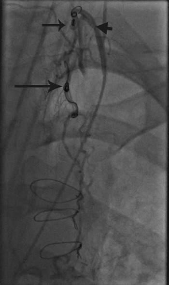
A 19-year-old male with a double inlet left ventricle, as well as transposition of the great arteries and pulmonary atresia, had previously undergone bilateral cavopulmonary anastomoses (1 month), and lateral tunnel Fontan with enlargement of a restrictive ventricular septal defect (9 years) followed by a 25 millimetre carbo-medics aortic valve replacement for severe aortic regurgitation (10 years). Most recently, he presented with large volume haemoptysis of 750 cubic centimetres. Despite 300–500 cubic centimetres per hour ongoing blood loss, neither his oxygenation nor haemodynamics were significantly affected. At cardiac catheterisation, a large leash of arterial vessels surrounding and bleeding into the left main bronchus was identified. The leash was supplied by bronchial arteries, right and left internal mammary collaterals, left inferior phrenic, multiple inferior thyrocervical branches, as well as pleural collateral vessels. These vessels were selectively cannulated as far distally as possible and embolised with non-detachable embolisation micro-coils (Cook Medical, Bloomington, Indiana, United States of America). Most direct access to the bleeding site was through a trifurcate-branching collateral arising from the left thyrocervical trunk, which contained a pre-existing non-occlusive proximal coil, making direct access to the bleeding vessels difficult (Fig 1). It was, however, possible to get distal to the coil and into the main supply vessel using a Progreat™ coaxial micro catheter (Terumo Medical, New Jersey, United States of America). A pulmonary artery angiogram confirmed the absence of arterial to venous shunts. Embolisations were carried out using a combination of 900 and 1100 micrometre Embozene™ particles (Celonova Biosciences, Peachtree City, Georgia, Atlanta, United States of America). Immediate follow-up angiography, however, demonstrated a direct bronchial artery to the left main bronchus fistula, accompanied by further large volume haemoptysis requiring left lung isolation with a double-lumen cuffed endotracheal tube.
Trifurcate-branching collateral arising from the left thyrocervical trunk which contains a pre-existing non-occlusive proximal coil (long arrow) with evidence of recanalisation of collateral distal to this point. Tip of catheter (arrow head) at the proximal end of the thyrocervical trunk and recently deployed coil (short arrow) within the proximal end of the collateral.

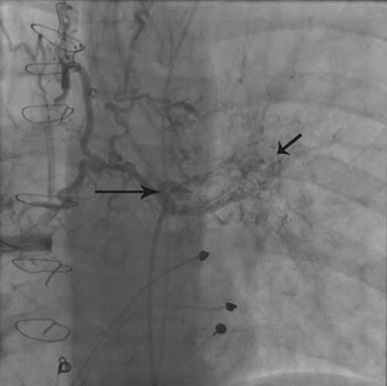
Now, the long and very tortuous bronchial artery system was targeted using a flow-directed micro catheter (Magic 1.2 F, BALT Extrusion, Montmorency, France) with the intention of glue embolising the abnormal peribronchial leash (Fig 2). Despite a distal position being achieved, attempts to get close to the bleeding site precipitated vessel dissection. Finally, the remaining visible feeder vessels were closed proximally with coils in an attempt to reduce arterial pressure to the bleeding focus. The patient was transferred back to the intensive care unit, having exhausted all endovascular treatment options at our disposal.
Large leash of arterial vessels surrounding and bleeding into the left main bronchus with a blush of contrast on angiography demonstrating a bleeding point (short arrow). Catheter within the descending aorta and catheter tip at the proximal end of the bronchial collateral (long arrow).

Rescue strategies such as lobectomy or pneumonectomy, though considered, were rejected as the Fontan circulation would potentially be compromised. Endobronchial stenting was not considered to be a safe option due to infection risk and mucus impaction, particularly in the context of plastic bronchitis often associated with a Fontan circulation. A novel surgical approach was undertaken through a left thoracotomy by which the left main bronchus was denuded of blood vessels through a process of diathermy and placement of ligaclips along its length under direct vision. The endobronchial tree was then sucked free of clot. His oxygenation improved rapidly and he was able to wean from the ventilator. At 2 weeks, he required drainage of a large left-sided haemothorax, which at surgical re-exploration was due to oozing points related to adhesional inflammation and neovascularisation. The bleeding points were diathermised. At 6 months, he is doing well and is back on full anticoagulation therapy. We plan to do an elective cardiac catheterisation at 1 year to assess new vessel formation.
Discussion
Massive haemoptysis in the Fontan circulation may be fatal. In the case presented above, percutaneous therapy failed to stop bleeding into the left bronchus. We undertook a successful novel surgical approach denuding culprit bronchial vessels under direct vision.
Large volume haemoptysis, that is, greater than 600 cubic centimetres over a 24-hour period, has many potential causes in the Fontan patient.Reference Deisenberg and Stayer3 These include arterio-venous malformation, anticoagulation, chest infection, and aorta-pulmonary collaterals due to neovascularisation. Nagla and Nagwa demonstrated significant activation of potent angiogenic factors such as vascular endothelial growth factor, platelet-derived endothelial growth factor/thymidine phosphorylase, and Leptin in cyanotic coronary heart disease.Reference El-Melegy and Mohamed4 Hypoxia causes tissue damage, releasing chemical mediators and cytokines such as the transcription factor Hypoxia inducible factor 1, which triggers vascular endothelial growth factor synthesis. In cyanotic coronary heart disease, vascular endothelial growth factor levels parallel the degree of cyanosis. Persistently elevated systemic venous pressures and associated venous endothelial cell stretch appear to drive angiogenesis despite the absence of cyanosis in Fontan patients.Reference Suda, Matsumura and Miyanish5
Of all the various strategies employed to treat haemoptysis, percutaneous embolisation is effective in up to 80% of cases.Reference Brinson, Noone and Mauro6, Reference Shigemura, Wan and Yu7 Coils should be deployed as distally as possible so as to isolate the bleeding point and not block future access. In fully anti-coagulated patients, clot formation is largely dependent on the mechanical occlusion properties of the coil. Unless densely packed, incomplete vessel obliteration is therefore common. In non-coronary heart disease patients, particulate embolisation is more commonly practised. This enables satisfactory devascularisation of the bleeding focus without compromising future access.Reference Shigemura, Wan and Yu7 When considered in the Fontan patient, pulmonary arterial to venous shunts and veno-venous shunts in the particulate embolisation trajectory should be excluded, and relatively larger particles should be used to avoid systemic embolisation.
Having exhausted all endovascular options, we employed a novel strategy of surgically devascularising the bleeding bronchus as a rescue therapy in preference to lobectomy or pneumonectomy. This had the desired therapeutic effect of removing the common pathway by which collateral vessels supplied the bleeding leash. Despite bronchial ischaemia and necrosis being a potential problem of this approach, we found instead evidence of extensive new vessel formation in the lung at 2 weeks, which suggest a high likelihood of future recurrence. It is tempting to consider monoclonal antibody to vascular endothelial growth factor, already in use in colorectal cancer and diabetic retinopathy in curbing new vessel formation.Reference Ferrara, Hillan and Novotny8
Conclusion
When conventional strategies fail to achieve haemodynamic stability and haemostasis in haemoptysis associated with the Fontan circulation, a direct lateral thoracotomy with denuding of the culprit bleeding bronchus approach can be successfully used in selective patients where the bleeding site is close to the main bronchi or in the proximal hilar regions.