Secundum atrial septal defect combined with valvular pulmonary stenosis is sometimes detected in children with CHD. Persistence of this complication has deleterious effects on right ventricular systolic function.Reference Xu, Liu, Zhao and Qin 1 Early intervention is sometimes needed to prevent cardiac dysfunction, and the traditional treatment is open-heart surgery. Although a previous report described the successful simultaneous percutaneous transcatheter closure of atrial septal defect and pulmonary valvuloplasty mainly in adult patients,Reference Medina, de Lezo, Delgado, Caballero, Segura and Romero 2 only few reports have described it in infants.
Here, we present a case of successful simultaneous transcatheter intervention for this complication in an infant.
Case report
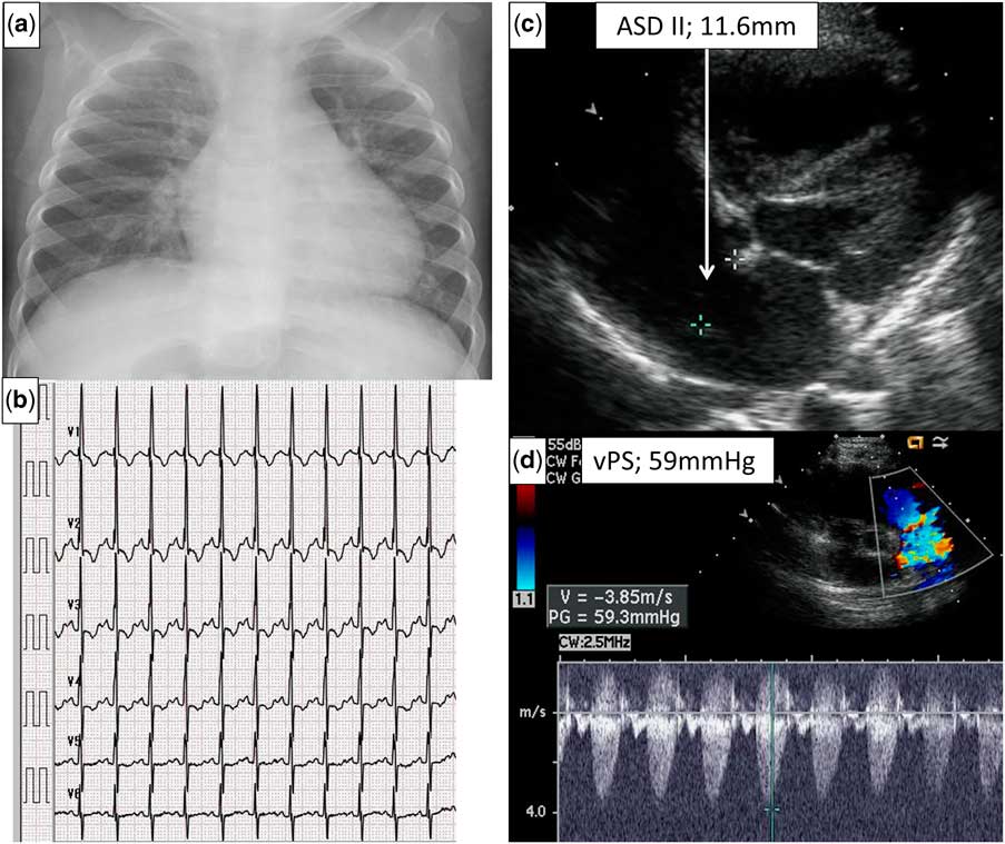
The patient was referred to our centre to undergo further examination for heart murmur at 10 months of age, weighing 8.5 kg. Chest radiography (Fig 1a) revealed slight pulmonary congestion, and electrocardiography showed ST-T change in the left precordial leads (Fig 1b). Transthoracic echocardiography revealed a secundum atrial septal defect of 11.6 mm in diameter with right atrial and ventricular dilation combined with pulmonary valve stenosis with a peak pressure gradient of 59 mmHg (Fig 1c and d). We considered that this patient had the potential myocardial damage owing to volume and pressure overload. We planned simultaneous transcatheter intervention to prevent cardiac dysfunction. Informed consent was obtained from the parents.
Examinations at first visit. ( a ) Chest radiograph showing slight cardiomegaly and pulmonary congestion. ( b ) Electrocardiogram showing ST-T change in the left precordial leads with right ventricle (RV) hypertrophy. ( c and d ) Transthoracic echocardiography (TTE) showing atrial septal defect (ASD) II of 11.6 mm in diameter, with right atrium and RV dilations combined with valvular pulmonary stenosis (vPS) with a peak pressure gradient of 59 mmHg at the pulmonary valve level.

A haemodynamic evaluation was performed before the catheter intervention. Cardiac catheterisation confirmed the echocardiographic findings with a peak pulmonary valve gradient of 39 mmHg and a significant left-to-right shunt (Qp/Qs, 2.1:1).
Transcatheter pulmonary valvuloplasty was performed first. The right ventriculography was performed to measure the pulmonary valve annulus diameters. We used a 15-mm/3-cm Tyshak balloon (NuMED, Hopkinton, New York, United States of America) with a diameter 1.2 times larger than the pulmonary valve annulus. The pressure gradient decreased from 39 to 18 mmHg after balloon dilation. Then, transcatheter closure of the atrial septal defect was performed.
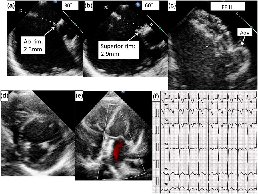
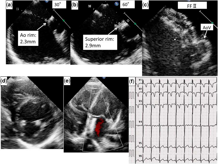
The location, size, and relationship of the defect with the surrounding tissue were assessed using trans-oesophageal echocardiography before the procedure. This echocardiogram revealed a defect of 12 mm in diameter with hypoplastic aortic (2.3 mm) and superior rims (2.9 mm) (Fig 2a and b), but the other rims were more than 5 mm. Balloon-stretched diameter of the defect was 14.5 mm. A transport sheath (9 F) was advanced over the guidewire into the left atrium. We chose a 15-mm Figulla Flex II occluder (Occlutech, Jena, Germany) at first, but transient atrioventricular block occurred. We sized down to a 12-mm occluder based on the echocardiogram. Before releasing the occluder, we performed a trans-oesophageal echocardiographic evaluation to assess the adequacy of its position and the stability of the device within the septum. After the occluder was confirmed to be touching the aorta but not impinging any structure, it was completely released (Fig 2c). The deployment was uneventful without complications. Aspirin therapy was administered for 6 months. Transthoracic echocardiography revealed the adequate position of the device, an estimated pressure gradient of 11 mmHg on the pulmonary valve level with trivial regurgitation, and round-shaped appearance of the interventricular septum at short-axis view 6 months after the intervention (Fig 2d and e). Electrocardiogram revealed a normal ST-T pattern on the left precordial leads but showed incomplete right bundle branch block (Fig. 2f).
Examinations during and after intervention. ( a and b ) The aortic and superior rims are hypoplastic, but the other rims are >5 mm. ( c ) The device touched the aortic valve (AoV) but caused no impingement. ( d and e ) trans-oesophageal echocardiography image showing the proper device position and no impingement to any structure. ( f ) Electrocardiographic image showing a normal ST-T pattern on the left precordial leads and no hypertrophy. FF II=Figulla Flex II.
Discussion
We describe the successful simultaneous transcatheter treatment of a secundum atrial septal defect complicated by valvular pulmonary stenosis in an infant.
Secundum atrial septal defect is the most common congenital cardiac lesion, but the association with valvular pulmonary stenosis is relatively rare.Reference van der Linde, Konings and Slager 3 When the two conditions are present simultaneously, significant left-to-right shunting is often prevented by the outflow obstruction, which protects the pulmonary vascular bed without premature damage until adulthood. However, persistence of this complication has deleterious effects on right ventricular systolic function.Reference Xu, Liu, Zhao and Qin 1 This fact indicates that total correction is needed.
Indications for valvular pulmonary stenosis are symptoms or a resting gradient ≥40 mmHg.Reference Silvilairat, Cabalka, Cetta, Hagler and O’Leary 4 Atrial septal defect closure is also recommended for patients of any age who present with a haemodynamically significant defect (Qp/Qs>1.5). In this case, transthoracic echocardiography showed that moderate-sized atrial septal defect with right atrial and ventricular dilation coexisted with valvular pulmonary stenosis with a 59-mmHg peak pressure gradient. We speculated that right ventricular pressure and volume overload influenced the left ventricle and caused the ST-T change in the electrocardiogram. Therefore, we considered that early intervention should be performed to prevent further myocardial damage.
Open-heart surgery has been considered the initial choice for this combination. On the other hand, the transcatheter technique for the treatment of CHD has considerably progressed in the past decade. Percutaneous transcatheter pulmonary valvuloplasty has become the initial choice for valvular pulmonary stenosis in all age groups, even in neonates.Reference Ahmadi and Sabri 5 Transcatheter closure of a secundum atrial septal defect has also become a feasible and effective method even in infants.Reference Petit, Justino, Pignatelli, Crystal, Payne and Ing 6 Furthermore, combining multiple interventions in a single catheterisation affords benefits to such patients, which obviates the need for a second interventional procedure.Reference urkay, Abdullah and Celal 7 On the basis of these reports, we chose simultaneous catheter treatment instead of surgery.
Choosing a correct and reasonable sequence of treatments to ensure the safety of transcatheter interventional therapy is crucial. The repair that should be performed first is controversial, but some previous reports recommended performing percutaneous transcatheter pulmonary valvuloplasty before transcatheter closure of the atrial septal defect from a technical view, which would limit the possibility of potentially dislodging the atrial septal defect device. Furthermore, an increase in left-to-right shunting after the initial correction of pulmonic stenosis and if not treated will lead to right ventricular volume overload and its sequelae.Reference Vera, Nounou and Kern 8 We performed pulmonary valvuloplasty first and then atrial septal defect closure in the same session and we obtained successful results with no major complications. However, during follow-up, abnormal ST-T changes returned to normal pattern but the incomplete right bundle branch block emerged. We speculated that the pressure overload decreased but the influence of volume overload still remained. We need to continue to check this patient’s cardiac function.
In conclusion, simultaneous transcatheter correction is a safe and effective therapeutic option even in infants with atrial septal defect combined with valvular pulmonary stenosis.
Acknowledgements
The authors thank Dr Takashi Soga for her help with the trans-oesophageal echocardiographic image acquisition and helpful insights.
Financial Support
This research received no specific grant from any funding agency or from commercial or not-for-profit sectors.
Conflicts of Interest
None.
Ethical Standards
The authors assert that all procedures contributing to this work comply with the ethical standards of Showa university and with the Helsinki Declaration of 1975, as revised in 2008.