INTRODUCTION
The Monte dei Cappuccini Monastery stands in Torino (Italy), on a hill 283 m a.s.l, on the right bank of the Po River. Formerly it was a fortress called bastita Taurini (one of the many that bound the city perimeter), built in the XII century and occupied by a garrison taking orders from the lord of the Torino Castle, who was a deputy of the Prince and holder of executive power. In 1581, Carlo Emanuele I – Duke of Savoy, acquired the bastita and offered it to the Order of Friars Minor Capuchin, who officially have used it as a monastery since 1583 (see Figure 1).
(a) Map of the Bastita during the XIII–XIV centuries (Isella Reference Isella2013). The walls of the Romanesque church (XI century) are also visible. The arrow indicates the location of the burial site. (b) Perspective drawing of the Bastita during the XIII century (Isella Reference Isella2013). (c) Nowadays Monte dei Cappuccini Monastery view from the Po River shore.

In 1989, an atypical burial was discovered in this area, consisting of a skeleton and a few objects, during the study of another one found in the same area (belonging to Count Filippo d’Agliè 1604–1667; Isella Reference Isella2013). It soon became clear that the skeleton was even more ancient, possibly dating back to the late Middle Ages. Due to the particular religious context of the finding, a rigorous archaeological survey could not be made at the time of the exhumation. Only recently, the human remains and related objects were rendered available for radiocarbon (14C) dating and for anthropological and archaeometric investigations.
We performed a high-precision radiocarbon dating on several bone samples in order to reduce as much as possible the uncertainty associated with the radiocarbon age. We applied to the samples three different pretreatment techniques and compared the obtained results, using statistical tests to verify the compatibility of the measurements. The final set of radiocarbon ages have been used to evaluate the average value, whose calibration has been performed using also the available historical notes to fix a terminus ante quem point.
MATERIALS AND METHODS
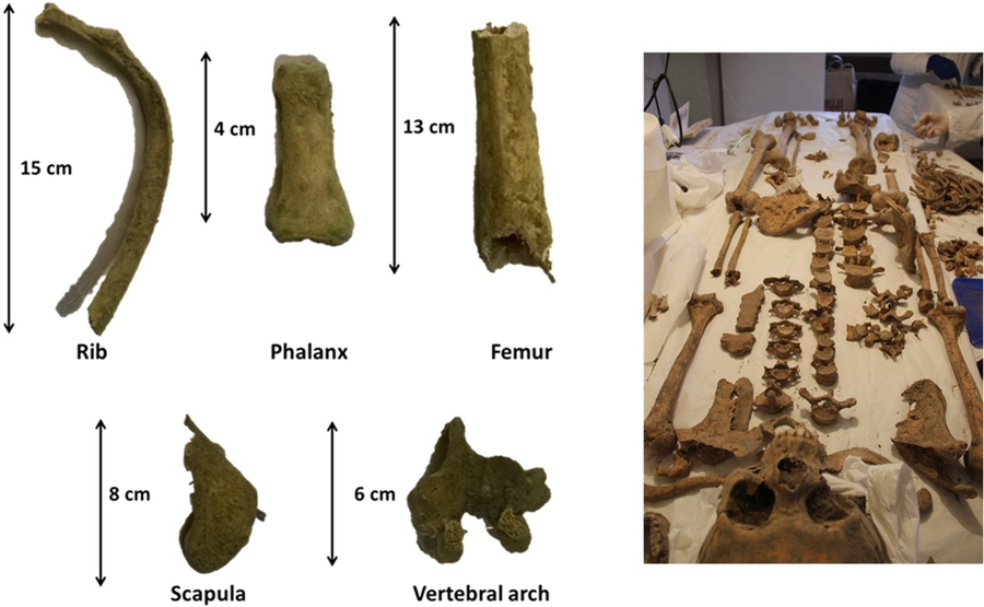
In the burial, a skeleton and five objects were found. The skeleton was complete, and the bones were in a good state of preservation (see Figure 2). The human remains were analyzed with regard to the standard anthropological criteria, using morphological and macroscopic methods (Buikstra and Ubelaker Reference Buikstra and Ubelaker1994; White and Folkens Reference White and Folkens2005). Details about the analysis of the skeleton are reported in Appendix A.
(a) Bone samples used for the radiocarbon dating; (b) picture of the skeleton found in the burial.

The objects found in the same burial context of the human remnants were (a) a metallic, truncated-conical neck, possibly representing the extremity of a powder horn used by harquebusiers or bombardiers in the XIV–XVI century, (b) a metallic lacefix, with a shape typical of the XIV–XVI century, (c) a wrought metallic fragment, possibly representing the portion of a dagger guard, which separates the hilt from the blade, (d) a rusty nail, with head wrapped in a sheath made of a scarcely compacted cluster ripped in half, with evident rust traces in those portions touching the nail, and (e) an orange-colored, squat cylindrical glass bead with an axial cavity filled by incoherent sediments. All these objects were characterized with a non-invasive protocol, by investigating their surfaces in areas unaffected by corrosion traces (so to avoid biasing the related results) with micro-X-ray fluorescence (μ-XRF). A µ-XRF Eagle III-XPL was used, equipped with a micro-focus air cooled X-Ray tube with a Rh anode (voltage: 40 kV; current: 1 mA), poly-capillary X-Ray optics and an EDS Si(Li) detector. The concentrations of the different elemental species were inferred by integrating the peak areas with the Vision-32 software package (EDAX), using quantitative correction by fundamental parameters with standard.
Radiocarbon Dating
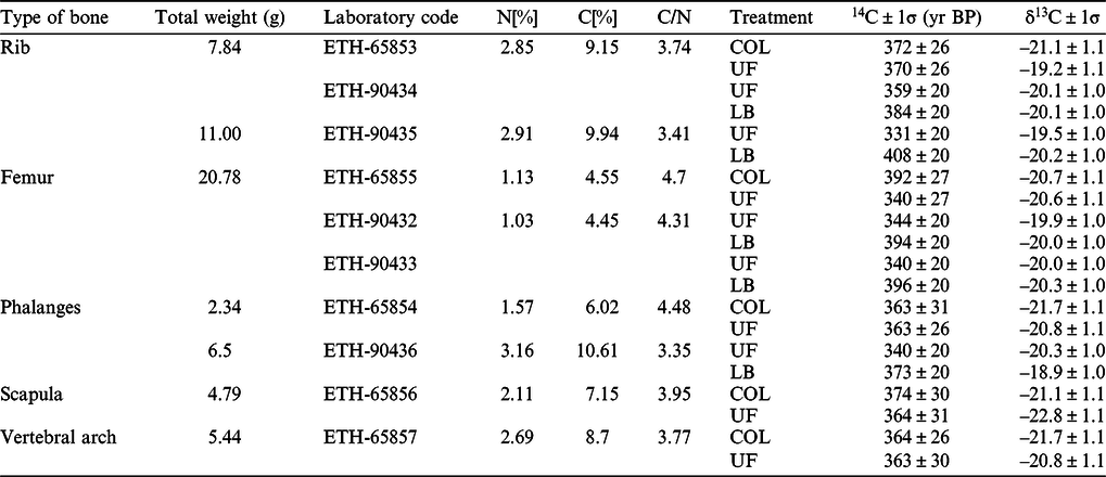
In order to obtain the highest possible dating precision, the analysis of multiple samples was planned. A large amount of material was available, and we selected five different types of bone (see Table 1 and Figure 2).
Types of bone, weight of the total material available for each sample—from which about 1.5–2 grams have been taken—and measured 14C ages (yr BP), obtained using the COL, UF and LB procedures, are reported.

The samples were cleaned with several ultrasonic baths in distilled water, dried in an oven at the temperature of 60°C and then powdered. Before collagen extraction, each sample (consisting in about 1.5–2 grams of material) was split in two parts, in order for the collagen to be extracted by using distinct procedures, namely the ultrafiltration (UF) and the collagen (COL; Hajdas et al. Reference Hajdas2008, Reference Hajdas, Michczynski, Bonani, Wacker and Furrer2009) methods. In addition, since for some samples enough material was available, the Longin-base (LB; Hajdas et al. Reference Hajdas2008) method was also applied to them. Therefore, we prepared five more samples for this second run of AMS measurement. Even in this case, we split every sample into two parts: the first one was treated with the UF method and the second one according to the LB procedure.
The UF pretreatment is based on the separation of peptide molecules (heavier) from the contaminant ones (smaller), by the gelatinization of the bone material and the use of ultra-filtration tubes. This procedure, which represents the standard pretreatment technique applied to bone samples, required about 1 g of material. Since the bones were well-preserved and quite recent, also the standard chemical treatment for organic matter (COL procedure) could be adopted for this specific case study. The main advantages of this pretreatment method lie in the fact that it requires small amounts of material (about 100 mg) and its main steps consist of simple acid (HCl) and base (NaOH) attacks. Finally, we applied to the femur, rib and phalanx samples also the Longin-base method, which consists in the dissolution of collagen in weak acid and in a short attack with a base solution before gelatinization.
The samples processed with the three pretreatment methods were converted into graphite targets using the automated graphitisation equipment (AGE), coupled with an elemental analyzer for the sample combustion. This fully automated system uses a molecular sieve trap (a column filled with zeolite material) to transfer the CO2 from the combustion to the graphitization reactor (Wacker et al. Reference Wacker, Nemec and Bourquin2010c). AMS 14C measurements were performed by using the 200 kV MIni CArbon DAting System (MICADAS) at ETH Zurich in Switzerland (Synal et al. Reference Synal, Stocker and Suter2007; Wacker et al. Reference Wacker, Bonani, Friedrich, Hajdas, Kromer, Nemec, Ruff, Suter, Synal and Vockenhuber2010b). Data evaluation was conducted using the AMS data reduction program BATS (Wacker et al. Reference Wacker, Christl and Synal2010a). The calibrated age was obtained by using the OxCal v4.3 software (Bronk Ramsey Reference Bronk Ramsey2009, Reference Bronk Ramsey2017) and the calibration dataset IntCal13 (Reimer et al. Reference Reimer, Bard, Bayliss, Beck, Blackwell, Bronk Ramsey, Buck, Cheng, Edwards, Friedrich, Grootes, Guilderson, Haflidason, Hajdas, Hatté, Heaton, Hoffmann, Hogg, Hughen, Kaiser, Kromer, Manning, Niu, Reimer, Richards, Scott, Southon, Staff, Turney and van der Plicht2013).
RESULTS AND DISCUSSION
Anthropological Examination of the Skeleton
The skeleton is well preserved in every portion. It belongs to a young adult male, aged approximately 20–30 years; the man’s stature in life is estimated at about 170 cm. Macroscopically, the bones do not show any pathological alterations and injuries, except for a well-defined enthesiopatic lesion caused by a hypertrophy of costoclavicular ligaments on both clavicles as a result of biomechanical stress.
Archaeometric Survey of the Objects
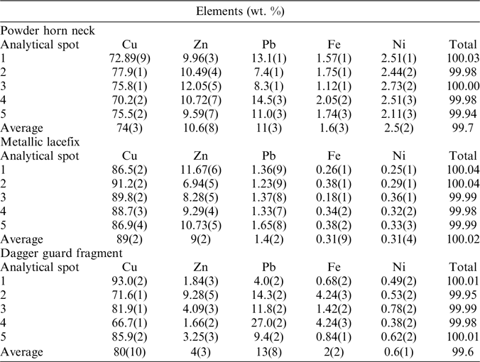
Three objects (the powder horn neck, the lacefix and the dagger guard), when analyzed by μ-XRF, show a predominant presence of Cu (70–90 wt %), with subordinate Zn (≤ 10 wt %) and Pb (1–10 wt %). Such a composition (see Table 3a, in the Appendix B) is typical of a “tombac”—a Medieval to post-Medieval brass alloy at times identified as “leaded brass” (Hložek et al. Reference Hložek, Komoróczy and Trojek2012; Gener et al. Reference Gener, Montero-Ruiz, Murillo-Barroso, Manzano and Vallejo2014). Such an evidence accounts for the pertinence of these findings to the studied burial context.
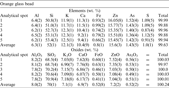
Scarce information can be obtained from the nail composition (mostly Fe, with a sedimentary cap), while the characterization of the orange bead is troublesome. μ-XRF showed quite an inhomogeneous chemistry, hardly consistent with that of an ancient glass—especially for what concerns the nature of the chromophores/opacifiers (e.g., the absence of Fe and Cu; see Table 3b in the Appendix B; Capra et al. Reference Capra, Pederiva and Dal Maschio2005; Charalambous et al. Reference Charalambous, Sakalis, Kantiranis, Papadopoulou, Tsirliganis and Stratis2010). Its contextualization in the studied burial context remains, therefore, dubious.
Radiocarbon Dating of the Bones
Compatibility Test of the Results
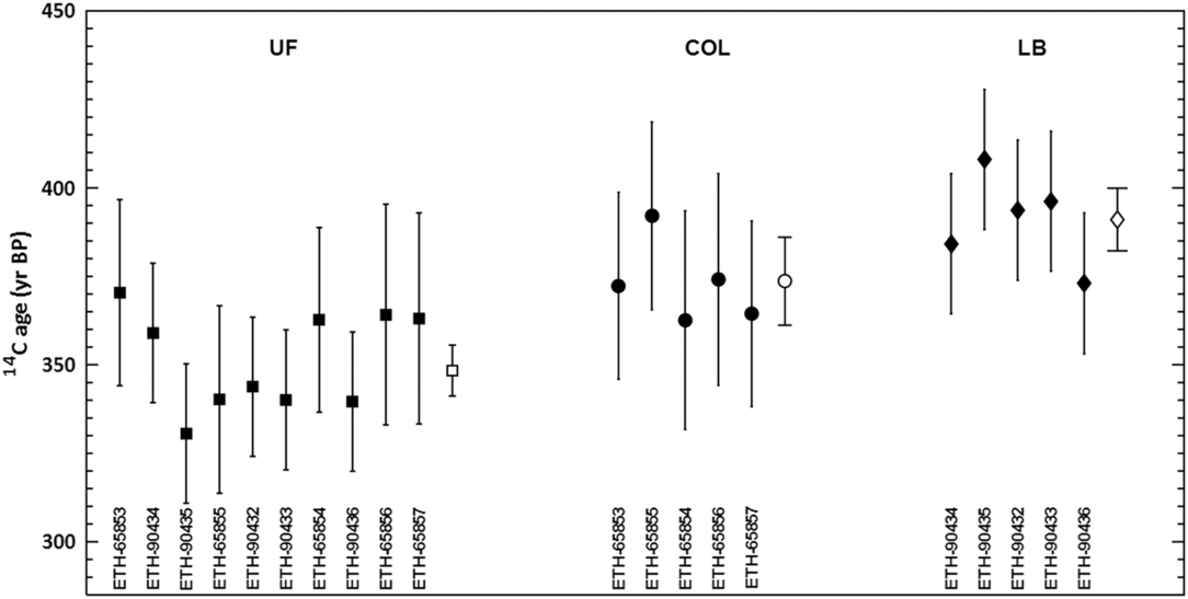
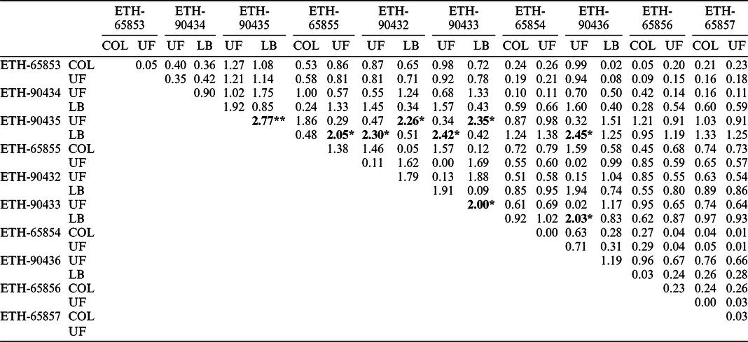
The ages obtained from the measurements of all the samples are reported in Table 1 and shown in Figure 3. It is clearly visible that, for all 10 samples, the values obtained with UF and COL treatments are compatible in 1σ range, while the LB method always returns radiocarbon ages older than those deriving from the UF procedure. In particular, the ages of sample ETH-90435 using UF and LB methods have a probability (P) lower than 1% to be compatible, according to the t-test (see e.g. Long and Rippeteau Reference Long and Rippeteau1974), as shown in Table 2. Therefore, we exclude these two ages from the set of measurements, hence considering the values which cannot be rejected at 99% confidence level (c.l.) The resulting weighted average is 366 ± 5 yr BP.
Radiocarbon ages of the samples listed in Table 1 treated with the ultrafiltration (UF; squares), collagen (COL; dots) and Longin-base (LB; diamonds) procedures. 1σ-error bars are also shown. The empty square, circle, and diamond represent the weighted average evaluated with the measurements obtained from the UF, COL, and LB methods, respectively.

Results from the t-test applied to each pair of radiocarbon age values measured in order to test their compatibility. Bold values refer to pair of measurements with a significant disagreement. * Indicates pairs with less than 5% of probability (P) to be compatible; ** indicates P < 1% (Long and Rippeteau Reference Long and Rippeteau1974).

We notice that in Table 2 other values show a significant disagreement (P < 5%); in these cases, the pairs are always formed by samples treated with the UF and the LB procedures, respectively. In all samples, the ages from the LB method are older than those from the UF ones: this evidence raises doubts about the suitability of the applied LB procedure as a pretreatment method for bone samples.
If we consider the averaged values corresponding to each pretreatment method, we can notice that the mean value from the UF data (349 ± 7 yr BP; empty square in Figure 3) and the LB one (391 ± 9 yr BP; empty diamond) are not compatible in 2σ-range and the t-test returns a really high significant disagreement (t = 3.7). This reinforces our conclusion about the anomalous older ages obtained with LB procedure. On the contrary, the average UF value and the COL one (374 ± 12 yr BP; empty circle in Figure 3) are compatible in 2σ-range and the t-test confirms that their disagreement is not relevant (t = 1.76), as previously demonstrated by the result obtained from the tests performed with each pair of measurements.
Radiocarbon Age Calibration
In order to perform the calibration of the final radiocarbon age obtained from the set of values with probability of compatibility higher than 1% (366 ± 5 yr BP), we considered the available historical evidence. The area of such a discovery had been occupied, since the monks’ settlement, by a trail crossing the monastery N/S—hardly to be destined to host a burial (see Figure 1). However, such an area was reputed to formerly host the cemetery of the bastita fortress. The surviving local chronicles—issued only after the Monastery was established, in 1583—say nothing about the burial. Therefore, the skeleton should be more ancient than the monastery itself—and possibly referred to a period when the fortress stood in its place.
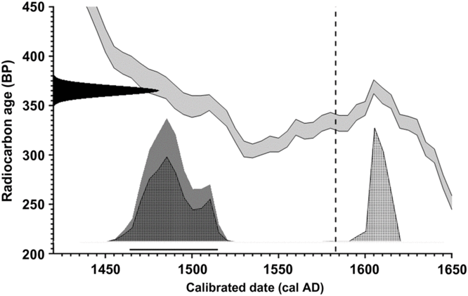
This conclusion has been also supported by the archaeometric survey of the objects, since at least three of them resulted to be compatible with the Medieval period. For this reason, we calibrated the radiocarbon age considering the 1583 AD date as a terminus ante quem (TAQ) point. According to this calibration, the death of this man occurred in the time interval between 1464 and 1515 cal AD (at 95.4% c.l., Figure 4). We can notice in Figure 4 that the unmodeled calibration of the radiocarbon age would have led to two possible time intervals (at 95.4% c.l.), namely 1463–1515 and 1600–1619 cal AD. Therefore, thanks to the synergy between accurate radiocarbon and archeometric analyses and historical surveys the uncertainty associated to the age of the burial has been noticeably reduced to the late XVth–early XVI century.
Calibration result of the mean value (366 ± 5 BP) of compatible radiocarbon ages reported in Table 1. Calibration was performed with the OxCal v4.3 software (Bronk Ramsey Reference Bronk Ramsey2009, Reference Bronk Ramsey2017) and the calibration dataset IntCal13 (Reimer et al. Reference Reimer, Bard, Bayliss, Beck, Blackwell, Bronk Ramsey, Buck, Cheng, Edwards, Friedrich, Grootes, Guilderson, Haflidason, Hajdas, Hatté, Heaton, Hoffmann, Hogg, Hughen, Kaiser, Kromer, Manning, Niu, Reimer, Richards, Scott, Southon, Staff, Turney and van der Plicht2013, grey band) and the date 1583 AD as terminus ante quem point (dotted line). The result of this calibration is described by the dark-grey area. The horizontal lines indicate the calibrated time intervals corresponding to 95.4% c.l. (1464–1515 cal AD). Dotted areas indicate the unmodeled age calibration, from which two possible time intervals would have been obtained at 95.4% c.l.

In the XV century, a period of social instability affected the Savoy dukedom and the city of Torino, which also concerned the kingdom of France. The most relevant episode which affected the fortress in this period is its settlement by Phillip of Savoy, lieutenant of the king of France Louis XI. Once in Torino, he aimed at disposing of the artillery stockpiled in the bastita in order to declare war on the duke of Milan, but the Cismountain Council and the lord of the Torino Castle tried to oppose. Phillip resolved such an issue in blood. The dating of the human remains is consistent with the information inferred from the local chronicles (Barbero Reference Barbero1995, Reference Barbero2002; Benedetto Reference Benedetto1997; Isella Reference Isella2013). As far as the identity of the buried man is concerned, this is still unknown. On the basis of this results, we cannot exclude that the burial might be related to the sequence of truculent events that occurred in the bastita Taurini in 1476, when Phillip of Savoy suppressed the attempts of resistance by probably executing any soldier of the garrison unwilling to recognize his leadership. Besides, the skeleton had been interred reversed with respect to the usual orientation (dictated by Medieval Christian ritual habit), namely with the head pointing east and the feet west. Moreover, the corpse was prone—his face towards the ground, instead of supine. This evidence could be interpreted as signs of scorn and contempt, reserved to subjects responsible and condemned for political crimes (i.e., high treason and rebellion). Finally, it has been also considered that only those persons appointed of particular rights and power (i.e., the prince or his dignitaries) could be buried inside the fortress, while all other interments were destined to the closest public cemeteries. These facts highlight the necessity of a deeper investigation about the identity of than man and the causes of its death.
CONCLUSIONS
The 14C dating of the skeleton of a young adult man, found in the upper garden of Monte dei Cappuccini (Torino, Italy), has been performed to clarify the age of the finds, which otherwise lacked a proper archaeological analysis of the burial site. The statistical reduction of the uncertainty associated with the dating has been crucial to restrict and define, as much as possible, the historical period in which the man lived. The samples have been pre-processed using three different methods, namely the ultrafiltration, the “collagen” and the Longin-base ones.
An addition of historical evidence and the use of a terminus ante quem point narrowed the calibrated time interval (at 95.4% c.l.) between 1464 and 1515 cal AD. Some objects found in the same burial context of the skeleton were studied with a non-destructive archaeometric approach, so to evaluate their feasible pertinence with the studied setting.
The most interesting result we obtained in this analysis is the statistically older age associated with the samples treated with the Longin-base method, with respect to those processed with the ultrafiltration one. The statistical significance of the disagreement between these methods has been supported by several measurements on the same individual. Nevertheless, since we described a single case study, other evidence is required to test the general validity of our conclusions.
ACKNOWLEDGMENTS
We wish to thank Maria Belen Röttig and Mantana Maurer for their precious technical assistance. Roberto Cossio and Emanuele Costa are thanked for their precious help in collecting XRF and SEM-EDS data, respectively. We would like to thank Padre Franco of the Monte dei Cappuccini Monastery for his kindness in welcoming us at the monastery and allowing us to perform this study.
APPENDIX A Anthropological Analysis
Estimation of sex has been based on the assessment of dimorphic characteristics of the skull and pelvis, such as: nuchal crest, mastoid process, supraorbital ridge and glabella, mental eminence (cranial traits); general morphology of os coxa, sacrum and pubic portion including observation of ventral arch, subpubic concavity, ischiopubic ramus, shape and depth of greater sciatic notch, pre-auricular sulcus (pelvis traits). Criteria for aging required the evaluation of dentition (teeth development and wear patterns), cranial sutures closure, surface changes at the pubic symphysis, auricular surface of the ilium and sternal rib ends. Observations of several late fusing skeletal elements (such as the epiphysis of clavicle, the iliac crest of the os coxae) were also considered for distinguishing young and adult from older individual. Regarding anthropometric evaluation, standard measurements were performed in order to evaluate stature; the skeletal elements analyzed included bone maximum lengths (humerus, radius ulna, femur, and tibia). Paleopathological observation was performed in order to identify any skeletal lesions.
The size and general robustness of the different part of the skull and post-cranial bones are compatible with a male sex diagnosis. Regarding the dentition, all teeth were erupted, except M3, and show low signs of wear on the occlusal surface. For what concerns the postcranial skeleton, the developmental status of all preserved bones corresponded to a young adult individual. All the epiphyses of the postcranial skeleton were fused, providing a minimum age-at-death of at least 18 years. In the clavicle, the sternal epiphysis was joined to the shaft, but the junction was still clearly defined by a line, so this individual was probably below 30 years old. According to dental evidence, the remains belonged to a young adult male approximately 20–30 years old. The man’s stature in life could be estimated at about 170 cm. From a pathological perspective, it presented musculoskeletal stress markers (enthesopaties) at the attachment site of the costo-clavicular ligament on both clavicles—clear indicators of occupational stress.
APPENDIX B Archaeometric Survey
Chemical composition, as resulting by μ-XRF, of the three objects (powder horn neck, lacefix, and dagger guard fragment) made of a brass alloy, expressed by normalizing at 100 the wt % of the main constituent metals.

Chemical composition, as resulting by μ-XRF, of the orange glass bead expressed by normalizing at 100 the wt. % of both the main elements and oxides.