INTRODUCTION
Several species, mostly of the genus Brachidontes Swainson 1840, dominate intertidal mussel beds on rocky shores, along Uruguay, Argentina and Chile. Brachidontes darwinianus (d'Orbigny 1846) occurs from Brazil to Uruguay (Rios, Reference Rios1994). Brachidontes rodriguezii (d'Orbigny, 1846) distribute from Uruguay to north Patagonia along the Argentinean coast (Adami et al., Reference Adami, Tablado and López Gappa2004, Reference Adami, Tablado and Sodor2008) and Brachidontes purpuratus (Lamarck, 1819) from north Patagonia extending northwards along the south-eastern Pacific to northern Peru (Coan & Valentich-Scott, Reference Coan and Valentich-Scott2012). According to Scarabino (Reference Scarabino1977), there is a replacement of species along the Atlantic coast, and this could be due to sea surface temperature. There is a transition zone between 40°S and 44°S where B. rodriguezii and B. purpuratus coexist (Balech & Ehrlich, Reference Balech and Ehrlich2008). The systematics of both species, based on external conchological characters and geographic distribution, have been discussed due to the phenotypic shell diversity (Van der Molen et al., Reference Van Der Molen, Marquez, Idaszkin and Adami2012; Adami et al., Reference Adami, Pastorino and Orenzans2013). According to Adami et al. (Reference Adami, Pastorino and Orenzans2013) the comparison of morphological features of the valves allows differentiation between both species, although most of the characters considered in the classification of this species seem to show ecophenotypic variation.
Brachidontes rodriguezii and B. purpuratus are considered dominant species on hard substrates along the Argentinean coast, nevertheless some reproductive and morphological aspects remain unknown. In bivalves, sperm morphological traits have been used in systematic studies to differentiate closely related species (Introíni et al., Reference Introíni, Magalhães, Aguiar-Jr, Quaresma, Lino- Neto and Recco-Pimentel2004, Reference Introíni, Magalhães, Fortunato and Recco-Pimentel2009; Drozdov et al., Reference Drozdov, Sharina and Tyurin2009). Sperm ultrastructure appears to be a highly preserved structure at species level, providing a useful asset in phylogenetic analyses (Garrido & Gallardo, Reference Garrido and Gallardo1996; Drozdov & Reunov, Reference Drozdov and Reunov1997; Kafanov & Drozdov, Reference Kafanov and Drozdov1998; Healy et al., Reference Healy, Keys, Daddon, Harper, Taylor and Crame2000; Gladyshev & Drozdov, Reference Gladyshev and Drozdov2002; Bieler et al., Reference Bieler, Mikkelsen, Collins, Glover, González, Graf, Harper, Healy, Kawauchi, Sharma, Staubach, Strong, Taylor, Tëmkin, Zardus, Clark, Guzmán, Mcintyre, Sharp and Giribet2014). The aim of this study is to describe the spermatogenesis of B. rodriguezii and the sperm ultrastructure of both Brachidontes species. We anticipate our study will contribute to highlighting the systematics of both species by means of electron microscopy.
MATERIALS AND METHODS
Reproductively mature males of Brachidontes rodriguezii were collected in Villa Gesell (37°16′S 56°53′W). Specimens of Brachidontes purpuratus were collected in Ushuaia (54°29′S 68°25′W). After removal from the shell, protocols for light and electron microscopy were followed. For light microscopy, pieces of the mantle were fixed in Bouin's solution, embedded in resin and stained with hematoxylin-eosin. For transmission electron microscopy, pieces of the mantle were fixed in modified Truby (2.5% glutaraldehyde in 0.1 M sodium phosphate buffer CaCl2) for 4 h at 4°C and subsequently washed thoroughly either in sucrose-adjusted cacodylate buffer or in CaCl2-adjusted phosphate buffer. Subsequently the tissue pieces were placed in a 1% solution of osmium tetroxide (in 0.1 M cacodylate or phosphate buffer) for 1.5 h and again washed in buffer. Tissues were dehydrated using an ascending series of ethanol (from 20% to absolute ethanol), then placed first in a 1:1 ethanol : propylene oxide solution for 15 min and finally embedded in Araldite resin. Ultra-thin sections were cut using either a Reichert or an LKB IV ultra microtome and stained with uranyl acetate and lead citrate. All sections were examined and photographed using Zeiss (Oberkochen, Germany) EM 109 T, Hitachi 300 and Jeol 1010 transmission electron microscopes operated at 75–80 kV. For scanning electron microscopy, small pieces of testis of B. rodriguezii were dehydrated and observed.
RESULTS
Structure of the gonad
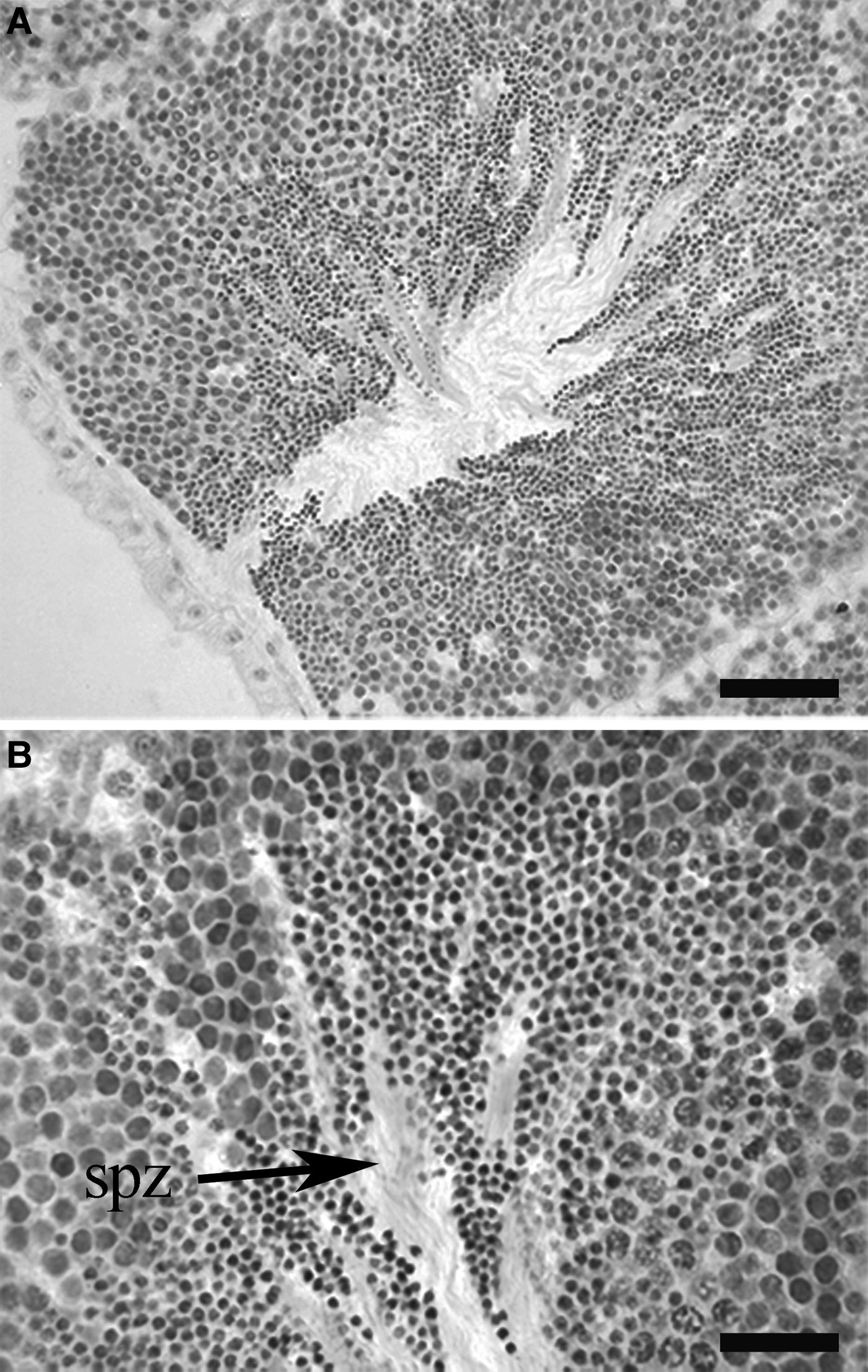
The gonad is a simple structure consisting of acini surrounded by connective tissue. Acini are observed in both mantle lobes, dorsal to the digestive gland, and also within the visceral mass in a posterior position. In transverse section, each acinus contains peripherally arranged spermatogenic cells surrounding a central lumen. Most acini showed all stages of development in Brachidontes rodriguezii. Each acinus contains a variety of developing sperm stages, which are distributed in a centripetal pattern from the inner acinus wall to the lumen (Figure 1A, B). Spermatogonia are located nearest the inner wall, while spermatocytes and spermatids are positioned between the spermatogonia and the acinus lumen. Spermatozoa are almost exclusively confined to the lumen and usually grouped (Figure 1B).
Gonad of B. rodriguezii mature male. (A) Detail of the acini in the mantle lobe. (B) Spermatogenic cells and spermatozoa within the acinus. spz, spermatozoa. Scale bars: A, 50 μm; B, 20 μm.

Spermatogenesis ultrastructure of B. rodriguezii
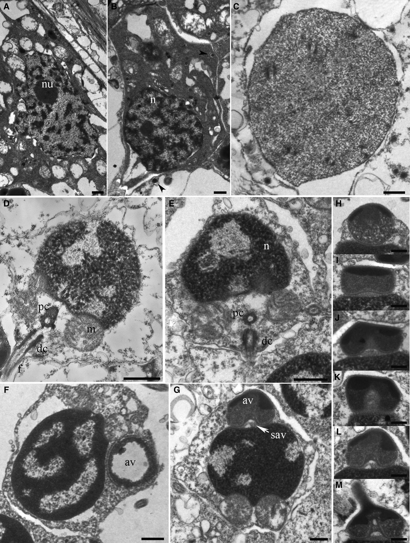
The spermatogenic epithelium consists of spermatogonia, spermatocytes, spermatids and spermatozoa, distributed in a centripetal pattern from the inner acinus wall to the lumen. Spermatogonia are located in the peripheral region of the acinus, the nucleus contains a single nucleolus and dispersed chromatin. The cytoplasm of spermatogonia contains proacrosomal vesicles. Two types were recognized, one closer to the acinus wall, with an elongated morphology and a more abundant cytoplasm (Figure 2A), and a second type, further into the acinus with spherical nuclei and little cytoplasm (Figure 2B). Spermatocytes with synaptonemal complex and diffuse chromatin were observed (Figure 2C). At early stages, spermatids are spherical cells. Further differentiation results in heterochromatin condensation and a slightly anterior-posterior elongation of the nucleus. Their nuclei contain diffuse patches of heterochromatin and some invaginations of the nuclei envelope appear. Spherical mitochondria appear in a posterior nuclear depression or fossa. Inside the nuclear fossa there are two centrioles, the proximal centriole, deep into the fossa and an outer, the distal centriole (Figure 2D, E). A simple flagellum rises from the distal centriole. During spermiogenesis, the acrosomal complex, consisting of the acrosomal vesicle and the periacrosmal region (between the vesicle and the nucleus) differentiates. The acrosomal vesicle turns from an oval structure to a conical one (the inner side of the acrosome becomes concave) containing electron-dense material. The anterior portion of the acrosomal vesicle contains electron-dense matter, whereas the basal part of the cone is comparatively less electron-dense (Figure 2H–M). The subacrosomal region contains fibrillar material. No electron dense content differentiation was observed at mature spermatozoa acrosomal vesicle.
Spermatogenesis in B. rodriguezii mature male. (A, B) Spermatogonia with nucleolus and proacrosomal vesicles (arrow head). (C) Spermatocyte. (D) Spermatid developing showing a detail of the mid piece with spherical mitochondria, proximal centriole and the distal centriole. (E) Spermatid showing an advance degree of condensation of the chromatin inside the nucleus. (F) Spermatid at early stage during spermiogenesis with the acrosomal vesicle cavity filled up with a ring of periacrosomal material. (G) Acrosomal vesicle and subacrosomal vesicle at the apical part of spermatid. (H–M) Stages of acrosomal vesicle formation during spermiogenesis. nu, nucleolus; m. mitochondria; pc, proximal centriole; dc, distal centriole; n, nucleus; av, acrosomal vesicle; sav, subacrosomal vesicle. Scale bars: 0.5 μm.
Sperm ultrastructure
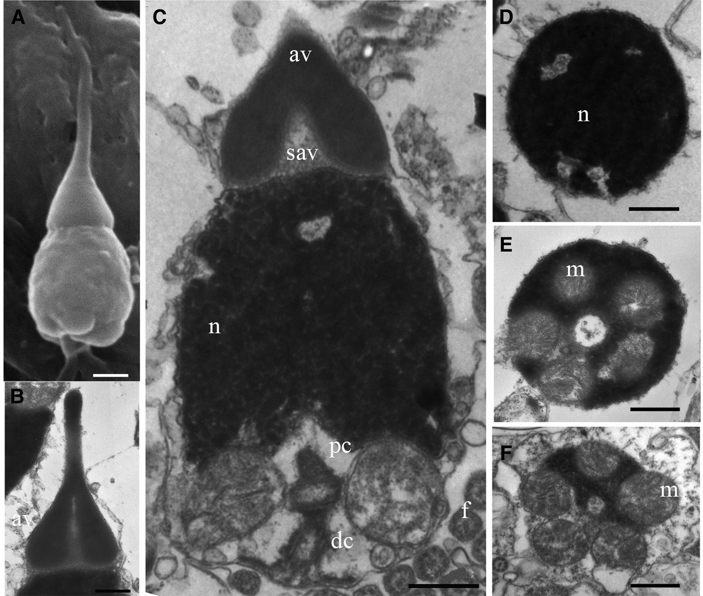
The sperm of B. rodriguezii is characterized by a barrel-shaped nucleus, capped by a conical acrosome with an anterior extension (Figure 3A). The acrosomal vesicle contains electron-dense material and the subacrosomal region consists of fibrillar material (Figure 3B). At the end of spermiogenesis, heterochromatin is electron-dense, homogeneous and compact. Some invaginations of the nuclear envelope (lacunae) are observed. These electron-lucent regions are randomly distributed (Figure 3C, D). The mid piece consists of a pair of centrioles with an orthogonal arrangement and five spherical mitochondria surrounding both centrioles (Figure 3E, F). Note that the mitochondria appear extremely closer to the nuclear envelope (Figure 3C, E). A simple flagellum rises from the distal centriole.
Sperm morphology in B. rodriguezii mature male. (A) Nucleus and acrosomal vesicle, general view by SEM. (B) Longitudinal section of sperm acrosomal vesicle and subacrosomal region. (C) Longitudinal section of a late spermatid with nucleus and mid piece showing the proximal centriole, the distal centriole. (D) Transverse section of the nucleus with nuclear lacunae. (E) Transverse section of the upper portion of the mid piece showing the spherical mitochondria. (F) Transverse section of the mid piece. av, acrosomal vesicle; n, nucleus; pc, proximal centriole; dc, distal centriole; m, mitochondria. Scale bars: 0.5 μm.
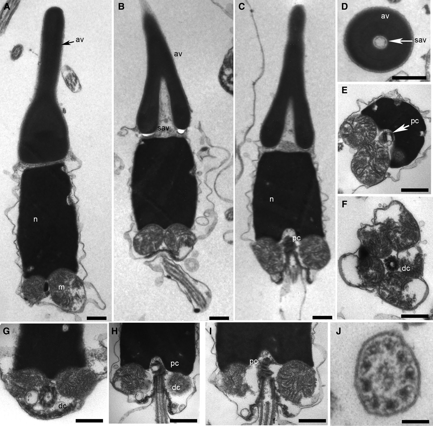
In Brachidontes purpuratus the apical portion of the nucleus is capped by a conical and elongated acrosome (Figure 4A–C). The acrosomal vesicle contains electron-dense material. The sub-acrosomal region consists of fibrillar material with a heterogeneous distribution (Figure 4B–D). Note that the apical portion content of the sub-acrosomal vesicle is less dense than the basal portion. Heterochromatin was electron-dense, homogeneous and compact, some invaginations of the nucleus envelope were observed (Figure 4E). The mid piece consisted of five spherical mitochondria surrounding a pair of centrioles, the proximal centriole and the distal centriole (Figure 4F–I). Posterior to the distal centriole there is a membrane. A simple flagellum rises from the distal centriole (Figure 4H–I). The flagellum exhibits the typical 9+2 microtubule structure, with 9 double outer tubules plus 2 single central tubules (Figure 4J).
Sperm morphology in B. purpuratus mature male. (A) Longitudinal sections of the sperm showing nucleus, acrosomal vesicle and spherical mitochondria. (B) Longitudinal section of nucleus and acrosomal vesicle and subacrosomal vesicle. (C) Longitudinal section of acrosomal complex, nucleus and mid piece showing the proximal centriole. (D) Transverse section of the acrosomal complex. (E) Transverse sections of the mid piece region showing proximal centriole. (F) Transverse sections of the mid piece region showing distal centriole. (G) Oblique section of the distal centriole. (h) Longitudinal section of the mid piece. (I) Longitudinal section of the mid piece, the pair of centrioles and the flagellum. (J). Transverse section of the flagellum. n, nucleus; av, acrosomal vesicle; m, mitochondria; sav, subacrosomal vesicle; pc, proximal centriole; dc, distal centriole; f, flagellum. Scale bars: A–I, 0.5 μm; J, 0.1 μm.
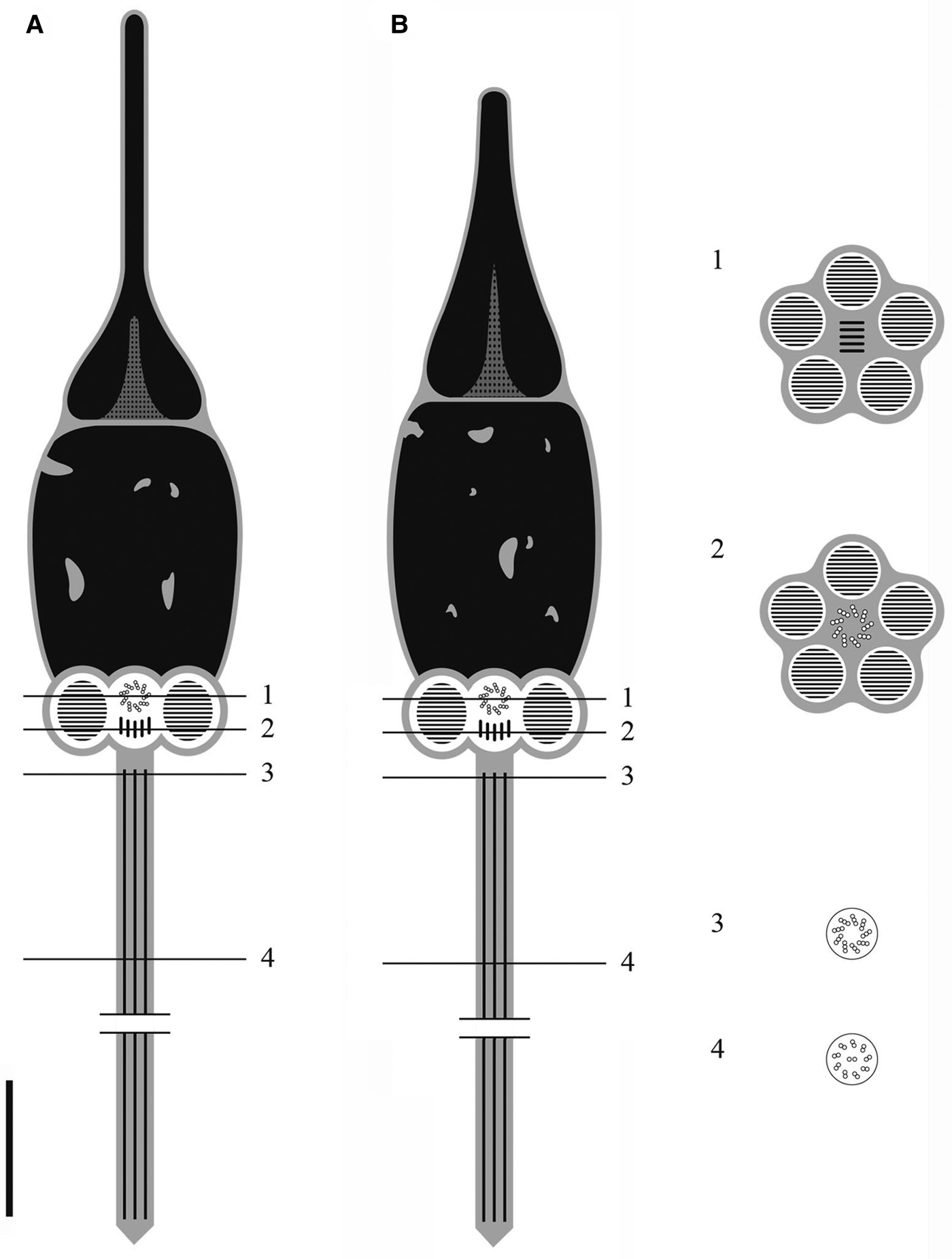
The different morphological aspects of B. rodriguezii and B. purpuratus are summarized in a diagram (Figure 5).
Diagram of sperm morphology in both species of Brachidontes. (A). B. rodriguezii. (B). B. purpuratus. Transversal sections of the sperm 1. Middle piece at proximal centriole level. 2. Mid piece at distal centriole level. 3. Basal region of axoneme. 4. Flagellum. Scale bar: 1 μm.
DISCUSSION
Sperm ultrastructure features have been used as tools in studies of taxonomic and phylogenetic relationships of Bivalvia (Popham, Reference Popham1979; Franzén, Reference Franzén1983; Drozdov & Reunov, Reference Drozdov and Reunov1997; Healy et al., Reference Healy, Keys, Daddon, Harper, Taylor and Crame2000; Drozdov et al., Reference Drozdov, Sharina and Tyurin2009). Several sperm ultrastructure studies were performed within Mytilidae (Kafanov & Drozdov, Reference Kafanov and Drozdov1998; Eckelbarger & Young, Reference Eckelbarger and Young1999; Introíni et al., Reference Introíni, Magalhães, Aguiar-Jr, Quaresma, Lino- Neto and Recco-Pimentel2004, Reference Introíni, Magalhães, Fortunato and Recco-Pimentel2009, Reference Introíni, Martins Maester, Pereira Leite and Recco-Pimentel2010; Desouky, Reference Desouky2009). Analysis of bivalve relationships integrating morphological characters and molecular markers were recently performed. The information on morphological characters showed that sperm ultrastructure characters are among the best morphological features to diagnose bivalve clades. Indeed, of those character systems (shell characters, larval, gills, muscles, foot, pedal gland characters, sperm ultrastructure, alimentary system and others) sperm ultrastructure and external shell characters included a greater proportion (almost 50%) of informative characters (Bieler et al., Reference Bieler, Mikkelsen, Collins, Glover, González, Graf, Harper, Healy, Kawauchi, Sharma, Staubach, Strong, Taylor, Tëmkin, Zardus, Clark, Guzmán, Mcintyre, Sharp and Giribet2014).
The environment where fertilization occurs and other factors, such as type, longevity of sperm and storage conditions before fertilization, have an important influence on sperm morphology (Franzén, Reference Franzén1955, Reference Franzén1956). Spermatozoa of Mytilidae are generally of the primitive or ect-aquasperm type, often found in invertebrates that release their gametes into the water and reproduce by external fertilization (Jamieson & Rouse, Reference Jamieson and Rouse1989; Kafanov & Drozdov, Reference Kafanov and Drozdov1998; Eckelbarger & Young, Reference Eckelbarger and Young1999; Reunov et al., Reference Reunov, Au and Wu1999). Sperm structure within Mytilidae shows a conical acrosomal complex, with a varying degree of complexity; Bathymodiolus presents a simple acrosomal vesicle while Mytilus exhibits a complex acrosomal vesicle (Popham, Reference Popham1979; Eckelbarger & Young, Reference Eckelbarger and Young1999; Healy et al., Reference Healy, Keys, Daddon, Harper, Taylor and Crame2000). The nucleus of the primitive sperm type shape is usually ovoid, and the mid piece generally presents five spherical mitochondria; some species exhibit little variation in mitochondria number, from four to five, exceptionally Modiolus within Mytilidae shows a range from 12 to 14 midpiece mitochondria (Kafanov & Drozdov, Reference Kafanov and Drozdov1998; Healy et al., Reference Healy, Keys, Daddon, Harper, Taylor and Crame2000; Gladyshev & Drozdov, Reference Gladyshev and Drozdov2002).
Description of Brachidontes rodriguezii sperm morphology resembles the sperm morphology described for another species of the genus. For Brachidontes solisianus and Brachidontes darwinianus Introíni et al. (Reference Introíni, Magalhães, Aguiar-Jr, Quaresma, Lino- Neto and Recco-Pimentel2004) mentioned a primitive sperm type with little differences between both species; B. solisianus showed a longer anterior projection of the acrosomal vesicle and an ovoid nucleus meanwhile B. darwinianus presented a shorter acrosomal complex and a spherical nucleus. In Brachidontes semistriatus (Krauss, 1848) sperm morphology is similar to the species mentioned above (Reunov & Hodgson, Reference Reunov and Hodgson1994). Bernard et al. (Reference Bernard, Davies and Hodgson1988) mentioned a similar sperm ultrastructure for Brachidontes virgiliae, although the acrosomal complex seems shorter and the mid piece showed six spherical mitochondria. Characterization of the sperm morphology of the species that belong to the same genus contributes to identification when morphological traits (like shell morphology) are easily misinterpreted (Hodgson & Bernard, Reference Hodgson and Bernard1986). The most variable organelle appears to be the acrosome, displaying diversity in shape and size (Garrido & Gallardo, Reference Garrido and Gallardo1996; Introíni et al., Reference Introíni, Magalhães, Aguiar-Jr, Quaresma, Lino- Neto and Recco-Pimentel2004, Reference Introíni, Magalhães, Fortunato and Recco-Pimentel2009). The acrosome vesicle is the first structure that makes contact with the oocyte, so it is expected to be highly modified, and may lead to reproduction isolation mechanisms (Drozdov et al., Reference Drozdov, Sharina and Tyurin2009).
Garrido & Gallardo (Reference Garrido and Gallardo1996) described Perumytilus purpuratus sperm morphology for the first time. Briones et al. (Reference Briones, Guiñez, Garrido, Oyarzún, Toro and Pérez2012) described sperm polymorphism in three different locations for Perumytilus purpuratus. The sperm morphology described herein for B. purpuratus was coincident with the description made by Briones et al. (Reference Briones, Guiñez, Garrido, Oyarzún, Toro and Pérez2012) for P. purpuratus in Valdivia, south of Chile. They also mentioned the existence of two different sperm morphologies associated with two different genetic clades for P. purpuratus along Chilean shores. One variety was seen in the north and central region, with a smaller acrosome length and acrosome/head ratio also smaller than the type described for the south region type. Different hypotheses were suggested to explain this phenomenon. One hypothesis is of two cryptic species, if sperm features are considered being species specific; one at the north and centre of the coast where currents and larval transport could lead to a homogenization of the population, and another species at the south. Another hypothesis is that an incipient process of speciation could be occurring (Briones et al., Reference Briones, Guiñez, Garrido, Oyarzún, Toro and Pérez2012). Probably there is a replacement of species along the south Pacific coast due to sea surface temperature as with the replacement of species of the genus Brachidontes along the south Atlantic (Scarabino, Reference Scarabino1977). Studies considering sperm morphology and genetic variation along the distribution areas of B. rodriguezii and B. purpuratus along south Atlantic coast should be done.
It is also possible that B. purpuratus presents more than one sperm morphotype along the south-western Atlantic coast, as Briones et al. (Reference Briones, Guiñez, Garrido, Oyarzún, Toro and Pérez2012) suggest for the south-east Pacific coast. Reunov et al. (Reference Reunov, Au and Wu1999) proposed sperm polymorphism for Perna viridis, however the only difference between both sperm morphotypes was the orientation of the sperm flagellum. Vekhova et al. (Reference Vekhova, Reunov, Alexandrova, Reunova and Adrianov2013) described sperm polymorphism for Mytilus trossulus and Crenomytilus grayanus. The differences between the sperm morphotypes were in the shape of the acrosome. The authors suggest that there is a relationship between the number of morphotypes and the adaptive capability of both species. When the variation of morphological types of spermatozoa within species is small, authors consider there is heteromorphism of these cells, however, there is a possibility of cryptic species or an incipient speciation process when there are marked differences in morphology of spermatozoa (Briones et al., Reference Briones, Guiñez, Garrido, Oyarzún, Toro and Pérez2012; Vekhova et al., Reference Vekhova, Reunov, Alexandrova, Reunova and Adrianov2013).
Spermatogenesis in Brachidontes rodriguezii is very similar to that reported in other mytilids. During early spermatogenesis, proacrosomal vesicles appear. Heterochromatin condensation and a slightly anterior-posterior elongation of the nucleus occur while the acrosomal complex differentiates during spermiogenesis (Bernard et al., Reference Bernard, Davies and Hodgson1988; Reunov et al., Reference Reunov, Au and Wu1999; Eckelbarger & Young, Reference Eckelbarger and Young1999; Kádar et al., Reference Kádar, Lobo Da Cunha, Santos and Dando2006; Yurchenko & Vaschenko, Reference Yurchenko and Vaschenko2010).
The systematics of Brachidontes purpuratus were recently under discussion. Some authors consider this species under the genus Perumytilus (Garrido & Gallardo, Reference Garrido and Gallardo1996; Oyarzún Cabañas et al., Reference Oyarzún Cabañas, Toro, Jaramillo, Guiñez, Briones and Astorga2010; Briones et al., Reference Briones, Guiñez, Garrido, Oyarzún, Toro and Pérez2012). According to Adami et al. (Reference Adami, Pastorino and Orenzans2013) there are morphological features of the shell that allowed discriminating between both species. Van der Molen et al. (Reference Van Der Molen, Marquez, Idaszkin and Adami2012) proposed that a morphometric analysis of the shells could be a useful tool for discriminating between both Brachidontes species. Molecular studies recently have shown there is a clear difference between both species, and also reconfirm the distribution pattern where B. purpuratus is confined to cold temperate waters, B. rodriguezii occurs in warm temperate waters and both species coexist in a transition zone (41°−43°S) (Trovant et al., Reference Trovant, Ruzzante, Basso and Orenzans2013). Also, according to Trovant et al. (Reference Trovant, Ruzzante, Basso and Orenzans2013) there is a significant level of genetic differentiation observed in B. rodriguezii along its distribution. This could be due to the irregularity of coastal features. However in the transition zone, B. rodriguezii and B. pupuratus show marked genetic differences (Trovant et al., Reference Trovant, Ruzzante, Basso and Orenzans2013). These differences suggest that there may be reproduction isolation mechanisms operating. We suggest these mechanisms can include the morphology of the sperm and consequently the differentiated fertilization of the oocytes. Many authors suggest making a revision of the genus, and also recommended keeping both species under the genus Brachidontes because the morphological traits are easily to recognize (Van der Molen et al., Reference Van Der Molen, Marquez, Idaszkin and Adami2012; Adami et al., Reference Adami, Pastorino and Orenzans2013; Trovant et al., Reference Trovant, Ruzzante, Basso and Orenzans2013).
Further studies considering sperm morphology and genetic variation along the distribution areas of B. rodriguezii and B. purpuratus could help to complete the molecular phylogeny and the associated sperm morphology.
ACKNOWLEDGEMENTS
We would like to thank P.E. Penchaszadeh for constant support and advice in the knowledge of our studies of this intertidal species. Carlos Bertoli with TEM assistance and Nicolás Torroglosa for his contribution in the images.
FINANCIAL SUPPORT
This work was partially supported by PIP 0253, UBACyT X273.