INTRODUCTION
Leishmania (Kinetoplastida, Trypanosomatidae) are protozoan parasites responsible for leishmaniasis, a disease characterized by two major clinical manifestations: (1) cutaneous leishmaniasis (CL), which can lead to disfiguring lesions and (2) visceral leishmaniasis (VL), a systemic disease which is fatal in the absence of treatment. According to a recent estimate, there are approximately 0·2 to 0·4 million new VL cases and 0·7 to 1·2 million CL cases every year (Alvar et al. Reference Alvar, Vélezm, Bern, Herrero, Desjeux, Cano, Jannin and den Boer2012). The disease is endemic worldwide in tropical and sub-tropical regions, including in Southern Europe (Dujardin et al. Reference Dujardin, Campino, Cañavate, Dedet, Gradoni, Soteriadou, Mazeris, Ozbel and Boelaert2008). It is noteworthy that infection with Leishmania does not necessarily lead to disease and, depending on the regions and the species, asymptomatic Leishmania carriers are more abundant than leishmaniasis patients (Ostyn et al. 2011). Co-infection with HIV is an increasing feature and may lead to a clinical activation of the parasites that is extremely difficult to manage.
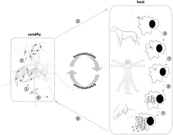
The parasite is transmitted by sandflies to a broad range of mammals, including humans, in which they develop within macrophages and other cells of the reticulo-endothelial system. During their life cycle (Fig. 1) the parasites are thus exposed to very different environments in which they are present in different life stages. In the sandfly (and this can be mimicked by in vitro culture), the parasite is first a procyclic promastigote, a replicating flagellated form, while later on it stops dividing and undergoes a series of molecular modifications that will ‘programme’ it for its future intracellular life (metacyclic promastigote). After transmission to a mammalian host, the parasite is phagocytosed by macrophages and it is able to survive and replicate within the phagolysosome as a small, non-flagellated parasite, the amastigote. These morphological transformations of the parasites are characterized by significant changes in gene and protein expression (Alcolea et al. Reference Alcolea, Alonso, Gómez, Moreno, Domínguez, Parro and Larraga2010; Tsigankov et al. Reference Tsigankov, Gherardini, Helmer-Citterich and Zilberstein2012), hence extreme care is required in the biological stage of the parasite when comparing different strains at molecular level.
Life cycle of Leishmania. In the vector, the sandfly, Leishmania grows as extracellular promastigotes, which will multiply (1) and undergo a transformation into metacyclic (infectious) forms (2). When an infected sandfly takes a blood meal, promastigotes will be injected in the host, which can be humans, dogs, rodents, etc., depending on the infecting Leishmania species (3). Metacyclic promastigotes will successfully infect host cells (4) and transform into intracellular amastigotes (5). These amastigotes will multiply (6) and eventually infect other neighbouring host cells (7). When another sandfly takes a blood meal, it will also imbibe amastigotes and/or amastigote containing host cells (8) which will transform into promastigotes in the sandfly gut (9) and enable a continuation of Leishmania's life cycle.

In nature, different scenarios of this life cycle can be encountered, from zoonotic cycles in the Amazonian jungle, in which humans are accidental hosts, to anthroponotic cycles where humans are thought to be the only host. This, together with the multiplicity of biotopes in which the parasite can be found, has led to a significant diversification of Leishmania parasites, highlighted among others by the clinical polymorphism. Consequently, the taxonomy of the genus Leishmania is rather complex and is still a subject of controversy (see Van der Auwera et al. Reference Van der Auwera, Fraga, Montalvo and Dujardin2011). Leishmania is considered to reproduce essentially in a clonal way, but automictic and allomictic recombination may occur (Rougeron et al. Reference Rougeron, De Meeûs, Hide, Waleckx, Bermudez, Arevalo, Llanos-Cuentas, Dujardin, De Doncker, Le Ray, Ayala and Bañuls2009). In the absence of obligate sexuality, the species definition in Leishmania is thus ambiguous and the current taxonomy is based on a phylogenetic definition of species and describes 14 medically relevant species.
Leishmaniasis, like other infectious diseases, is the result of an evolutionary arms race between pathogens and their hosts, which after millennia has often led to a mutual non-destruction pact resulting in peaceful cohabitation as long as a biological equilibrium is maintained. However, medical interventions and other ecological interferences may lead to new episodes in the competition between pathogens and their hosts, through the development of drug resistance. Recent developments in ‘omics technologies provide us a new perspective on this phenomenon. In the present review, we aim first to describe and clearly define the concept of drug resistance, then to review the advances brought by (post-)genomic approaches for its understanding, and finally to discuss how to translate these findings into tools for improving patient health.
CHEMOTHERAPEUTIC ARSENAL: THE NEED TO SAFEGUARD THE FEW AVAILABLE DRUGS
In the absence of an effective vaccine (Singh and Sundar, Reference Singh and Sundar2012), the control of leishmaniasis is essentially dependent on chemotherapy and vector control (Singh et al. Reference Singh, Kumar and Sing2012). However, the arsenal of drugs currently available is limited and drug efficacy is sooner or later jeopardized by drug resistance. For more than half a century antimonials (SSG) successfully constituted the first-line treatment of leishmaniasis. However, it showed severe side effects including pancreatitis, cardiac and renal toxicity (Sundar and Rai, Reference Sundar and Rai2002). This, together with the high treatment failure rate (up to 65%) and the emergence of resistance in the State of Bihar, India, made the drug obsolete in the Indian subcontinent (Sundar et al. Reference Sundar, More, Singh, Singh, Sharma, Makharia, Kumar and Murray2000). However, it is still used in first line in Africa and Latin America. Miltefosine (MIL), an alkylphosphocholine originally developed as an anticancer drug, has been used since 2005 in first line for the oral treatment of VL in the Indian subcontinent (Sundar et al. Reference Sundar, Mondal, Rijal, Batthacharya, Ghalib, Kroeger, Boelaert, Desjeux, Richter-Airijoki and Harms2008, Government of India Guidelines), but has shown variable efficacy in other clinical forms (Minodier and Parola, Reference Minodier and Parola2007). Its major limitation is teratogenicity, so that pregnancy is a contra-indication for treatment, ruling out women of childbearing age who constitute an estimated 25% of all VL patients (Olliaro et al. Reference Olliaro, Guerin, Gerstl, Haaskjold, Rottingen and Sundar2005). The long half-life (150 h) of the drug and its misuse may facilitate the emergence of drug resistance. Alarming rates of increased MIL-treatment failure have already been reported in India (Sundar et al. Reference Sundar, Singh, Rai, Prajapati, Singh, Ostyn, Boelaert, Dujardin and Chakravarty2012), Bangladesh (Rahman et al. Reference Rahman, Ahmed, Faiz, Chowdhury, Islam, Sayeedur, Rahman, Hossain, Bangali, Ahmad, Islam, Mascie-Taylor, Berman and Arana2011) and Nepal (Rijal et al. 2013), but this could not (yet) be related with clear-cut MIL resistance of the infecting parasite. The polyene macrolide antibiotic amphotericin B (AmB) has proven to be highly effective for the treatment of VL caused by SSG-resistant Leishmania donovani (SSG-R). It is an unpleasant drug because of its toxic side effects and the need for hospitalization of the patient during the 4 week course of treatment. However, expensive lipid-associated formulations of this drug have been made (Ambisome®) and have a reduced toxicity and an extended plasma half-life, therefore possibly requiring only a single infusion (Matlashewski et al. Reference Matlashewski, Arana, Kroeger, Battacharya, Sundar, Das, Sinha, Rijal, Mondal, Zilberstein and Alvar2011). Treatment failure and relapse during amphotericin B treatment occur rarely, except among HIV-infected patients (mainly due to relapse and reinfection) (Lachaud et al. Reference Lachaud, Bourgeois, Plourde, Leprohon, Bastien and Ouellette2009). The anti-leishmanial activity of the aminoglycoside paromomycin (PMM) was already known in the 1960s (Kellina, Reference Kellina1961; Reference NealNeal, 1968), and although PMM shows an efficacy of 95% and is relatively cheap, it is still under phase IV clinical trials – a reflection of the limited funding available. PMM is unlikely to ever become a monotherapy for VL, due to the risk of resistance emergence (Hendrickx et al. Reference Hendrickx, Inocêncio da Luz, Bhandari, Kuypers, Shaw, Lonchamp, Salotra, Carter, Sundar, Rijal, Dujardin, Cos and Maes2012) and the existence of other treatment regimens available (MIL, single dose liposomal AmB and combination schemes). Combination therapy may reduce both the treatment duration and the chance on the emergence of drug-resistant parasites while still guaranteeing an excellent efficacy, as shown by the first clinical trial of different combination regimens against VL in the Indian subcontinent (Sundar et al. Reference Sundar, Sinha, Rai, Verma, Nawin, Aalam, Chakravarty, Vaillant, Verma, Pandey, Kumari, Lal, Arora, Sharma, Ellis, Strub-Wourgaft, Balasegaram, Olliaro, Das and Modabber2011).
Although combination therapy is supposed to slow down the emergence of resistance, it may still be challenged by double (or cross) resistance as shown by recent experimental work (García-Hernández et al. Reference García-Hernández, Manzano, Castanys and Gamarro2012). Since leishmaniasis, like other neglected tropical diseases, affects the poorest people in the poorest regions of the world, the interest of the pharmaceutical industry in developing new drugs has been somewhat limited. Patients’ need-driven initiatives were launched (see for instance www.dndi.org) to counter this neglect. However, it will take several more years before the rare new lead compounds, if any, reach patients, and experience from the past suggests that resistance to these new drugs will emerge sooner or later. To safeguard the efficacy of current drugs and guide the development of new ones, it is thus of utmost importance to acquire a deep understanding of the phenomenon of drug resistance and its link with treatment outcome.
TREATMENT FAILURE, INNATE AND ACQUIRED DRUG-RESISTANCE: THE NEED FOR CLEAR DEFINITIONS
Understanding drug resistance requires first of all clear conceptual definitions, as there is often confusion of terms in the literature. On one hand, an important distinction is needed between treatment failure and drug resistance. Treatment failure is the clinical phenotype: it occurs when a patient does not respond to a drug, or when the treatment was not effective in the long run, resulting in non-response (persistence of symptoms at the end of therapy) or relapse (re-occurrence of the disease in an earlier apparently successfully treated patient). In general, treatment failure can occur due to several treatment-related (e.g. compliance, dosage, quality), parasite-related (e.g. intrinsic or acquired drug resistance) or host-related (e.g. pharmacogenetics, immune response) factors (Croft et al. Reference Croft, Sundar and Fairlamb2006). In contrast, drug resistance is a pathogen's phenotype resulting in its capacity to survive a given drug concentration, as measured with an in vitro susceptibility assay (see below). On the other hand, a clear distinction is also required between intrinsic and acquired resistance, (1) the former resulting from a biochemical feature of the pathogen, independent and prior to contact with the drug that allows it to tolerate that drug, while (2) acquired resistance is an adaptation selected through contact with the drug. Given the biochemical variation between the constitutive species, it is not surprising that different species may react differently to various anti-leishmanial therapeutic compounds (Croft et al. Reference Croft, Sundar and Fairlamb2006). Most of the data in this regard are gathered from clinical observations, but in vitro susceptibility assays have confirmed the occurrence of significant intrinsic species-specific differences. For instance, using parasites collected before the implementation of MIL, Yardley et al. (Reference Yardley, Croft, De Doncker, Dujardin, Koirala, Rijal, Miranda, Llanos-Cuentas and Chappuis2005) showed a significantly higher tolerance of Leishmania braziliensis to MIL than L. donovani. This also shows that any study on drug resistance in Leishmania must take into account species diversity and that results obtained with one species or particular strain cannot a priori be extrapolated to another one.
The current standard tool to evaluate whether an organism is resistant to a drug is exposing it to the drug under controlled conditions, and comparing its response with other known susceptible organisms. In the case of SSG, this involves a laborious in vitro test where intracellular amastigotes are exposed to SSG (Croft et al. Reference Croft, Yardley and Kendrick2002). The necessity to test the intracellular form of the parasite, and thus using a rather complex in vitro system with different cell types, is due to the intrinsic insensitivity of the extracellular promastigote stage of Leishmania to SSG (Vermeersch et al. Reference Vermeersch, da Luz, Toté, Timmermans, Cos and Maes2009). In contrast, for MIL, studies have shown that in vitro amastigote and promastigote susceptibilities are correlated, indicating that screening of large sets of strains for MIL resistance might also be done at the easier-to-assess promastigote level (Kulshrestha et al. Reference Kulshrestha, Bhandari, Mukhopadhyay, Ramesh, Sundar, Maes, Dujardin, Roy and Salotra2013). Besides being time-consuming and far from standardized, in vitro assays (even those running on intracellular amastigotes) also constitute a reductionist representation of the natural conditions to which these organisms are naturally exposed in treated patients. In case of acquired resistance to SSG, this is especially highlighted by the low predictive value of the current in vitro SSG susceptibility test for treatment outcome of the infected patient: in Nepal, strains that were defined in vitro to be SSG-R were found both in patients that were cured with SSG as in patients where SSG treatment had failed (Rijal et al. Reference Rijal, Yardley, Chappuis, Decuypere, Khanal, Singh, Boelaert, De Doncker, Croft and Dujardin2007). In view of the strong dependence of the mode of action of SSG on host factors, this is most likely due to the lack of immunogenic factors in the current gold standard SSG susceptibility test. Such scenarios might also apply for other (new) drugs. In this context, there are many expectations from molecular tools to complement and improve the in vitro tests. This requires elucidation of the molecular mechanisms leading to resistance, a task that must take into consideration the limits of the currently defined phenotypes: a marker of (in vitro defined) drug resistance could be a priori different from a marker of treatment failure.
GENOMIC FEATURES OF LEISHMANIA
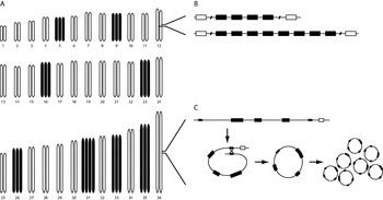
Before addressing the genomic approaches used for understanding drug resistance in Leishmania, an overview of the major features of its genome is required. The genomes of several sequenced Leishmania species (Leishmania major, L. infantum, L. braziliensis, L. donovani, L. mexicana and L. tarentolae) are relatively well conserved, with a high degree of synteny (conservation of gene order) and few species-specific genes, in contrast to their phenotypic diversity (Ivens et al. Reference Ivens, Peacock, Worthey, Murphy, Aggarwal, Berriman, Sisk, Rajandream, Adlem, Aert, Anupama, Apostolou, Attipoe, Bason, Bauser, Beck, Beverley, Bianchettin, Borzym, Bothe, Bruschi, Collins, Cadag, Ciarloni, Clayton, Coulson, Cronin, Cruz, Davies, De Gaudenzi, Dobson, Duesterhoeft, Fazelina, Fosker, Frasch, Fraser, Fuchs, Gabel, Goble, Goffeau, Harris, Hertz-Fowler, Hilbert, Horn, Huang, Klages, Knights, Kube, Larke, Litvin, Lord, Louie, Marra, Masuy, Matthews, Michaeli, Mottram, Müller-Auer, Munden, Nelson, Norbertczak, Oliver, O'Neil, Pentony, Pohl, Price, Purnelle, Quail, Rabbinowitsch, Reinhardt, Rieger, Rinta, Robben, Robertson, Ruiz, Rutter, Saunders, Schäfer, Schein, Schwartz, Seeger, Seyler, Sharp, Shin, Sivam, Squares, Squares, Tosato, Vogt, Volckaert, Wambutt, Warren, Wedler, Woodward, Zhou, Zimmermann, Smith, Blackwell, Stuart, Barrell and Myler2005; Peacock et al. Reference Peacock, Seeger, Harris, Murphy, Ruiz, Quail, Peters, Adlem, Tivey, Aslett, Kerhornou, Ivens, Fraser, Rajandream, Carver, Norbertczak, Chillingworth, Hance, Jagels, Moule, Ormond, Rutter, Squares, Whitehead, Rabbinowitsch, Arrowsmith, White, Thurston, Bringaud, Baldauf, Faulconbridge, Jeffares, Depledge, Oyola, Hilley, Brito, Tosi, Barrell, Cruz, Mottram, Smith and Berriman2007; Downing et al. Reference Downing, Imamura, Decuypere, Clark, Coombs, Cotton, Hilley, De Doncker, Maes, Mottram, Quail, Rijal, Sanders, Schönian, Stark, Sundar, Vanaerschot, Hertz-Fowler, Dujardin and Berriman2011; Rogers et al. Reference Rogers, Hilley, Dickens, Wilkes, Bates, Depledge, Harris, Her, Herzyk, Imamura, Otto, Sanders, Seeger, Dujardin, Berriman, Smith, Hertz-Fowler and Mottram2011; Raymond et al. Reference Raymond, Boisvert, Roy, Ritt, Légaré, Isnard, Stanke, Olivier, Tremblay, Papadopoulou, Ouellette and Corbeil2012). Genetic variation is usually measured as sequence divergence, but in the case of Leishmania, structural polymorphisms such as gene and chromosome copy number differences are a major source of genetic variation (Fig. 2). Inter- and intraspecific chromosome size polymorphisms, as already observed in early karyotyping experiments, are the result of expansions or contractions of gene family arrays or other tandem repeats (Pagès et al. Reference Pagès, Bastien, Veas, Rossi, Bellis, Wincker, Rioux and Roizès1989; Inga et al. Reference Inga, De Doncker, Gomez, Lopez, Garcia, Le Ray, Arevalo and Dujardin1998). In addition to tandem duplications or deletions of genes, Leishmania is also able to produce extra-chromosomal, linear or circular amplicons (episomes) of chromosome fragments containing one or more genes by homologous recombination (Ouellette et al. Reference Ouellette, Hettema, Wüst, Fase-Fowler and Borst1991; Grondin et al. Reference Grondin, Papadopoulou and Ouellette1993; Leprohon et al. Reference Leprohon, Légaré, Raymond, Madore, Hardiman, Corbeil and Ouellette2009). The number of chromosomes has long been the subject of debate, as it was unclear whether Leishmania is diploid or aneuploid, but recent genome and karyotype studies of several Leishmania species, strains and clones have found compelling evidence for aneuploidy (Downing et al. Reference Downing, Imamura, Decuypere, Clark, Coombs, Cotton, Hilley, De Doncker, Maes, Mottram, Quail, Rijal, Sanders, Schönian, Stark, Sundar, Vanaerschot, Hertz-Fowler, Dujardin and Berriman2011; Rogers et al. Reference Rogers, Hilley, Dickens, Wilkes, Bates, Depledge, Harris, Her, Herzyk, Imamura, Otto, Sanders, Seeger, Dujardin, Berriman, Smith, Hertz-Fowler and Mottram2011; Sterkers et al. Reference Sterkers, Lachaud, Crobu, Bastien and Pagès2011). The presence of different chromosome copy numbers between species, between strains and even between cells of the same population confirmed that aneuploidy is a constitutive feature of this parasite (reviewed by Mannaert et al. Reference Mannaert, Downing, Imamura and Dujardin2012 and by Sterkers et al. Reference Sterkers, Lachaud, Bourgeois, Crobu, Bastien and Pagès2012). This structural variation can be regarded as a gene dosage adjustment system in the absence of transcriptional regulation. Trypanosomatid genes are organized in unidirectional, polycistronic transcription units, which are post-transcriptionally spliced and polyadenylated, and regulation of gene expression likely occurs by mRNA degradation, translational control or protein degradation (Clayton and Shapira, Reference Clayton and Shapira2007). The ability to adjust the copy number of genes or entire chromosomes may provide a selective advantage, enabling the parasite to adapt to changing environments, e.g. in case of drug pressure (Mannaert et al. Reference Mannaert, Downing, Imamura and Dujardin2012). Both sequence and structural polymorphisms could contribute to an altered tolerance of the parasite to drugs (Leprohon et al. Reference Leprohon, Légaré, Raymond, Madore, Hardiman, Corbeil and Ouellette2009; Billal et al. Reference Billal, Feng, Leprohon, Légaré and Ouellette2011). Therefore, it is highly important to identify all genetic changes, single nucleotide polymorphisms (SNPs), copy number variations (CNVs) and ploidy changes that can be linked to the phenotype of interest of the parasite. These changes can be found by comparing drug-resistant with drug- sensitive Leishmania strains, by targeting specific genes of which the involvement in drug resistance is known or suspected, or by using a whole genome approach to identify all possible changes.
Structural diversity of the Leishmania genome. (A) Different Leishmania species show aneuploidy. This example represents the karyotype of the L. donovani reference strain, one of many circulating aneuploid karyotypes. Most chromosomes are disomic (white), but trisomy and tetrasomy (black) are common. (B) The number of gene copies can vary between species, between strains of the same species and even between homologous chromosomes of one strain, causing chromosome size polymorphism. (C) Extra-chromosomal amplification of genes on circular episomes can occur through homologous recombination between direct repeats (zigzag lines). Episomes can be replicated, and copy numbers can differ between strains or species. Genes are represented by boxes, amplified genes (B and C) are shown in black.

TARGETED APPROACHES FOR UNDERSTANDING DRUG RESISTANCE
When specific genetic targets are suspected to be involved in drug resistance, one can focus on that target using a variety of tests of which the majority are based, at least in part, on the polymerase chain reaction (PCR). As such, the genetic sequence of that target, its expression at RNA or protein level and/or its activity through reporter assays might be evaluated. Searching for single nucleotide polymorphisms (SNPs) in the DNA sequence of specific enzymes, transporters or other biologically active proteins that are specifically targeted by a drug or used for its import or export, has proven to be a successful approach for in vitro induced miltefosine-resistant (MIL-R) strains. Mutations in LdMT, an inwards translocator of MIL, or its beta subunit LdRos3 were found to be responsible for in vitro-induced MIL resistance (reviewed by Pérez-Victoria et al. Reference Pérez-Victoria, Sanchez-Canete, Seifert, Croft, Sundar, Castanys and Gamarro2006). In a single natural L. infantum isolate from an Algerian HIV-co-infected VL patient in France, a mutation in the same LdMT was also found (Cojean et al. Reference Cojean, Houzé, Haouchine, Huteau, Lariven, Hubert, Michard, Bories, Pratlong, Le Bras, Loiseau and Matheron2012). However, such variation was not observed in dozens of clinical L. donovani strains isolated from Indian and Nepalese patients that relapsed after MIL treatment (Bhandari et al. Reference Bhandari, Kulshrestha, Deep, Stark, Prajapati, Ramesh, Sundar, Schonian, Dujardin and Salotra2012), highlighting that natural drug resistance can be more complex than in vitro induced resistance. Similarly, the gene expression of LdMT and LdRos3 was comparable between pre-treatment, post-treatment and relapse isolates of Nepalese and Indian patients treated with MIL (Bhandari et al. Reference Bhandari, Kulshrestha, Deep, Stark, Prajapati, Ramesh, Sundar, Schonian, Dujardin and Salotra2012) despite earlier reports that an in vitro-induced lower expression of these genes can contribute to MIL resistance (Sánchez-Cañete et al. Reference Sánchez-Cañete, Carvalho, Pérez-Victoria, Gamarro and Castanys2009). Analysing the genetic sequence of specific enzymes and transporters thought to be related to the transport and action mechanism of SSG (Fig. 3) in clinical SSG-S and SSG-R isolates proved equally unsuccessful as for studies on MIL resistance, showing no genetic variation in the selected targets that could be related to the SSG-R phenotype (Mandal et al. Reference Mandal, Maharjan, Singh, Chatterjee and Madhubala2010; Decuypere et al. Reference Decuypere, Vanaerschot, Brunker, Imamura, Muller, Khanal, Rijal, Dujardin and Coombs2012). Assessing the expression of these genes with real-time quantitative PCR (Q-PCR) proved more informative with, for instance, different candidate marker genes involved in thiol metabolism showing a possible link with SSG resistance. Interestingly, the eventual genes of interest differed between Leishmania species (Decuypere et al. Reference Decuypere, Rijal, Yardley, De Doncker, Laurent, Khanal, Chappuis and Dujardin2005, Reference Decuypere, Vanaerschot, Rijal, Yardley, Maes, De Doncker, Chappuis and Dujardin2008, Reference Decuypere, Vanaerschot, Brunker, Imamura, Muller, Khanal, Rijal, Dujardin and Coombs2012; Carter et al. Reference Carter, Hutchison, Henriquez, Légaré, Ouellette, Roberts and Mullen2006; Adaui et al. Reference Adaui, Castillo, Zimic, Gutierrez, Decuypere, Vanaerschot, De Doncker, Schnorbusch, Maes, Van der Auwera, Maes, Llanos-Cuentas, Arevalo and Dujardin2011a, Reference Adaui, Schnorbusch, Zimic, Gutierrez, Decuypere, Vanaerschot, De Docker, Maes, Llanos-Cuentas, Chappuis, Arevalo and Dujardinb; Mukhopadhyay et al. Reference Mukhopadhyay, Mukherjee, Mukherjee, Naskar, Mondal, Decuypere, Ostyn, Prajapati, Sundar, Dujardin and Roy2011; Kumar et al. Reference Kumar, Singh, Bhandari, Kulshrestha, Negi and Salotra2012). In addition, variability was also found between studies on strains of the same Leishmania species of the same geographical region. These apparent inconsistencies can at least partly be explained by SSG resistance having a multifactorial origin (Downing et al. Reference Downing, Imamura, Decuypere, Clark, Coombs, Cotton, Hilley, De Doncker, Maes, Mottram, Quail, Rijal, Sanders, Schönian, Stark, Sundar, Vanaerschot, Hertz-Fowler, Dujardin and Berriman2011) and by findings showing that the parasite's genetic background is an important contributor to determine which SSG resistance mechanism applies (Decuypere et al. Reference Decuypere, Vanaerschot, Brunker, Imamura, Muller, Khanal, Rijal, Dujardin and Coombs2012).
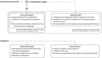
The mode of action of two major drugs against leishmaniasis. * The indirect action of pentavalent antimonials has been shown to be indispensable for the drug's in vivo action, while the importance of MIL's indirect action mechanism is still under debate. The action mechanisms of pentavalent antimonials and MIL are more thoroughly reviewed in Vanaerschot et al. (Reference Vanaerschot, Decuypere, Berg, Roy and Dujardin2012) and Dorlo et al. (Reference Dorlo, Balasegaram, Beijnen and de Vries2012), respectively.

Importantly, the mode of action of anti-leishmanial drugs may imply direct and indirect mechanisms, the latter through the host cell (Fig. 3). Hence, the parasite can also express its drug resistance through manipulation of the host. This is especially true for SSG-R L. donovani, which have shown to modulate the host to (1) reduce the presence of the drug in the host cell and (2) minimize SSG's immuno-modulating action (reviewed by Vanaerschot et al. Reference Vanaerschot, Decuypere, Berg, Roy and Dujardin2012). Various targeted reporter assays have demonstrated that SSG-R L. donovani induces IL-10 in its host cells, and that this is responsible for a higher gene expression of the host cell's multidrug resistance-associated protein 1 (MRP1) and permeability glycoprotein (P-gp), which can export SSG out of the host cell (Mukherjee et al. Reference Mukherjee, Mukhopadhyay, Bannerjee, Chowdhury, Mukherjee, Naskar, Allam, Chakravortty, Sundar, Dujardin and Roy2013). Also the gene expression of a key enzyme in glutathione synthesis was downregulated in SSG-R L. donovani infected host cells, causing a lower activation of SSG into its direct toxic form in the host cell (Carter et al. Reference Carter, Hutchison, Henriquez, Légaré, Ouellette, Roberts and Mullen2006; Haldar et al. Reference Haldar, Yadav, Singhai, Bisht, Singh, Bhaumid, Basu, Sen and Roy2010). Targeting the expression of cytokines can also significantly contribute to a better understanding of drug resistance and treatment failure for drugs where the host factors are, or are suspected to be, important for the mode of action (Thakur et al. Reference Thakur, Mitra and Narayan2003; Narayan et al. Reference Narayan, Bimal, Singh, Gupta, Singh, Sinha and Das2009; Costa et al. Reference Costa, Carregaro, Lima-Júnior, Silva, Milanezi, Cardoso, Giudice, de Jesus, Carvalho, Almeida and Silva2011).
UNTARGETED GENOMIC APPROACHES FOR UNDERSTANDING DRUG RESISTANCE
Since drug resistance in Leishmania can be multifactorial (Ashutosh et al. Reference Ashutosh, Sundar and Goyal2007; Chawla et al. Reference Chawla, Jhingran, Panigrahi, Stuart and Madhubala2011), without a priori knowledge an untargeted approach would be most suitable to detect the genetic changes leading to resistance. In the pre-genomics era, drug resistance studies were mainly limited to a handful of genes with suspected involvement in resistance, but the advent of genomics and next-generation sequencing technologies has lifted this research field to a new level. Before genome sequencing became well established, functional cloning has proven valuable to detect genes involved in drug resistance (reviewed by Clos and Choudhury, Reference Clos and Choudhury2006). This method was used to identify the MIL transporter LdMT as responsible gene in MIL resistance by functional rescue: MIL-R parasites were transfected with cosmids from genomic DNA from a susceptible L. infantum line, and only the parasites transfected with the ‘sensitive’ LdMT regained sensitivity to MIL (Pérez-Victoria et al. Reference Pérez-Victoria, Gamarro, Ouellette and Castanys2003). A similar approach was used to identify another gene, P299, as a MIL marker that also provides protection against SSG (Choudhury et al. Reference Choudhury, Zander, Kube, Reinhardt and Clos2008).
Since drug resistance often appears to be mediated by gene amplification (Beverley, Reference Beverley1991), it is desirable to use a method that can detect copy number variations in the entire genome, such as full genome microarrays, comparative genome hybridization arrays and whole genome sequencing (WGS). WGS has the advantage of being able to detect mutations, copy number variations and ploidy changes simultaneously. A study of 17 clinical L. donovani lines with differential in vitro SSG susceptibility identified SNP and structural markers that were associated with SSG resistance, including 34 SNPs only present in four resistant lines, positive selection in genes associated with drug resistance, differential gene copy numbers and an episome containing an acid phosphatase and the MAPK1 gene (Downing et al. Reference Downing, Imamura, Decuypere, Clark, Coombs, Cotton, Hilley, De Doncker, Maes, Mottram, Quail, Rijal, Sanders, Schönian, Stark, Sundar, Vanaerschot, Hertz-Fowler, Dujardin and Berriman2011). In a full genome microarray-based gene expression profiling study, the same MAPK1 gene was also found overexpressed in SSG-R clinical L. donovani strains, together with histones H1, H2A and H4 (Singh et al. Reference Singh, Kumar, Duncan, Nakhasi and Salotra2010). Another study found no ploidy or copy number changes related to resistance in the genome of multiple independent L. major lines with experimentally induced MIL resistance, but identified mutations in the LdMT gene and in a putative pyridoxal kinase gene (PK) in all resistant mutants (Coelho et al. Reference Coelho, Boisvert, Mukherjee, Leprohon, Corbeil and Ouellette2012). Full genome microarrays are mostly used for gene expression profiling of different stages in the life cycle of Leishmania, but they are also useful for drug resistance studies to detect genes that are differentially expressed in drug-resistant and sensitive strains. Since regulation of gene expression is mainly post-transcriptional, changes in mRNA are often, though not always, the result of gene amplifications, deletions, or ploidy changes. Underlying genomic changes can be confirmed by Southern blot or comparative genome hybridization. Modulated expression of several chromosomes, gene overexpression and downregulation in experimental methotrexate-resistant L. major and L. infantum lines (MTX-R) and in experimental SSG-R L. infantum lines were the result of aneuploidy, circular gene amplification (dihydrofolate reductase-thymidylate synthase, MTX-R; ABC protein MRPA, SSG-R), linear gene amplification (pteridine reductase 1, MTX-R), and gene deletion (folate transporter FT1, MTX-R), respectively (Ubeda et al. Reference Ubeda, Légaré, Raymond, Ouameur, Boisvert, Rigault, Corbeil, Tremblay, Olivier, Papadopoulou and Ouellette2008; Leprohon et al. Reference Leprohon, Légaré, Raymond, Madore, Hardiman, Corbeil and Ouellette2009). In SSG-R mutants of L. amazonensis, higher expression levels of the MRPA locus were due to the presence of a linear amplicon or by aneuploidy of the whole chromosome (do Monte-Neto et al. Reference do Monte-Neto, Coelho, Raymond, Légaré, Corbeil, Melo, Frézard and Ouellette2011). Other overexpressed genes were related to thiol metabolism, but some did not have a direct link with drug resistance (nucleobase transporter NT3). In many cases, the differentially expressed or mutated genes are unknown or their involvement in drug resistance is unclear. Even if a link with drug resistance is more obvious, their contribution should be validated experimentally.
POST-GENOMIC APPROACHES TO COMPLEMENT GENOMIC DATA
Exploiting genome information to unravel pathogen biology has become the prime objective of numerous post-genomic studies. The metabolome has a privileged position in a systems biology context: it is situated at the other end of the modern ‘dogma’ of molecular biology, with information flowing from the genome, via the transcriptome and the proteome to the metabolome (Breitling et al. Reference Breitling, Bakker, Barret, Decuypere, Dujardin and Suhre2012). The metabolome is considered as the closest representation of the phenotype and represents the collection of metabolites, which are the final products of gene expression. Metabolite levels are not easily predicted by protein or transcript levels due to the highly non-local control structure of the metabolic network (e.g. enzyme inhibitors) and thus require separate analysis (Scheltema et al. Reference Scheltema, Decuypere, t'Kindt, Dujardin, Coombs and Breitling2010). Moreover, genome, trancriptome and proteome studies have limitations to functionally annotate identified sequences, whereas the metabolome consists of fewer components that are key actors of universal cellular processes underlying the relevance of this ‘omics platform (Scheltema et al. Reference Scheltema, Decuypere, t'Kindt, Dujardin, Coombs and Breitling2010). In trypanosomatids such as Trypanosoma and Leishmania, metabolomics are of particular interest because the gene expression is regulated at the post-transcriptional level (De Gaudenzi et al. Reference De Gaudenzi, Noé, Campo, Frasch and Cassola2011; Requena, Reference Requena2011) and hence genome and transcriptome studies might have certain limitations when studying the rapid effects of drug treatment or the mechanisms of drug resistance (Berg et al. Reference Berg, Vanaerschot, Jankevics, Cuypers, Breitling and Dujardin2012). For the moment, there is no consensus concerning the relative importance of changes in mRNA levels during parasite development (Lahav et al. Reference Lahav, Sivam, Volpin, Ronen, Tsigankov, Green, Holland, Kuzyk, Borchers, Zilberstein and Myler2011): some microarray analysis studies indicated the occurrence of substantial changes during promastigote to amastigote differentiation (>10%, Almeida et al. Reference Almeida, Gilmartin, McCann, Norrish, Ivens, Lawson, Levick, Smith, Dyall, Vetrie, Freeman, Coulson, Sampaio, Schneider and Blackwell2004; Saxena et al. Reference Saxena, Lahav, Holland, Anupama, Huang, Volpin, Myler and Zilberstein2007), whereas other studies suggested that transcription is constitutive throughout the parasite life and that there is a surprisingly low level of differentially expressed stage-specific genes (0·2–5% of total genes) (Akopyants et al. Reference Akopyants, Matlib, Bukanova, Smeds, Brownstein, Stormo and Beverley2004; Cohen-Freue et al. Reference Cohen-Freue, Holzer, Forney and McMaster2007; Haile and Papadopoulou, Reference Haile and Papadopoulou2007; Leifso et al. Reference Leifso, Cohen-Freue, Dogra, Murray and McMaster2007). Within this limited set of genes, a dramatic downregulation of gene expression from stationary phase promastigotes to amastigotes was observed, highlighting the hypothesis of a pre-adaptation at metacyclic promastigote stage for intracellular survival as amastigote (Alcolea et al. Reference Alcolea, Alonso, Gómez, Moreno, Domínguez, Parro and Larraga2010). Quantitative proteomics analyses showed a weak correlation to gene expression (20–30% correlation between changes in mRNA and protein levels, Lahav et al. Reference Lahav, Sivam, Volpin, Ronen, Tsigankov, Green, Holland, Kuzyk, Borchers, Zilberstein and Myler2011; Kramer, Reference Kramer2012) and have so far – similar to transcriptomics studies – mainly focused on the identification of stage-specific changes rather than on the investigation of resistance mechanisms, although useful contributions have been made to study SSG resistance (El Fadili et al. Reference El Fadili, Drummelsmith, Roy, Jardim and Ouelette2009; Singh et al. Reference Singh, Almeida, Kothari, Kumar, Mandal, Chatterjee, Venkatachalam, Govind, Mandal and Sundar2007; Walker et al. Reference Walker, Gongora, Vasquez, Drummelsmith, Burchmore, Roy, Ouelette, Gomez and Saravia2012). Compared to transcriptomics, proteomics studies revealed a higher level of differentially expressed stage-specific proteins for different Leishmania species (5–12%, L. donovani by Bente et al. Reference Bente, Harder, Wiesgigl, Heukeshoven, Gelhaus, Krause, Clos and Bruchhaus2003; L. mexicana by Nugent et al. Reference Nugent, Karsani, Wait, Tempero and Smith2004; L. infantum by Acestor et al. Reference Acestor, Masina, Walker, Saravia, Fasel and Quadroni2002), suggesting that Leishmania protein expression levels are likely regulated at the posttranscriptional or translational level (Cohen-Freue et al. Reference Cohen-Freue, Holzer, Forney and McMaster2007; Lahav et al. Reference Lahav, Sivam, Volpin, Ronen, Tsigankov, Green, Holland, Kuzyk, Borchers, Zilberstein and Myler2011). The metabolome will be affected to an even greater degree, due to the exponential effect of changes on the proteomic level (changes of a single enzyme will affect multiple metabolites) and due to the high connectivity in the metabolic network. A metabolomics study by Silva and colleagues showed that there is a distinct variation in the metabolome during development in culture of L. donovani promastigotes: 26% of the total number of putatively identified metabolites was statistically different (Silva et al. Reference Silva, Cordeiro-da-Silva and Coombs2011).
Although the technological developments are very recent and still being optimized, current liquid chromatography mass spectrometry LC-MS (Hilic-Orbitrap) platforms enable the metabolic profiling of multiple strains, thereby paving the way for large-scale integrative ‘omics studies (e.g. diversity studies) (reviewed by Berg et al. Reference Berg, Vanaerschot, Jankevics, Cuypers, Breitling and Dujardin2012). In metabolomics studies, powerful biochemical, statistical, computational and bioinformatics tools are combined to generate a unique and much-needed dimension to better understand the parasite biology (Lakshmanan et al. Reference Lakshmanan, Rhee and Daily2011). For example, hypothesizing that gene dosage plays a major role in rapid evolutionary adaptation and phenotypic diversity of the parasite, metabolomics is currently used to exploit the impact of structural genome variation on the metabolic profile. Fig. 4 illustrates the complementarity of metabolomics and genomics. A recent metabolomics study (Berg, unpublished results) on three strains with a different SSG-susceptibility clearly reveals dramatic metabolic differences (1) between the different strains, (2) between different promastigote growth stages within the same strain, and (3) when a resistant strain is maintained under drug pressure. Parallel WGS of these strains showed that SNP analysis only allows distinction between strains. No distinction can be made at SNP level within a given strain between life stages or under drug pressure, while genome structure does show some differences. The functional impact of these structural changes at genome level is yet unclear, but we recommend the following: (1) genome structure should definitively be integrated into genome studies and (2) a particular attention should be paid to the choice of the life stage, not only for metabolomics studies, but also for genomics.
Principal component analyses (PCA) based on a parallel metabolomics and genomics study of 3 L. donovani strains at two different growth stages of the promastigote life form or under SbIII drug pressure. S: strain sensitive against both trivalent and pentavalent antimonials SbIII-S/SbV-S; I: strain sensitive against trivalent antimonials but resistant against pentavalent antimonials SbIII-S/SbV-R; R: strain resistant against both trivalent and pentavalent antimonials SbIII-R/SbV-R; Ri=resistant strain (R) grown under SbIII drug pressure for two weeks; LOG=logarithmic promastigote growth stage; STAT=stationary promastigote growth stage. (panel A) PCA plot of metabolomics results based on quantitative information of 326 putatively identified metabolites shows that separation is based on variability caused by (1) promastigote growth related differences (LOG strains cluster together and are clearly separated from STAT strains by PC1) and by (2) strains and drug pressure (S, I, R, Ri, separated by both PC1 and PC2). (panel B) PCA based on the SNPs determined with WGS shows a separation of the three strains. The two different growth stages of each strain (LOG, STAT) and the effect of SbIII drug pressure during two weeks cannot be distinguished based on SNPs only, the dots overlap for each strain. (panel C) PCA based on structural variation (local copy number variation+ploidy changes) determined with WGS shows a separation by strain and drug pressure, but small differences between the life stages of each individual strain (for S, I and Ri) can be distinguished as well.

Current metabolomics studies have focused on metabolic changes related to SSG resistance in stationary phase promastigotes of L. donovani and have revealed modifications in complete pathways of the SSG-R strains. For instance, changes in the glycerophospholipids might be responsible for the increased fluidity of the plasma membrane of SSG-R parasites (Mukhopadhyay et al. Reference Mukhopadhyay, Mukherjee, Mukherjee, Naskar, Mondal, Decuypere, Ostyn, Prajapati, Sundar, Dujardin and Roy2011), possibly affecting the transport of antimonials inside the parasitic cell (t'Kindt et al. Reference t'Kindt, Scheltema, Jankevics, Brunker, Rijal, Dujardin, Breitling, Watson, Coombs and Decuypere2010). SSG-R parasites also upregulate both the transsulfuration pathway (biosynthesis of cysteine) and the urea cycle (synthesis of putrescine), possibly leading to higher levels of glutathione and trypanothione and an enhanced protection against oxidative stress (Canuto et al. Reference Canuto, Castilho-Martins, Tavares, López-Gonzálvez, Rivas and Barbas2012; Berg, unpublished results). Last but not least, increased levels of amino acids and purine nucleosides in SSG-R strains might serve as a survival kit during the initial stage of infection in nutrient-poor host cells.
For a true systems biology approach of genomics and metabolomics, some issues still need to be tackled. The interpretation of the identified genetic diversity elements in relation to specific phenotypic diversity remains challenging. A certain fraction of the genomic variation remains silent at the metabolic level, further complicating the integration of both global molecular profiles. Also, metabolic signatures reveal little about the genetic regulatory networks causing the metabolomic phenotype variation of the pathogen population. With quantitative trait loci mapping (QTL mapping), hotspots with system-wide effects can be highlighted (Fu et al. Reference Fu, Keurentjes, Bouwmeester, America, Verstappen, Ward, Beale, de Vos, Dijkstra, Scheltema, Johannes, Koornneef, Vreugdenhil, Breitling and Jansen2009), allowing to detect specific metabolite levels that serve as phenotypic indicators of the existence of underlying genetic contributors (Huang et al. Reference Huang, Wuchty, Ferdig and Przytycka2009). Moreover, metabolic traits of the organism under study can be related to genetic polymorphisms in genome-wide association studies (GWAS), hereby throwing new light on the functions of certain genes (Homuth et al. Reference Homuth, Teumer, Volker and Nauck2012) and differentiating ‘driver’ mutations from biologically neutral ‘passenger’ changes.
EXPERIMENTAL VALIDATION OF GENES PUTATIVELY INVOLVED IN DRUG RESISTANCE
Functional analysis of differential genes in drug-resistant strains is essential to discriminate drivers from passengers and to understand their role in the resistance mechanism(s). Several genetic manipulation techniques, such as targeted gene replacement (knockout) or transfection with wild type or mutated genes (overexpression), are available for Leishmania. Unfortunately, gene knockdown by RNA interference is limited to L. braziliensis and other Viannia species, since no RNAi machinery is found in species of the Leishmania subgenus (Robinson and Beverley, Reference Robinson and Beverley2003; Peacock et al. Reference Peacock, Seeger, Harris, Murphy, Ruiz, Quail, Peters, Adlem, Tivey, Aslett, Kerhornou, Ivens, Fraser, Rajandream, Carver, Norbertczak, Chillingworth, Hance, Jagels, Moule, Ormond, Rutter, Squares, Whitehead, Rabbinowitsch, Arrowsmith, White, Thurston, Bringaud, Baldauf, Faulconbridge, Jeffares, Depledge, Oyola, Hilley, Brito, Tosi, Barrell, Cruz, Mottram, Smith and Berriman2007). Gene knockout can be complicated by the ability of Leishmania to amplify genes or whole chromosomes, resulting in failure to create homozygous knockouts of essential genes, but this can be overcome by episomal gene complementation and conditional knockout (Murta et al. Reference Murta, Vickers, Scott and Beverley2009; Morales et al. Reference Morales, Watanabe, Dacher, Chafey, Osorio y Fortéa, Scott, Beverley, Ommen, Clos, Hem, Lenormand, Rousselle, Namane and Späth2010). Due to differences in the environment, the mechanisms responsible for drug resistance in natural strains in the host are not necessarily the same as in experimental strains under laboratory conditions. Therefore, validation of identified genetic markers in naturally drug-resistant strains can be complicated. Even if genetic changes can be attributed to acquired drug resistance in experimental strains, translation to the field, where changing environments and host immunity add to the complex conditions, is not always straightforward.
TRANSLATION TO THE FIELD/IMPROVING HEALTH PRACTICES
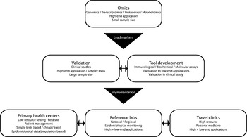
It is evident that generation of all the above-mentioned knowledge is only a first step towards improving the management of parasite resistance in practice. The next big challenge lies in implementing the acquired knowledge to the benefit of the patients and the populations at risk, outside the context of well-defined and well-funded clinical studies (summarized in Fig. 5). Here we need to make a clear distinction between poor and limited resource settings on the one hand, and highly equipped laboratories on the other hand.
Schematic overview of the steps involved in translating ‘omics knowledge into disease management. Identified potential markers (top) need validation in large-scale clinical trials. In parallel, simpler assays can be developed that can be validated and used in the clinical studies. Finally, all available tools need to be implemented at different levels (bottom).

In well-equipped clinical laboratories, which are typically found outside the impoverished endemic areas, one could revert to high-end applications for scoring resistance-related markers, both of a genetic or a biochemical nature. Even full-genome sequencing directly on clinical samples and automated extraction of relevant genomic information may in time be feasible. Alternatively, biochemical or genetic PCR-based assays are even easier to implement and would not require any investments beyond currently available assays routinely used in such laboratories. Nevertheless, centres dealing with few cases on an annual basis would probably outsource the resistance profile determination to avoid the heavy investment in validation studies, as pharmaceutical companies have little to gain by producing quality-controlled and ready-made kits for a disease that is of marginal importance in the developed world. Outsourcing of resistance profiling is also common practice for other infectious agents, such as HIV.
A bigger problem is represented by limited-resource laboratories, which often have no access to even simple techniques such as PCR. Unless one could translate the acquired knowledge into a cheap and simple prognostic test that could score for parasite resistance in a given patient, treating clinicians would probably have to rely on available epidemiological data. Such epidemiological data are however typically scarce, and keeping them up-to-date requires intensive monitoring programmes that continuously scout for emerging and innate resistance in circulating Leishmania strains in different regions and for different drugs. Such screening must rely on a representative sampling, both in human and – if relevant – animal hosts, and could make use of the aforementioned high-end applications, provided policy makers would be willing to invest in well-equipped regional or national laboratory infrastructure.
Whichever the context, it is clear that translating knowledge on Leishmania resistance and treatment failure into useful tools is not a trivial exercise, and involves stakeholders at every level: scientists, health-policy makers, governments, private partners and non-profit organizations. We make strong advocacy for involving these parties from the very early steps of development on, to ensure that our knowledge on parasite resistance does not accumulate into merely another scientific publication, but is used for the benefit of the fight against leishmaniasis.
CONCLUSION
In this most recent episode of the arms race with human hosts, Leishmania have demonstrated unique and complex adaptive skills. Genome plasticity offers the parasite multiple solutions to the stresses induced by drugs. This represents a challenge for any new drug implemented in the field and justifies a close monitoring of drug efficacy. Similarly, the capacity to induce drug resistance should be integrated at early stage in research and development pipelines, and be used to guide drug development. Untargeted genomic approaches offer an unprecedented support to understand and monitor drug resistance, but should be complemented by downstream ‘omics approaches, which are closer to the phenotype itself. The next challenges reside in (1) the integration between the different ‘omics layers, paving the way towards systems biology of drug resistance and (2) the translation of the findings for a direct benefit of the patients and the population at risk. Next-generation sequencing platforms are currently under continuous innovation, simplifying operational steps, allowing genome-wide studies on single cells and making the technology less expensive. Whole genomics and transcriptomics are thus becoming more and more accessible, which will undoubtedly contribute to a better understanding of biological phenomena such as drug resistance.
ACKNOWLEDGEMENTS
We thank Hideo Imamura for his contribution to Fig. 4.
FINANCIAL SUPPORT
Research of our group described in this review was funded by the GeMInI consortium (grant ITMA SOFI-B), the Research Foundation Flanders (FWO project G.0B81.12), the EC-FP7 project Kaladrug-R (contract 222895), the Inbev-Baillet Latour Fund (grant for M. B.) and the Belgian Cooperation (grant for G. V. D. A).