INTRODUCTION
Visceral leishmaniasis (VL) or kala-azar is a vector-borne parasitic disease resulting from infection of macrophages by the obligate intracellular parasite, Leishmania donovani. L. donovani is transmitted to humans by the sand fly (Phlebotomus spp.). The parasite exists as flagellated and motile promastigotes in the vector host and as non-motile and aflagellated amastigotes in host tissue macrophages. VL is considered as a major tropical disease by DNDi (www.dndi.org) with an estimated 500 000 new cases occurring annually. Approximately 90% of these occur in rural areas of India (especially Bihar), Nepal, Bangladesh, Sudan, and north-eastern Brazil in some of the world's poorest regions (Guerin et al. Reference Guerin, Olliaro, Sundar, Boelaert, Croft, Desjeux, Wasunna and Bryceson2002; Murray, Reference Murray2004). In 1999, there were 57 000 deaths reported due to VL, but the real number is thought to be significantly higher (www.dndi.org). Bihar state alone, accounts for approximately 90% of the estimated 200 000–250 000 annual new cases in India. Pentavalent antimonials (SbV) are the first line drugs for the treatment of VL in India since the 1940s. However, widespread primary failure (>65% unresponsiveness) to sodium stibogluconate (SSG) has been reported from North Bihar, India, which has been mainly attributed to the inappropriate, irregular and incomplete treatments with SSG leading to development of drug resistance (Croft et al. Reference Croft, Sundar and Fairlamb2006).
Both promastigote and amastigote forms of the parasite synthesize complex glycoconjugates, some of which are secreted, and others displayed, on the outer surface of the cell (McConville and Ferguson, Reference McConville and Ferguson1993; Mengeling et al. Reference Mengeling, Beverley and Turco1997; Montgomery et al. Reference Montgomery, Curtis and Handman2002; Foth et al. Reference Foth, Piani, Curtis, Ilg, McConville and Handman2002). Among these glycoconjugates, a family of phosphoglycan-modified molecules, comprising lipid-linked, protein-linked and free phosphoglycans are prominent. Leishmania produce a range of glycoconjugates containing phosphoglycan (PG) that includes membrane-bound lipophosphoglycan (LPG) and proteophosphoglycans (PPG), as well as secreted PG, and acid phosphatase (sAP) (Foth et al. Reference Foth, Piani, Curtis, Ilg, McConville and Handman2002). In addition, glycosylphosphatidylinositol (GPI)-anchored molecules are also displayed by Leishmania on its surface. These glycoconjugates have been shown to play important roles in parasite virulence both in vector and the mammalian host (Mengeling et al. Reference Mengeling, Beverley and Turco1997; Ilg et al. Reference Ilg, Craik, Currie, Multhaup and Bacic1998, Reference Ilg, Handman and Stierhof1999a; Piani et al. Reference Piani, Ilg, Elefanty, Curtis and Handman1999; Stierhof et al. Reference Stierhof, Bates, Jacobson, Rogers, Schlein, Handman and Ilg1999).
PPGs are mucin-like glycoproteins of Leishmania that are found as membrane bound and also as secreted forms. It has been shown that the biologically active phosphoglycan structures of lipophosphoglycan are also present on several proteophosphoglycans (Ilg et al. Reference Ilg, Handman, Ng, Stierhof and Bacic1999b; Ilg, Reference Ilg2000; Foth et al. Reference Foth, Piani, Curtis, Ilg, McConville and Handman2002). Since LPG seems to be absent from L. donovani amastigotes, PPG may play the important functions attributed to LPG in attachment of the parasites to host macrophages (Ilgoutz and McConville, Reference Ilgoutz and McConville2001). PPG is a product of both promastigotes (pPPG) and amastigotes (aPPG) as observed in the case of L. mexicana and L. major, the causative organisms for cutaneous leishmaniasis (CL), and has been demonstrated to have species and stage-specific functions (Ilg et al. Reference Ilg, Stierhof, McConville and Overath1995, Reference Ilg, Stierhof, Craik, Simpson, Handman and Bacic1996, Reference Ilg, Handman and Stierhof1999a; Gopfert et al. Reference Gopfert, Goehring, Klein and Ilg1999; Klein et al. Reference Klein, Gopfert, Goehring, Stierhof and Ilg1999; Ilg, Reference Ilg2000). No such characterization of PPG has been reported in the case of L. donovani. In this report we demonstrate that both promastigotes and amastigotes of L. donovani contain PPG which is differentially expressed in SSG-sensitive and resistant field isolates.
MATERIALS AND METHODS
Isolation and cultivation of clinical isolates of L. donovani
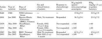
Field isolates were procured from patients admitted to the Kala-azar Medical Research Centre of the Institute of Medical Sciences, BHU, Varanasi and also from its affiliated hospital situated at Muzaffarpur, and were put into culture. Three were identified as SSG resistant (2039, 2041, and 2093) and 2 as SSG sensitive (2001, 2087) and were used for this study. The designation of responsive patients was based on the absence of fever, clinical improvement with reduction in spleen size and the absence of parasites in the splenic aspirate. Patients who showed the presence of parasites even after the SSG treatment were labelled as unresponsive cases. These patients were subsequently treated successfully with amphotericin B (Table 1). The biopsy materials were cultivated initially at 26°C in NNN-agar tubes and subsequently promastigotes were passaged in HEPES-buffered (pH 7·4) Medium 199 (Sigma, USA) with 10–20% heat-inactivated foetal bovine serum (HIFBS) at 25°C in 25 cm2 tissue culture flasks.
A brief description of clinical isolates/strain from Bihar (India) and evaluation of their chemo-sensitivity to sodium stibogluconate in vitro and in vivo

Parasite culture
L. donovani strain Dd8 (MHOM/IN/80/Dd8) was cultured in RPMI-1640 medium supplemented with 10% heat-inactivated foetal bovine serum (Sigma, USA), 100 U/ml penicillin (Sigma, USA) and 100 μg/ml streptomycin (Sigma, USA) at 26°C. The various field isolates (2001, 2039, 2041, 2087 and 2093) and L. major V121 were grown in Medium 199 supplemented with 10% heat-inactivated foetal bovine serum (Sigma, USA), 100 U/ml penicillin (Sigma, USA) and 100 μg/ml streptomycin (Sigma, USA) at 26°C.
Macrophage cell line culture and maintenance
The adherent mouse macrophage cell line J774A.1 was maintained in Dulbecco's modified Eagle's medium (Sigma, USA) supplemented with 10% heat-inactivated FBS, 100 U/ml penicillin and 100 μg/ml streptomycin at 37°C in 5% CO2 in a humidified atmosphere.
Animal host
Laboratory-bred male golden hamsters (Mesocricetus auratus) weighing 45–50 g were used as the experimental host. They were housed in climatically controlled rooms and fed with standard rodent food pellet (Lipton Ltd, Bombay) and water ad libitum. Experiments on the animals were performed following the guidelines of institutional animal ethics committee of the CDRI.
Infection to animals
Hamsters were infected intracardially with late log phase promastigotes of clinical isolates. Briefly, promastigotes were harvested by centrifugation at 2800 g for 15 min at 4°C, washed thrice with PBS and resuspended in PBS to a concentration of 1×10⁷ per 0·1 ml. Hamsters were inoculated intracardially with promastigotes in 0·1 ml of PBS. Parasite burden was assessed on days 20–25 post-infection by performing splenic biopsies as described previously (Sharma et al. Reference Sharma, Singh, Garg, Haq and Dube2004). L. donovani (Dd8) was passaged using spleen-derived amastigotes from heavily infected donor hamsters.
Isolation of amastigotes from macrophage cell line
J774A.1 macrophages (108 cells) in 50 ml culture flasks (Nunc) were infected with promastigotes at a multiplicity of infection of 10:1 (parasites/macrophage) and incubated at 37°C in 5% CO2 for 8–12 h after which the cells were washed thrice with phosphate-buffered saline (PBS, pH 7·2) and finally supplemented with complete medium. Infected macrophages were harvested using a cell scraper and centrifuged at 2000 g for 10 min and the pellet was resuspended in 1 ml of PBS and passed repeatedly through a 26-gauge sterile needle to facilitate the release of intracellular amastigotes by forced bursting of the macrophages. The amastigotes released from the macrophages were purified using Percoll (Sigma, USA) density-gradient centrifugation. Briefly, amastigotes released from the macrophages were centrifuged at 800 g for 10 min to remove tissue debris. The supernatant was centrifuged at 1600 g for 15 min at 4°C. The pellet was resuspended in 5 ml of 45% Percoll (Sigma, USA) in PBS-EDTA (2 mm) and was layered over a cushion of 2 ml of 90% Percoll. The gradient was centrifuged at 3500 g at 4°C for 1 h in a swing-out rotor. The amastigotes were collected from the interphase of the 45–90% step gradient and washed with PBS.
Isolation of amastigotes from infected spleen of hamsters
Infected hamsters were sacrificed after 60 days of infection and the spleen collected to isolate the amastigotes. Briefly, the spleen was homogenized in PBS and centrifuged at 800 g for 10 min to remove tissue debris. The supernatant was then centrifuged at 1600 g for 15 min at 4°C to collect the amastigotes in the pellet and these were purified by Percoll density-gradient centrifugation as described above.
Antibodies
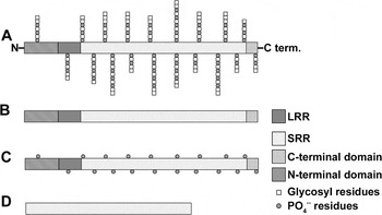
Normal rabbit serum was used as an antibody control for confocal microscopy. All other antibodies employed for this study were the kind gift of Dr Emanuela Handman, WEHI, Australia. The domains of PPG recognized by these antibodies are shown diagrammatically in Fig. 1. These polyclonal antibodies were raised in rabbits against various forms of PPG, namely, anti-deglycosylated and dephosphorylated native filamentous PPG (Panel B), anti-filamentous PPG treated with mild acid (Panel C) and anti-recombinant DNA-derived PPG repeats (APSSSS) (Panel D). Rabbit antiserum to L. donovani grp78 protein (from Dr Emanuela Handman, Australia) was used as loading control.
The schematic structure of PPG (Montgomery et al. Reference Montgomery, Curtis and Handman2002; Ilg et al. Reference Ilg, Stierhof, Craik, Simpson, Handman and Bacic1996) (A) and the domains of PPG recognized by the anti-deglycosylated and dephosphorylated native filamentous PPG (B), anti-filamentous PPG treated with mild acid (C) and anti-recombinant DNA-derived PPG repeats (APSSSS) sequence (D) antibodies. The diagram is not to scale. SRR: serine rich repeats, LRR: leucine rich repeats.

Immunofluorescence analysis by confocal microscopy
Late log phase promastigotes, spleen and macrophage-derived amastigotes were adhered on poly-L-lysine coated cover-slips and were fixed for 20 min in 4% paraformaldehyde in PBS (pH 7·2) and incubated with a rabbit polyclonal antiserum raised against deglycosylated and dephosphorylated PPG and detected with fluorescein isothiocyanate (FITC)-conjugated goat anti-rabbit IgG antibodies (Bangalore Genei, India). The stained parasites were examined in the mounting medium (90% glycerol in PBS supplemented with traces of paraphenyldiamine (PPD), and single or serial optical sections were collected with a confocal laser-scanning microscope (Bio-Rad, USA).
Immunofluorescence analysis by flow cytometry (FACS)
Late log phase promastigotes, spleen and macrophage-derived amastigotes were fixed for 20 min in 4% paraformaldehyde in PBS (pH 7·2) and incubated with a rabbit polyclonal antiserum raised against deglycosylated and dephosphorylated PPG (1:500). A set of fixed cells was kept untreated and served as controls. After washing thrice with PBS+BSA (0·5%) the cells were detected with fluorescein isothiocyanate (FITC)-conjugated goat anti-rabbit IgG antibodies (1:2000) (Bangalore Genei, India). Both sets of parasites were analysed in a FACS Calibur flow cytometer (BD Biosciences, USA) equipped with a 15 mV 488 nm air-cooled argon laser with excitation at 488 nm and emission at 515 nm. Ten thousand cells were acquired for each analysis. Multiparametric data were analysed by CellQuest software (Dube et al. Reference Dube, Singh, Sundar and Singh2005).
Western blotting
The whole cell lysate (300 μg/lane) or 107 promastigotes/lane of all the L. donovani strains along with L. major was separated on 2 sets of SDS-PAGE containing 6% stacking and 10% separating gels. One set of gels was stained with Coomassie blue and the other was used for transferring proteins to nitrocellulose (NC) membrane using a Hoefer Semi-dry transfer assembly at 0·8 mA/cm2 of NC membrane (Laemmli, Reference Laemmli1970; Towbin et al. Reference Towbin, Staehelin and Gordon1979). Successful transfer of the proteins was verified by the transfer of the pre-stained molecular weight standards (Fermentas, USA ). Following overnight blocking of non-specific binding sites in 5% skimmed milk, NC membrane was probed with different antibodies as mentioned above (all in 1/1000 dilutions) followed by incubation with horseradish peroxidase (HRP)-conjugated goat anti-rabbit antibodies (Bangalore Genei, India) (1/5000 dilution). After washing, protein band detection was performed using the Amersham ECL kit of enhanced chemiluminescence (Amersham Biosciences, Singapore). The same blot was stripped off each time and re-probed with each of the above-mentioned antibodies and later developed by chemiluminescence. Densitometry analysis of each film was carried out using an Alpha Imager 2200 (Bio-Rad, USA) for assessing the expression of PPGs. For densitometry calculations, the same band area was taken for band intensity and was normalized to grp-78.
Statistical analysis
All FACS and Western blotting experiments were performed in triplicate. The data relating to FACS and densitometry analysis are presented as mean±standard deviation (s.d.). The statistical significance of differences in percentage expression between resistant and sensitive isolates was analysed by Student's t-test using SigmaStat (version 2.03) software.
RESULTS
Localization of PPG in promastigotes and amastigotes
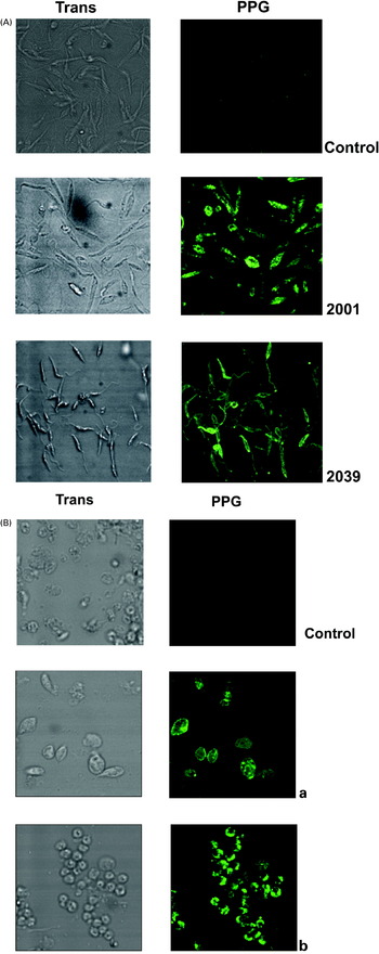
Confocal laser fluorescence microscopy was performed in promastigotes and amastigotes incubated with rabbit polyclonal antiserum, raised against deglycosylated and dephosphorylated PPG, that recognizes the Ala-Pro-Ser repeats in the proteophosphoglycan backbone, followed by FITC-labelled secondary antibody. The promastigotes of the drug-resistant as well as sensitive isolates exhibited bright green fluorescence throughout their outer surface, flagella and flagellar pocket (Fig. 2A) indicating the presence of PPG on their membrane surface. Proteophosphoglycan was also detected in amastigotes isolated from the spleen as well as from the macrophage cell line. As observed with the promastigotes, amastigotes of all the isolates also showed bright green fluorescence throughout their outer surface and flagellar pocket (Fig. 2B). The fluorescence was intense in both the stages of all the resistant isolates. The control antibodies (normal rabbit serum) used showed no fluorescence (Fig. 2A).
(A) Immunolocalization of PPG in promastigotes of representative strains of SSG-sensitive 2001 and SSG-resistant 2039 clinical isolates by confocal laser scanning microscopy. Normal rabbit serum used as antibody control. (B) Immunolocalization of PPG in amastigotes derived from macrophage cell line (a) and splenic macrophages (b) by confocal laser scanning microscopy.

Quantitative expression of PPG by FACS in promastigotes and amastigotes
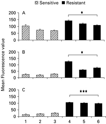
The FACS analysis of the promastigote and amastigote forms of all the clinical isolates was done to measure the intensity of fluorescence which is directly indicative of the amount of PPG present on the surface or flagellar pocket of the parasites. As described for confocal microscopical studies, the cells of both the stages were treated with antibody against deglycosylated and dephosphorylated PPG followed by FITC-labelled secondary antibody. The data revealed that the fluorescence value in promastigotes of the SSG-resistant strains (2039, 2041, 2093) was significantly higher (P<0·05) (Mean FL-1 values 141, 117 and 110, respectively) as compared to drug-sensitive strains Dd8, 2001 and 2087 strains, which possessed less PPG as evident by the low value of mean fluorescence values (101, 65 and 68, respectively) in these isolates (Fig. 3). The mean FL1-value in unstained parasites (control) was 2–3 units, which corresponds to non-specific auto-fluorescence of the parasites. Similarly, the mean FL-1 value in amastigotes isolated from J774A.1 macrophage cell line of the drug-resistant strains was significantly higher (P<0·05) as compared to drug-sensitive strain which showed a low mean fluorescence value (20, 19 and 28, respectively) in these isolates (Fig. 3). Correspondingly, the mean fluorescence value of the spleen-derived amastigotes of drug-resistant strains was also higher (Mean FL-1 value 119, 114 and 96, respectively) as compared to drug-sensitive strains (Mean FL-1 value 21, 22 and 27, respectively) and this difference was highly significant (P<0·001) (Fig. 3).
Quantitative analysis of PPGs in promastigotes (A), macrophage cell line-derived amastigotes (B) and splenic amastigotes form (C) of SSG-resistant and sensitive clinical isolates by FACS. Each experiment was performed in triplicate. Each bar represents the mean fluorescence value±s.d. Significance values indicate significant differences between sensitive and resistant groups (* P<0·05 and *** P<0·001). Strains are indicated as follows: (1) Dd8, (2) 2001, (3) 2087, (4) 2039, (5) 2041 and (6) 2093.

Expression of PPG in promastigotes of clinical isolates by Western blot
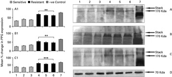
Immunoblot analysis of whole cell lysates of all the clinical isolates as well as L. major (as control or reference strain) with different polyclonal antibodies against various forms of PPGs of L. major, raised in rabbits, revealed the presence of PPGs in all SSG-sensitive and resistant strains (Fig. 4). PPG bands in all the L. donovani isolates were not present in the stacking gel as observed with L. major. Prominent PPG bands of approximately 170 KDa were observed in the separating gel in all the isolates including that of L. major. The intensity of bands was greater in resistant isolates as compared to sensitive ones. This was further confirmed by the densitometry analysis of the blot wherein the percentage change in expression of PPG was found to be significantly higher (P<0·01) in SSG-resistant strains using anti-deglycosylated and dephosphorylated native filamentous PPG. Similarly, the PPG expression was also significantly higher (P<0·01) against the antibody to the native filamentous PPG treated with mild acid. This variation was again highly significant (P<0·001) against the recombinant DNA-derived PPG repeats (APSSSS) (Fig. 4). The loading control indicated a similar abundance of the endoplasmic reticulum protein grp78.
Expression of Leishmania donovani PPGs in SSG-sensitive and resistant strains by immunoblotting using heterologous rabbit polyclonal antiserum raised against deglycosylated and dephosphorylated PPG (A), native filamentous PPG treated with mild acid (B), recombinant DNA-derived PPG repeats (APSSSS) (C) of L. major and grp 78 (loading control) (D) of L. donovani as primary antibody. The histograms represent the densitometry analysis showing the relative change in PPG expression±s.d. of each blot probed with heterologous rabbit polyclonal antiserum raised against deglycosylated and dephosphorylated PPG (A1), native filamentous PPG treated with mild acid (B1) and recombinant DNA-derived PPG repeats (APSSSS ) (C1) in comparison to control (grp-78). Significance values indicate the differences between sensitive and resistant groups (** P<0·01 and *** P<0·001). Lanes 1–7 were loaded with different Leishmania strains as follows: (1) Dd8, (2) 2001, (3) 2087, (4) 2039, (5) 2041, (6) 2093 and (7) L. major (as positive control).

DISCUSSION
The PPGs form a heterogeneous population of polypeptides of variable size, glycosylation and expression (Foth et al. Reference Foth, Piani, Curtis, Ilg, McConville and Handman2002). Their salient feature is a central repetitive domain of (Ala–Pro–Ser) in which most serine residues are phosphoglycosylated (Ilg, Reference Ilg2000). The N-terminal to the central repetitive domain is a non-repetitive sequence separated by a leucine-rich repeat (LRR) motif (Ilg et al. Reference Ilg, Handman, Ng, Stierhof and Bacic1999b; Montgomery et al. Reference Montgomery, Ilg, Thompson, Kobe and Handman2000) and the carboxy terminal consists of a second non-repetitive region terminating in a hydrophobic amino acid sequence compatible with GPI addition (Ilg et al. Reference Ilg, Handman and Stierhof1999a). PPGs are ubiquitous in all Leishmania. The conserved Ala–Pro–Ser repeats form major antigenic determinants since the antibodies to this domain recognize related molecules in all species examined, namely, L. major (VI21, Friedlin VI, LRC-L119), L. mexicana (LRC-L94), and L. donovani (LRC-L52) (Montgomery et al. Reference Montgomery, Ilg, Thompson, Kobe and Handman2000; Rogers et al. Reference Rogers, Ilg, Nikolaev, Ferguson and Bates2004).
Although the function of the various members of the PPG family is not known, there is evidence that PPG binds to macrophages and may be involved in parasite invasion (Montgomery et al. Reference Montgomery, Curtis and Handman2002; Kedzierski et al. Reference Kedzierski, Montgomery, Bullen, Curtis, Gardiner, Jimenez-Ruiz and Handman2004). Its presence in both promastigotes (pPPG) and amastigotes (aPPG) also suggests that it may play a role in the amastigote interaction with host macrophages. The presence of a PPG in a macrophage vesicle also supports this contention and suggests that it may also contribute to the parasite's survival in the infected macrophage (Piani et al. Reference Piani, Ilg, Elefanty, Curtis and Handman1999). There are no reports regarding the role of PPG in promastigotes and amastigotes of L. donovani, the causative agent of fatal VL, which has a different disease profile from that of CL. This study was initiated to examine the role(s) of PPGs in L. donovani. We have examined both the qualitative and quantitative detection of PPG in a few recent Indian clinical isolates, namely, SSG-resistant and sensitive strains. We have shown in our earlier work (Dube et al. Reference Dube, Singh, Sundar and Singh2005) on the clinical isolates in laboratory conditions, that the resistance of the clinical isolates was restricted to the SSG and did not lead to any cross-resistance against the other clinically relevant drugs. It is noteworthy that repeated passages of the resistant clinical isolates in hamsters and their responses to SSG established that they are truly resistant and retained their phenotypic characteristics. Thus, for the present study we have used the clinical isolates that have been maintained in our laboratory since 2000 (Dube et al. Reference Dube, Singh, Sundar and Singh2005).
Further, we have used antibodies raised against treated forms of fPPG of L. major e.g. anti-deglycosylated and dephosphorylated native filamentous PPG, anti-filamentous PPG treated with mild acid, and anti-recombinant DNA-derived PPG repeats (APSSSS). As is evident in Fig. 1B, the antibody recognizes only the peptide domain of PPG which is devoid of both phosphate and glycosyl residues. The glycans of fPPG were further removed by mild acid hydrolysis so that this anti-filamentous PPG recognize only the polypeptide backbone of fPPG but do not recognize the glycans present in PPG or LPG (Ilg et al. Reference Ilg, Stierhof, Craik, Simpson, Handman and Bacic1996; Montgomery et al. Reference Montgomery, Curtis and Handman2002; Rogers et al. Reference Rogers, Ilg, Nikolaev, Ferguson and Bates2004). In addition, since the major component of PPG has serine repeats we used anti-recombinant DNA-derived PPG repeats (APSSSS) which recognize the repetitive sequence (Montgomery et al. Reference Montgomery, Curtis and Handman2002).
Confocal microscopical analysis using antibody against dephosphorylated and deglycosylated PPG revealed the localization of PPG on the entire cell surface of promastigotes, including the flagella and flagellar pocket in all the isolates. The fluorescence was more intense at flagellar pocket/kinetoplast, which indicates the possibility of higher content of PPG at this location in the parasite. This is in agreement with the observations made by Foth et al. (Reference Foth, Piani, Curtis, Ilg, McConville and Handman2002) with L. major promastigotes. Amastigotes also showed fluorescence on their entire surface, although, in the case of L. major the precise localization of PPG was more difficult to determine by immunofluorescence, as observed earlier (Foth et al. Reference Foth, Piani, Curtis, Ilg, McConville and Handman2002). Interestingly, the fluorescence was observed to be more intense in resistant isolates as compared to the sensitive ones. This observation was further confirmed quantitatively by FACS, and provided additional evidence that PPG is found at a higher content in the promastigotes and amastigotes of all the drug-resistant isolates compared with the sensitive lines. The higher FL-1 value in promastigotes as compared to amastigotes in all the strains may be due to the difference in their size. It is interesting to note that the level of PPG in either resistant or sensitive isolates was independent of the time of isolation. The above observation was further confirmed by immunoblot analysis using the 3 antibodies raised against various forms of PPG of L. major. It was noteworthy that all these antibodies could only detect high molecular weight PPG in the stacking gel with L. major but not with the L. donovani isolates revealing a marked difference between the 2 species. Nevertheless, these antibodies could detect PPG in the separating gel in all the L. donovani isolates (170 kDa), as well as L. major strain, but there were differences in the PPG band pattern since there were 3 PPG bands just above 170 kDa in the case of the latter strain. These findings are in conformity with the work done earlier (Montgomery et al. Reference Montgomery, Curtis and Handman2002). A possible explanation may be the variation between different strains in the degree of glycosylation of the polypeptide, which may make it inaccessible to the antibody (Montgomery et al. Reference Montgomery, Curtis and Handman2002). Therefore, the precise nature of this material in L. donovani strains awaits further characterization.
The presence of a higher content of PPG in resistant isolates is quite intriguing and the precise reason for this is yet to be established. Some workers have speculated that the development of drug resistance in clinical isolates is probably due to the alterations in the plasma membrane (Kothari et al. Reference Kothari, Kumar, Sundar and Singh2007; Rakotomanga et al. Reference Rakotomanga, Saint-Pierre-Chazalet and Loiseau2005). These authors suggested that plasma membrane is the site at which drug is taken up by the cell, and any significant modification in its membrane composition may have an impact on drug-membrane interactions. Simultaneously, Singh et al. (Reference Singh, Almeida, Kothari, Kumar, Mandal, Chatterjee, Venkatachalam, Govind, Mandal and Sundar2007), by analysing differential gene expression in drug-resistant clinical isolates by microarray, have identified phosphoglycans as one of the cell surface molecules. In another study differences in lipid composition affecting the parasite membrane structure between wild type and parasites resistant to miltefosine, amphotericin B and atovaquone have been demonstrated (Perez-Victoria et al. Reference Perez-Victoria, Castanys and Gamarro2003). Our observations also further indicate that a change in PPG expression is a mechanism of resistance but at the same time we cannot exclude the possibility of other mechanisms acting in synergism, and there are chances that different isolates may respond to the drug pressure differently as host factors may also contribute in the ultimate phenotype.
Further, due to the absence of any marker of clinical antimony resistance in L. donovani isolates, several laboratories have generated and identified markers of SbV resistance (Sundar et al. Reference Sundar, Pai, Kumar, Pathak-Tripathi, Gam, Ray and Kenney2001; Singh et al. Reference Singh, Papadopoulou and Ouellette2001), but evidence of their existence in field isolates from refractory patients are yet to be found. In the light of the above observations, although the numbers of isolates are few, PPG may also be used as a marker for identifying drug-resistant clinical isolate. This is the first preliminary report indicating such variable expression in clinical isolates of L. donovani obtained from one endemic field of VL, hence further studies related to proteomics and genomics need to be performed to reach a conclusion as to whether PPG has any role in the mechanism of drug resistance.
Grateful acknowledgments are due to Mrs Joan Curtis and Dr Emanuela Handman, Division of Infection and Immunity, Walter Eliza Hall Institute of Medical Research (WEHI), Melbourne, Australia, for providing the anti-PPG antibodies to L. major, and to the Department of Biotechnology, New Delhi for granting a DBT Overseas Associateship to enable A. D. to carry out some of the work at WEHI. We are extremely thankful to Mr Ramesh C. Nayak and Mr Ravi K Lella for their technical help in carrying out confocal analysis, to Mr Ali Kauser for editing the photographs/figures and to Drs Nikhil Kumar and Neeloo Singh for their critical comments on the manuscript. Financial assistance to M. S., N. S. and S. K. G. by the Council of Scientific and Industrial Research, New Delhi is also gratefully acknowledged. This is CDRI communication No. 6821.