Introduction
Recurrent aphthous stomatitis is a common oral mucosal disease characterised by painful, multiple, recurrent, small, oval or round necrotic ulcers. It is the most common oral ulcerative disease affecting 10–20 per cent of the general population.Reference Jurge, Kuffer, Scully and Porter1 Recurrent aphthous stomatitis typically occurs between 10 and 30 years of age and recurs at varying intervals throughout life. Generally, as the patient ages, it recurs with decreasing frequency and severity.Reference Ślebioda, Szponar and Kowalska2 Its aetiology and pathogenesis are not entirely clear. Nutritional, familial, hormonal, allergic, psychological, traumatic and infectious factors, and inflammatory response are suspected to be among the aetiological factors associated with recurrent aphthous stomatitis.Reference Jurge, Kuffer, Scully and Porter1–Reference Greenberg and Pinto3 Histopathological change in the pre-ulcerative stage includes epithelium infiltration by mononuclear (lymphocytic) cells. Recurrent aphthous stomatitis is thought to either occur idiopathically or as a result of several triggering factors.Reference Pedersen and Ryder4
Calprotectin, also known as myeloid-related protein 8, myeloid-related protein 14 or calgranulin, is an S100 protein and is an inflammation marker found in neutrophils and monocytes.Reference Ozler5 The S100 proteins are a family of calcium-modulated proteins. Calprotectin, which has bacteriostatic properties, is released together with the neutrophil activation of monocytes and epithelial adhesion, and it can be detected in the blood as an inflammatory marker.Reference El-Rıfai, Moskaluk, Abdrabbo, Harper, Yoshida and Riggins6 The functions of calprotectin are mostly associated with active inflammatory processes such as antibacterial defence mechanisms or allograft rejection, T helper cell type 1 mediated responses, or autoimmune reactions.Reference Stříž and Trebichavský7 In many autoimmune and inflammatory diseases such as endometrium, larynx, lung and stomach cancers, calprotectin levels increase.Reference El-Rıfai, Moskaluk, Abdrabbo, Harper, Yoshida and Riggins6–Reference Dale and Brandtzaeg8
This study aims to compare the serum calprotectin concentrations between patients with recurrent aphthous stomatitis and healthy individuals, and to determine whether calprotectin may be a prognostic indicator of ulcer activity.
Materials and methods
This study, which was a prospective, cross-sectional and historic cohort study, was approved by the local ethics committee and carried out in accordance with the Declaration of Helsinki (project number: E-18-2287).
A total of 79 patients with recurrent aphthous stomatitis without three or more recurrent systemic symptoms in a year were included in the study, and three groups were formed based on the presence of active lesions. The groups were matched according to age and gender. The three groups comprised: (1) an active lesion group of 26 patients with recurrent aphthous stomatitis who were in the ulcer activation period; (2) an inactive lesion group of 26 patients with recurrent aphthous stomatitis who were not in the ulcer activation period; and (3) a control group of 27 healthy volunteers without recurrent aphthous stomatitis.
Patients with any systemic or autoimmune disease, history of neoplasm, Behcet's disease, acute trauma, history of active infection, and patients who smoked, drank alcohol, were pregnant or lactating were excluded from the study.
A detailed medical history, age, gender, serum calprotectin value, neutrophil–lymphocyte ratio, platelet–lymphocyte ratio, and neutrophil, leukocyte, mean platelet volume and lymphocyte values were recorded for all patients after standard examination was performed. Serum calprotectin levels and neutrophil–lymphocyte ratio, platelet–lymphocyte ratio and mean platelet volume values were also compared between the groups. Furthermore, the relationship between oral ulcer activity and the calprotectin, neutrophil–lymphocyte ratio, platelet–lymphocyte ratio and mean platelet volume levels was also investigated.
Serum calprotectin values were examined in the venous blood drawn from the patients. Cytometry scattergrams were obtained within 2 hours after blood sampling. Calprotectin measurement was performed in the ChemWell® 2900 (Algen) device using the Human Calprotectin (Elabscience, Texas, USA) enzyme-linked immunosorbent assay kit. The results were reported in nanograms per millilitre. Intra-assay and inter-assay coefficient of variation (CVA; per cent CVA) values were determined to be 3.92 and 4.76, respectively. All samples were studied in pairs, and the mean values of these two studies were used for statistical analysis.
A composite index was used to determine the ulcer activity in patients with acute aphthous stomatitis. The composite index survey is designed to have five items and three subscales. In the index, the number of ulcers, pain status, and functional changes in taste, speech and chewing during the previous month were included. Scoring was done according to the presence of aphthous stomatitis in the composite index, pain status and functional status. Oral ulcer activity was identified as active or inactive according to the presence or absence of oral ulcers. One point was given if presence of an ulcer during the previous month was noted, and no point was given if there was no ulcer.
The visual analogue scale (VAS) was used to determine the pain status associated with an oral ulcer during the previous month.Reference Mumcu, Sur, Inanc, Karacayli, Cimilli and Sisman9 The patients were asked to mark the severity of the ulcer pain they experienced in the previous month on a scale with a distance of 0–100 mm. For VAS scores between 0 and 2, 2.1 and 4, 4.1 and 6, 6.1 and 8, and 8.1 and above, the composite index score was calculated as 0, 1, 3, 4 and 5, respectively.
In order to determine functional status, the patients were individually asked how the ulcers affected their tasting, speaking and eating–drinking status during the previous month. The answers were coded using a Likert-type scale with values determined as 0 = never, 1 = little time, 2 = part of the time, 3 = most of the time and 4 = always. As a result, the composite index score (0–10) was identified as the sum of the subscale scores of the oral ulcer activity during the previous month (0–1) and the pain status associated with oral ulcers as evaluated by the categorised VAS (0–5). The functional disease score was evaluated using a 5-point Likert-type scale.
Results
There were 26 patients in the active lesion group, consisting of 13 males (50 per cent) and 13 females (50 per cent). There were 26 patients in the inactive lesion group, consisting of 13 males (50 per cent) and 13 females (50 per cent). There were 27 patients in the control group, consisting of 13 males (48.15 per cent) and 14 females (51.85 per cent). The mean age (± standard deviation (SD)) of the active, inactive and control groups was 43.35 ± 14.74, 39.80 ± 14.12 and 40.80 ± 14.12 years, respectively. There was no statistically significant difference between the groups in terms of gender and age (p = 0.180 and p = 0.725, respectively).
No significant difference was seen among the three groups in terms of haemoglobin, white blood cell, platelet, neutrophil and lymphocyte count (p > 0.05) (Table 1).
Comparison of demographic data and laboratory parameters among groups

SD = standard deviation
The platelet–lymphocyte ratio averages (± SD) of the groups were found to be 127.81 ± 30 in the active lesion group, 127.4 ± 31 in the inactive lesion group and 119.4 ± 26.8 in the control group. A significant difference was not determined when comparing the platelet–lymphocyte ratio averages of the three groups (p = 0.5).
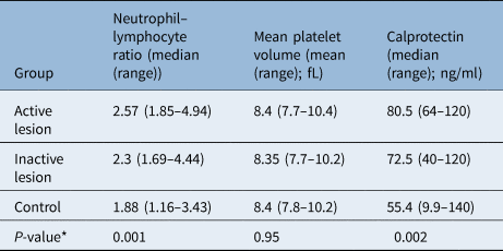
The median neutrophil–lymphocyte ratio, mean platelet volume and calprotectin values of the groups are presented in Table 2. Although there was no significant difference among the three groups in terms of mean platelet volume values (p = 0.95), a statistically significant difference was observed in the neutrophil–lymphocyte ratio and calprotectin values (p = 0.001 and p = 0.002, respectively).
The median neutrophil–lymphocyte ratio, mean platelet volume and calprotectin values of the groups

*of Kruskal–Wallis analysis.
In the post-hoc tests performed to determine which groups differed from each other in terms of neutrophil–lymphocyte ratio values, it was observed that the median neutrophil–lymphocyte ratio values of both the active and inactive lesion groups were statistically significantly higher compared with the control group (p = 0.017 and p < 0.001, respectively). However, a statistically significant difference was not detected between the neutrophil–lymphocyte ratio values of the active and inactive lesion groups (p = 0.08).
Post-hoc tests were performed to identify which among the groups showed a difference in terms of calprotectin value. Although there was no statistically significant difference between the median calprotectin value of the inactive lesion group and control group (p = 0.033), a significantly higher median calprotectin value was observed in the active lesion group than in the control group (p = 0.002). Furthermore, the median calprotectin value of the active lesion group was statistically significantly higher than that of the inactive lesion group (p = 0.017).
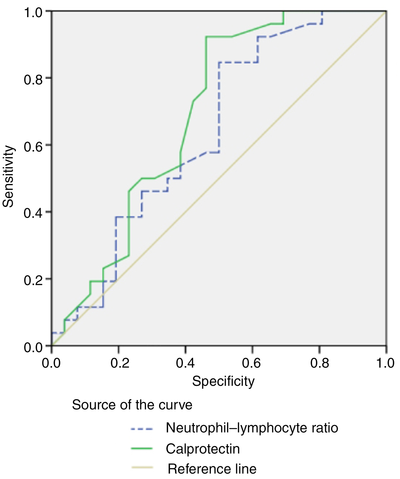
In the receiver operating characteristic analysis, the diagnostic features of the calprotectin and neutrophil–lymphocyte ratio values were tested for their ability to predict the presence of active lesions. The area under the calprotectin curve was 0.692 (p = 0.017), and the area under the neutrophil–lymphocyte ratio curve was determined to be 0.641 (p = 0.08) (Figure 1).
Receiver operating characteristic analysis of the calprotectin and neutrophil–lymphocyte ratio values.

The demographic data and laboratory values of the groups are presented in Table 1. The median neutrophil–lymphocyte ratio, mean platelet volume and calprotectin values of the groups are presented in Table 2.
Statistical analysis
Statistical analysis was performed in order to test whether there was a difference in terms of neutrophil–lymphocyte ratio, mean platelet volume, calprotectin and platelet–lymphocyte ratio among the three groups categorised according to the presence of active lesions.
The Kolmogorov–Smirnov normality test was applied to evaluate the suitability of the groups to a normal distribution. Based on the results of this test, it was determined that the platelet–lymphocyte ratio values of the groups were distributed normally (p = 0.19), but neutrophil–lymphocyte ratio, mean platelet volume and calprotectin values did not comply with a normal distribution (p < 0.05). Therefore, the difference among the groups in terms of platelet–lymphocyte ratio value was determined using the one-way analysis of variance test, and the difference in terms of neutrophil–lymphocyte ratio, mean platelet volume and calprotectin values was tested using the Kruskal–Wallis analysis.
The parametric test results were presented as mean ± SD, and the results of the non-parametric tests were presented as median (minimum–maximum). The Mann–Whitney U test was used as a post-hoc test. The diagnostic decisive features of calprotectin and neutrophil–lymphocyte ratio values in predicting the presence of active lesions were tested using the receiver operating characteristic curve analysis. All statistical analyses were performed using SPSS® (version 16) statistical software. When evaluating the area under the curve in the receiver operating characteristic analysis, the diagnostic value of the test was considered to be statistically significant with a type-1 error level below 5 per cent. A p-value below 0.05 was considered statistically significant. Furthermore, because the Bonferroni correction (triplet combination for three groups) was used in post-hoc tests, the p-value was considered to be significant when it was less than 0.017 (0.05/3 = 0.017).
Discussion
Recurrent aphthous stomatitis is defined as recurrent ulceration of the oral mucosa. Recurrent aphthous stomatitis is a widespread disease, but its aetiology and pathogenesis are still not clear.Reference Greenberg and Pinto3 Histopathological changes illustrate an inflammatory process mainly involving non-keratinised mucosa. In the inflammatory response, neutrophil and monocyte concentrations elevate, whereas lymphocyte concentrations in the peripheral blood decrease. Oedema develops followed by keratinocyte invasion and then localised vasculitis develops. Before healing and regeneration happen in the epithelium, localised inflammation, infiltrated by neutrophils, lymphocytes and plasma cells, occurs.Reference Natah, Häyrinen-Immonen, Hietanen, Patinen, Malmström and Savilahti10 The hypersensitivity of T lymphocytes to various antigens has been argued to play a significant role in the pathogenesis of recurrent aphthous stomatitis. However, it is thought that immunological mechanisms play an essential role in the pathogenesis of recurrent aphthous stomatitis.Reference Porter and Scully11
Calprotectin is an antimicrobial protein found mainly in the cytoplasm of neutrophils. It is secreted extracellularly by stimulated neutrophils and monocytes or released as a result of cell disruption or death.Reference Boussac and Garin12 Elevated serum calprotectin levels may occur in different immune and immunopathological reactions, especially in acute inflammation or T helper cell type 1 mediated responses. However, calprotectin has been demonstrated to inhibit matrix metalloproteinase, which plays a role in the growth and spread of cancer cells.Reference Ozler5,Reference El-Rıfai, Moskaluk, Abdrabbo, Harper, Yoshida and Riggins6 In some respects, the sensitivity and dynamics of calprotectin may be superior to conventional inflammatory markers such as C-reactive protein.Reference Stříž and Trebichavský7 However, it may give rise to inaccurate data because it can also increase in other inflammatory diseases.
Some studies on calprotectin have demonstrated that it could be a biomarker that is useful for the early diagnosis of various cancers. Moreover, it is thought that the calprotectin concentration increases because of an inflammatory response in cancerous tissues. Calprotectin has been found to be a valuable index that can be used to predict clinical outcomes of inflammatory diseases in oncology. In their study, Topuz et al. found a higher serum calprotectin concentration in patients with laryngeal carcinoma than in patients with benign laryngeal pathology and in healthy controls. They reported that calprotectin is a biomarker that could be used for the diagnosis, treatment and follow-up evaluation of laryngeal cancer.Reference Topuz, Binnetoglu, Yumusakhuylu, Sarı, Baglam and Gerin13 Many studies have demonstrated that the calprotectin gene expression increases in some tumour tissues such as gastric cancer, pulmonary adenocarcinoma, hepatocellular carcinoma and invasive ductal breast cancer.Reference Arai, Teratani, Kuruto-Niwa, Yamada and Nozawa14,Reference Arai, Yamada and Nozawa15
Various studies in the literature have demonstrated that the neutrophil–lymphocyte ratio is also a new biomarker that suggests the presence of inflammation. Ozler and Akoglu reported that neutrophil and inflammatory marker levels in patients with recurrent aphthous stomatitis are higher compared with those of the healthy population.Reference Ozler and Akoglu16 In the same study, a positive correlation was found between the neutrophil–lymphocyte ratio and ulcer activation period. In the present study, the median neutrophil–lymphocyte ratio values of both the active and inactive lesion groups were observed to be statistically significantly higher than those of the control group. However, a statistically significant difference was not determined between the neutrophil–lymphocyte ratio values of the active lesion group and inactive lesion group. These results suggest that neutrophil–lymphocyte ratio is associated with the presence of recurrent aphthous stomatitis rather than the presence of active lesions.
In the present study, while no statistically significant difference was determined between the median calprotectin value of the inactive lesion group and the control group, the median calprotectin value of the active lesion group was found to be significantly higher than that of the control group. At the same time, the median calprotectin value of the active lesion group was found to be statistically significantly higher than that of the inactive lesion group. In the receiver operating characteristic analysis, which was established to test the capacity of calprotectin and neutrophil–lymphocyte ratio in demonstrating the presence of active lesions, both tests were found to be significant. However, while the calprotectin area under the curve was 0.692, the area of neutrophil–lymphocyte ratio was 0.641. With these results, it was concluded that calprotectin is a more significant biomarker than neutrophil–lymphocyte ratio in determining presence of active lesions. Furthermore, having a higher calprotectin value in the active lesion group than that of the inactive lesion group and control group shows the relationship of calprotectin to the presence of active lesions.
It has been argued that platelets play an important role in inflammatory processes. Ekiz et al. demonstrated that the mean platelet volume and erythrocyte sedimentation rate were significantly higher in patients with recurrent aphthous stomatitis than in the controls.Reference Ekiz, Balta, Sen, Rifaioglu, Ergin and Balta17 In the present study, it was determined that mean platelet volume values did not change significantly among the three groups categorised according to the presence of active aphthous ulcer lesions.
Recent studies have reported that platelet–lymphocyte ratio may also be a new biomarker that may indicate the presence of inflammation and its severity.Reference Alan, Tuna and Türkoğlu18 Alan et al. reported higher platelet–lymphocyte ratio values in individuals with Behcet's disease than in healthy individuals.Reference Alan, Tuna and Türkoğlu18 In the present study, no statistically significant difference in terms of platelet–lymphocyte ratio was seen among the three groups categorised according to the presence of active lesions.
Kristinsson et al. evaluated faecal calprotectin levels for inflammatory bowel disease.Reference Kristinsson, Roseth, Fagerhol, Aadland, Schjonsby and Bormer19 In addition, calprotectin levels in saliva may be predictive for the prognosis of recurrent aphthous stomatitis. Building on this approach, it would be worthwhile to look at saliva calprotectin levels in patients with recurrent aphthous stomatitis. This may be a further putative use for the growing field of salivary diagnostics.
The main limitation of this study was the relatively small study population. To the best of our knowledge, there are no studies on serum calprotectin concentration in patients with recurrent aphthous stomatitis. This study aimed to compare serum calprotectin concentration in patients with recurrent aphthous stomatitis and healthy individuals and to consider calprotectin as a marker for diagnosis and prognosis in the ulcer activation period. Examination of the serum calprotectin value may make it possible to predict the response to treatment before starting treatment in patients with recurrent aphthous stomatitis.
• Calprotectin is a biomarker for inflammation in neutrophils and monocytes
• Calprotectin increases in many autoimmune and inflammatory diseases
• Recurrent aphthous stomatitis is a common disease of oral mucosa
• This study compares the serum calprotectin concentrations in patients with recurrent aphthous stomatitis and active ulcer lesions
• Calprotectin is a new prognostic biomarker that can be used for the diagnosis, treatment and follow-up evaluation of recurrent aphthous stomatitis
Conclusion
Calprotectin is a new prognostic biomarker that can be used for the diagnosis, treatment and follow-up evaluation of recurrent aphthous stomatitis, especially in the ulcer activation period. However, in order to understand the efficacy of using calprotectin as a prognostic biomarker in patients with recurrent aphthous stomatitis, there is a need for studies with more extensive patient participation.
Competing interests
None declared