INTRODUCTION
The nematode genus Strongyloides consists of small intestinal parasites of various vertebrates (Speare, Reference Speare and Grove1989; Dorris et al. Reference Dorris, Viney and Blaxter2002; Viney and Lok, Reference Viney and Lok2007). The life cycle of Strongyloides spp. (Fig. 1A) has been reviewed and discussed recently (Viney and Lok, Reference Viney and Lok2007; Streit, Reference Streit2008). In brief, parasitic worms, which are all female, live in the small intestines of their respective hosts. They produce female and male progeny by mitotic parthenogenesis. The young females can develop into infective third-stage larvae (L3i) that infect a new host (homogonic or direct development). Alternatively the females, like all the males, develop into free-living adults that reproduce sexually (heterogonic or indirect development). With very few exceptions (Yamada et al. Reference Yamada, Matsuda, Nakazawa and Arizono1991; Streit, Reference Streit2008), the entire progeny produced by the free-living Strongyloides sp. consists of females that develop into L3i.
Life cycles of Strongyloides ratti (A) and Parastrongyloides trichosuri (B). A also applies for the other species of Strongyloides mentioned in this publication except: (i) in S. papillosus and S. vituli embryonated eggs are passed and the L1s hatch within a short time outside of the host and (ii) in S. stercoralis a fraction of the larvae develop into L3i within the host (autoinfective L3i). For further explanation see text.

The presence of easily accessible free-living adults and a number of techniques and resources facilitating experimental work recently developed for multiple species of Strongyloides and for their relative Parastrongyloides trichosuri (Viney et al. Reference Viney, Green, Brooks and Grant2002; Grant et al. Reference Grant, Skinner, Howes, Grant, Shuttleworth, Heath and Shoemaker2006a, Reference Grant, Stasiuk, Newton-Howes, Ralston, Bisset, Heath and Shoemakerb; Eberhardt et al. Reference Eberhardt, Mayer and Streit2007; Nemetschke et al. Reference Nemetschke, Eberhardt, Viney and Streit2010b; Shao et al. Reference Shao, Li, Nolan, Massey, Pearce and Lok2012) render this group of parasites highly attractive for genetic analysis.
In all species of Strongyloides investigated thus far, the sex of the progeny produced by the parthenogenetic parasitic females is determined by an environmental stimulus (environmental sex determination, ESD) such that an increasing immune response of the host against the worms leads to a higher proportion of males (reviewed in Streit, Reference Streit2008). Nevertheless, in Strongyloides ratti (a parasite of rats) and Strongyloides stercoralis (a parasite of humans and dogs), X chromosomes exist (Nigon and Roman, Reference Nigon and Roman1952; Hammond and Robinson, Reference Hammond and Robinson1994; Harvey and Viney, Reference Harvey and Viney2001) (Fig. 2). In these species both sexes have two pairs of autosomes, and the females have two but the males only one X chromosome (environmentally controlled XX/XO sex determination). Therefore in these species 2n = 6 in females and 2n = 5 in males. In Strongyloides papillosus (a parasite of sheep) the genetic material homologous to the chromosomes I and X of S. ratti is combined in one chromosome (Nemetschke et al. Reference Nemetschke, Eberhardt, Hertzberg and Streit2010a). In males an XX/XO system appears to be functionally restored through a sex-specific chromatin diminution event that creates a hemizygous region corresponding to the X chromosome in S. ratti (Albertson et al. Reference Albertson, Nwaorgu and Sulston1979; Nemetschke et al. Reference Nemetschke, Eberhardt, Hertzberg and Streit2010a) (Fig. 2). Since in the process an internal portion of one chromosome is eliminated and both ends are retained as separate chromosomes this leads to 2n = 5 in males while in females 2n = 4. Nemetschke et al. (Reference Nemetschke, Eberhardt, Hertzberg and Streit2010a) proposed that the different sex-determining systems within the genus Strongyloides may represent different intermediate steps in the transition from XX/XO genetic sex determination (GSD) to ESD with no genetic differences between the sexes. This hypothesis assumes that the situation in S. ratti is ancestral and chromatin diminution in S. papillosus is derived. However, there is no strong evidence to support or reject this assumption. Knowing the chromosome numbers and the sex-determining systems of closely related species whose phylogenetic position with respect to S. ratti and S. papillosus is clear, could shed some light on this question (Fig. 2). Therefore, we analysed Strongyloides vituli, a species clearly much more closely related to S. papillosus than S. ratti (Eberhardt et al. Reference Eberhardt, Mayer, Bonfoh and Streit2008) and P. trichosuri, which is an outgroup to the genus Strongyloides.
Phylogenetic relationships and chromosomes in the species discussed. Chromosomes/genomic regions present in two copies in both sexes are depicted in black (print version) or blue (online version), chromosomes/genomic regions present in two copies in females and in only one copy in males are depicted in grey (print) or red (online version). For Strongyloides stercoralis it is likely but it has not been demonstrated that the X chromosome is homologous to the X chromosome in S. ratti. For S. vituli the chromosomes are drawn identical to S. papillosus although the exact topology is not known (see discussion). The arrow indicates the postulated X chromosome to chromosome I fusion, which led to the reduction of the haploid chromosome number from three to two. 1According to Eberhardt et al. (Reference Eberhardt, Mayer, Bonfoh and Streit2008). 2The references for the chromosomes are for: S. papillosus: Albertson et al. (Reference Albertson, Nwaorgu and Sulston1979), Nemetschke et al. (Reference Nemetschke, Eberhardt, Hertzberg and Streit2010a); S. vituli: this publication; S. stercoralis: Hammond and Robinson (Reference Hammond and Robinson1994); S. ratti: Harvey and Viney (Reference Harvey and Viney2001), Nigon and Roman (Reference Nigon and Roman1952); P. trichosuri: this publication.
Strongyloides vituli is a parasite of cattle and is most closely related to S. papillosus (Eberhardt et al. Reference Eberhardt, Mayer, Bonfoh and Streit2008). Although Brumpt (Reference Brumpt1921) considered the Strongyloides in cattle a species different from S. papillosus, and instated the name S. vituli, most later authors treated all Strongyloides in domestic ruminants as S. papillosus. Only recently, molecular diagnostics revealed that in cattle frequently two species of Strongyloides co-exist (Eberhardt et al. Reference Eberhardt, Mayer, Bonfoh and Streit2008). One of them is more abundant and differs in its 18S RNA sequence from S. papillosus found in sheep. For this species the name S. vituli was reactivated (Eberhardt et al. Reference Eberhardt, Mayer, Bonfoh and Streit2008). The other species is presumably S. papillosus, which is the only species of Strongyloides described in sheep. Here we show that S. vituli females have two pairs of chromosomes (2n = 4) and that loci homologous to loci undergoing male-specific chromatin diminution in S. papillosus are also hemizygous in S. vituli males.
The genus Parastrongyloides is closely related to Strongyloides spp. (Dorris et al. Reference Dorris, Viney and Blaxter2002) and P. trichosuri, a parasite of Australian brushtail possums, is well established as a laboratory system (Grant et al. Reference Grant, Skinner, Howes, Grant, Shuttleworth, Heath and Shoemaker2006a, Reference Grant, Stasiuk, Newton-Howes, Ralston, Bisset, Heath and Shoemakerb). Like Strongyloides spp. P. trichosuri can also undergo parasitic and free-living life cycles (Fig. 1B). Unlike Strongyloides spp., this species is also dioecious in the parasitic generations and it can undergo a large, if not unlimited number of consecutive free-living cycles (Grant et al. Reference Grant, Stasiuk, Newton-Howes, Ralston, Bisset, Heath and Shoemaker2006b).
Here we show cytologically and genetically that P. trichosuri has three pairs of chromosomes and employs XX/XO sex determination (2n = 6 in females, 2n = 5 in males). We confirm that, unlike all species of Strongyloides studied so far, P. trichosuri reproduces sexually not only in the free-living but also in the parasitic generation.
Taken together our results strongly suggest that XX/XO sex determination with a haploid chromosome complement of two autosomes and one X chromosome (n = 3, 2n = 6 in females, 2n = 5 in males) is ancestral in the Strongyloididae.
MATERIALS AND METHODS
Culturing and manipulating Strongyloides sp. and P. trichosuri
Strongyloides ratti and S. papillosus were maintained as described earlier (Viney et al. Reference Viney, Matthews and Walliker1992; Eberhardt et al. Reference Eberhardt, Mayer and Streit2007; Nemetschke et al. Reference Nemetschke, Eberhardt, Viney and Streit2010b). Parastrongyloides trichosuri was maintained in continuous free-living cycles (Grant et al. Reference Grant, Stasiuk, Newton-Howes, Ralston, Bisset, Heath and Shoemaker2006b) on NGM plates (Stiernagle, Reference Stiernagle and Hope1999) with a piece of autoclaved rabbit feces supplemented with Escherichia coli OP 50 bacteria.
Strongyloides vituli isolates
Feces from a total of seven fattening bulls were collected between May and July 2012 in Tübingen-Unterjesingen, Germany (48°31′11″N 8°58′06″E). The feces were cultivated for 2 days and the worms isolated by the Baerman technique (as described by Eberhardt et al. Reference Eberhardt, Mayer and Streit2007; Nemetschke et al. Reference Nemetschke, Eberhardt, Viney and Streit2010b).
Parastrongyloides trichosuri isolates
Two re-isolates from the isolate deliberately released at Kahurangi, New Zealand (Cowan et al. Reference Cowan, Ralston, Heath and Grant2006) and one isolate from Brisbane Australia were used. The isolate Brisbane was isolated by collection of feces deposited by wild, free-ranging brushtail possums on the parapet of a pedestrian bridge in the Walton Bridge Reserve, The Gap, Brisbane (27°26′S, 152°52′E), a suburban park in which brushtail possums had been observed frequently. The faeces were <12 h old at the time of collection.
Parasitic P. trichosuri were obtained at necropsy of free-ranging wild brushtail possums captured from Hunterville, New Zealand (39°53′S 175°39′E). The anterior third of the small intestine was removed at the point of capture and kept on ice for 8–10 h. Once in the laboratory, parasites were isolated from the small intestine according to Grant et al. (Reference Grant, Stasiuk, Newton-Howes, Ralston, Bisset, Heath and Shoemaker2006b). Briefly, the intestinal sample was slit along the longitudinal dimension in warm saline and the internal (mucosal) surface rubbed gently with a fingertip to release the parasites. These were then collected individually from the saline with the aid of a dissecting microscope into a second dish of warm saline. Individual male and female adults were then picked separately from this preparation. The females were cut behind the pharynx and the posterior portions containing the uterus were placed individually on culture plates over night. Hatched progeny was collected individually for genotyping.
Parastrongyloides trichosuri sequences
DNA fragments homologous to known molecular genetic markers of S. ratti (Nemetschke et al. Reference Nemetschke, Eberhardt, Viney and Streit2010b) were isolated by BLAST searching the est_others database at NCBI (http://blast.ncbi.nlm.nih.gov/Blast.cgi) followed by PCR amplification of the corresponding genomic sequence from P. trichosuri genomic DNA. Alternatively the corresponding DNA sequences were isolated by degenerate PCR.
Generation of molecular markers
Molecular markers were generated as described (Eberhardt et al. Reference Eberhardt, Mayer and Streit2007; Nemetschke et al. Reference Nemetschke, Eberhardt, Viney and Streit2010b). For S. vituli the same PCR primers as for S. papillosus were used. All markers used are listed in Suppl. file 1 – in Online version only.
Single worm lysis and genotyping
Genotyping of individual worms was done as described (Eberhardt et al. Reference Eberhardt, Mayer and Streit2007; Nemetschke et al. Reference Nemetschke, Eberhardt, Viney and Streit2010b).
DAPI staining/microscopy
Adult worms were fixed with ice-cold methanol followed by rehydration through a concentration series of methanol in PBS (80%, 60%, 40%, 20%, 0% [twice]). Then the worms were mounted on polylysine-coated slides in 10 μL of Vectashield containing 1 μg mL−1 DAPI (4′,6-diamidino-2-phenylindole).
RESULTS
Strongyloides vituli has two pairs of chromosomes and likely undergoes male-specific chromatin diminution
We isolated 96 free-living female and 169 free-living male Strongyloides sp. from 2-day-old bovine fecal cultures from a total of seven fattening bulls (see Materials and methods). The cultures also contained a high number of L3i from the direct cycle and we isolated 282 of them. Sixteen females were set aside for cytological observations. Of the other females, the males and the L3i a portion of the small ribosomal subunit RNA (SSU) was sequenced to determine the species (cf. Eberhardt et al. Reference Eberhardt, Mayer, Bonfoh and Streit2008). 277 (98·2%) of the L3is were S. vituli, the remaining five (1·8%) were S. papillosus. Among the free-living females 71 (88·75%) were S. vituli and nine (11·3%) were S. papillosus and among the free-living males 38 (22·5%) were S. vituli and 131 (77·5%) were S. papillosus. These numbers confirm the observations by Eberhardt et al. (Reference Eberhardt, Mayer, Bonfoh and Streit2008) that S. papillosus in cattle tends to form much higher proportions of males and heterogonic females than S. vituli. None of the in total 531 worms genotyped contained both versions of the SSU sequence confirming that these two species, although present in the same host, do not normally hybridize.
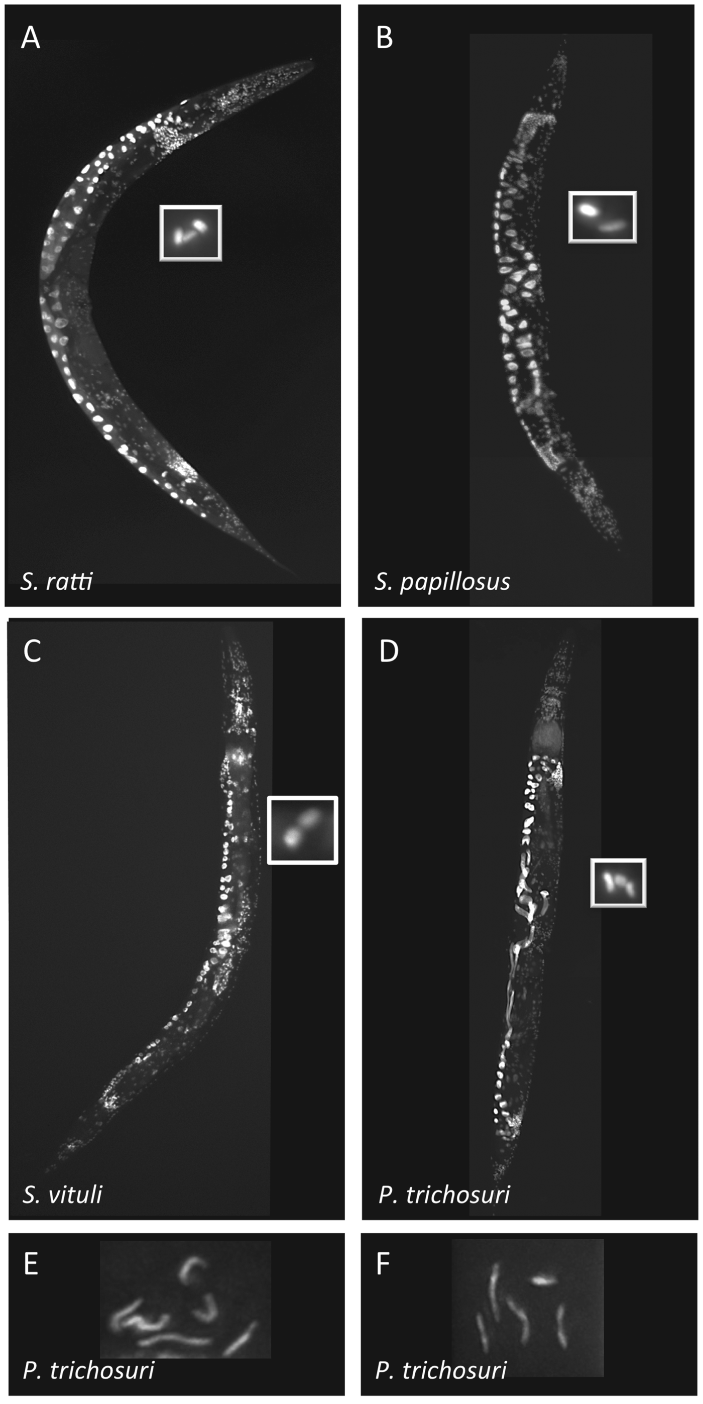
The 16 females mentioned above were stained with DAPI and meiotic chromosomes counted (Fig. 3C). In all cases the number of chromosomes was two bivalents (n = 2). We managed to recover from the microscope slide and determine the SSU sequence for five of the 16 females. Four of them were S. vituli, one was S. papillosus. From these results we conclude that the haploid number of chromosomes in S. vituli is two, as in S. papillosus.
Chromosome numbers in different species of Strongyloides and in Parastrongyloides trichosuri. A–D: DAPI stained free-living females of the species indicated are shown at low (10×) magnification. Boxed are meiotic (bivalent) chromosomes in oocytes at high (100×) magnification. E,F: mitotic chromosomes from P. trichosuri embryos are shown. Shown is one particular focal plane. Stacks of images were recorded and the chromosomes reconstructed in three dimensions. This allowed unambiguous counting of six chromosomes as in E or five chromosomes as in F.
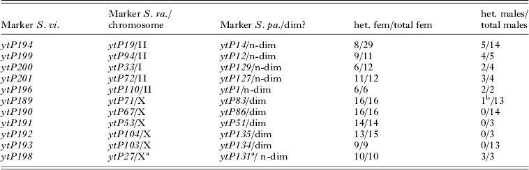
Next, we amplified 10 S. vituli genomic regions (Suppl. File 1 – in Online version only) that contained single nucleotide polymorphisms (SNPs) homologous to regions in S. ratti and S. papillosus for which the chromosomal locations were known: five SNP markers that are autosomal in S. ratti and do not undergo chromatin diminution in S. papillosus and five SNP markers that are homologous to markers that are located on the X chromosome in S. ratti and do undergo chromatin diminution in S. papillosus (cf. Nemetschke et al. Reference Nemetschke, Eberhardt, Hertzberg and Streit2010a, Reference Nemetschke, Eberhardt, Viney and Streitb). For the five S. ratti autosomal markers we found S. vituli males and females that were heterozygous for any two alleles, indicating that both sexes have two copies of these loci (Table 1), whereas for the five S. ratti X-linked markers we never observed heterozygous males, while heterozygous females were common (except for a single marker in a single animal; Table 1). Although for some of the markers the number of males genotyped is too small to reach a definitive conclusion for the markers concerned, taken together our results clearly indicate that S. vituli males have a hemizygous region, which is homologous to the region undergoing chromatin diminution in S. papillosus. Most probably S. vituli males undergo sex-specific chromatin diminution, as do the males of S. papillosus.
Strongyloides vituli males have a hemizygous region
Columns: Marker S. vi. – marker in S. vituli (see Suppl. File 1 – in Online version only); Marker S. ra./chromosome – number of the homologous marker in S. ratti/chromosome the marker is located on (Nemetschke et al. Reference Nemetschke, Eberhardt, Hertzberg and Streit2010a, Reference Nemetschke, Eberhardt, Viney and Streitb); Marker S. pa./dim? – number of the homologous marker in S. papillosus/indication if the marker undergoes sex-specific chromatin diminution (dim) or not (n-dim) (Nemetschke et al. Reference Nemetschke, Eberhardt, Hertzberg and Streit2010a), het. fem/total fem – number of heterozygous S. vituli females found/total number of S. vituli females analysed; het. males/total males – number of heterozygous S. vituli males found/total number of S. vituli males analysed, markers located in a hemizygous region can never be heterozygous.
a This is the only marker found by Nemetschke et al. (Reference Nemetschke, Eberhardt, Hertzberg and Streit2010a) to be X-chromosomal in S. ratti but not undergo chromatin diminution in S. papillosus.
b This single animal may indicate that this particular marker does not undergo diminution in all males or it may be a rare triploid animal, which we do observe occasionally (unpublished) or it may be a PCR artefact.
Parastrongyloides trichosuri has three pairs of chromosomes
First, we stained free-living females of P. trichosuri with DAPI and counted the meiotic chromosomes (Fig. 3D). As in S. ratti (Fig. 3A), we found three bivalents (n = 3) in P. trichosuri. In embryos derived from a continuously free-living culture we found two different karyotypes, one with six chromosomes (2n = 6, Fig. 3E) and one with five chromosomes (2n = 5, Fig. 3F). These observations showed that the haploid number of chromosomes in P. trichosuri is three (n = 3) and suggested that it employs chromosomal sex determination.
Parastrongyloides trichosuri employs XX/XO sex determination and the X chromosome is homologous to the S. ratti X chromosome
We isolated 19 sequence polymorphisms (Suppl. File 1 – in Online version only) in genes for which the chromosomal locations of putative S. ratti orthologues were known (Nemetschke et al. Reference Nemetschke, Eberhardt, Viney and Streit2010b) and analysed their inheritance in crosses between single free-living males and one or two females per cross (Table 2, Suppl. Table 1S – in Online version only and data not shown). For the six markers that are X-linked in S. ratti, the results of all P. trichosuri crosses were consistent with the female progeny receiving two copies, one from either parent, and the male progeny inheriting only one allele from the mother (sex-linked inheritance). All 13 markers whose S. ratti homologues are located on an autosome behaved as autosomal markers in P. trichosuri. These findings demonstrate that in the free-living generation P. trichosuri employs XX/XO sex determination and that the X chromosomes in these two species are homologous.
Autosomal and sex-linked loci in free-living Parastrongyloides trichosuri

As an example the results of one cross for one autosomal and one X-linked marker are given. For more crosses and markers see Suppl. Table 1S – in Online version only. Columns: Marker P. tr. – marker in P. trichosuri (see Suppl. File 1 – in Online version only); Marker S. ra./chromosome – number of the homologous marker in S. ratti/chromosome the marker is located on (Nemetschke et al. Reference Nemetschke, Eberhardt, Viney and Streit2010b).
a Listed is the result of the sequencing reaction at one of the polymorphic sites of the marker. If two bases are present the individual was heterozygous, if only one base is present the individual was homo- or hemizygous.
b Given are [number of progeny with this particular genotype] × [genotype].
Reproduction in the parasitic generation of P. trichosuri is sexual
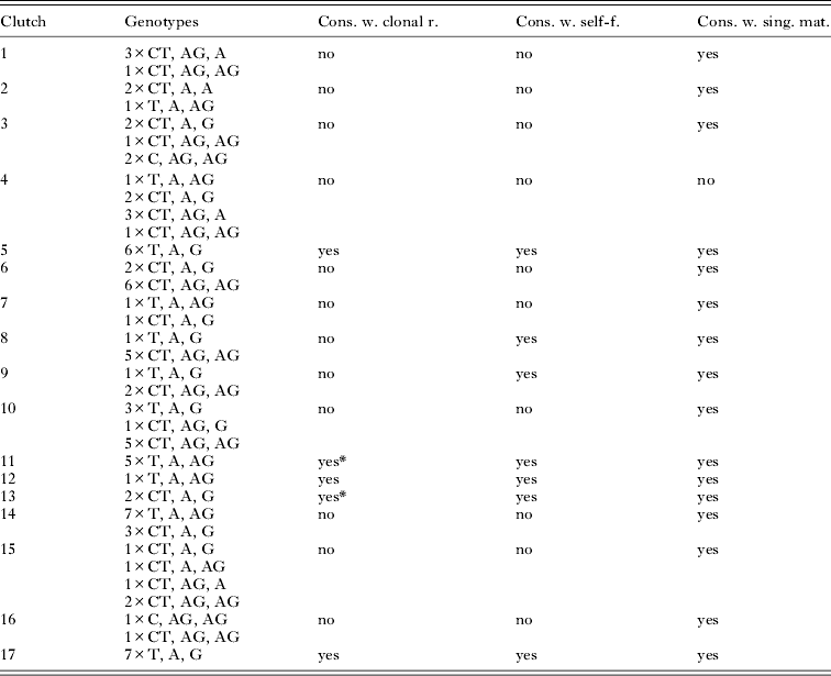
In Strongyloides sp. the parasitic generation reproduces by mitotic parthenogenesis (genetically demonstrated for S. ratti (Viney, Reference Viney1994; Nemetschke et al. Reference Nemetschke, Eberhardt, Viney and Streit2010b) and for S. papillosus (Nemetschke et al. Reference Nemetschke, Eberhardt, Hertzberg and Streit2010a)) and, at least in most species, parasitic males do not exist (Streit, Reference Streit2008). In contrast, parasitic males are present in P. trichosuri (Mackerras, Reference Mackerras1959). Grant et al. (Reference Grant, Stasiuk, Newton-Howes, Ralston, Bisset, Heath and Shoemaker2006b) have shown that two genetically different strains of P. trichosuri do hybridize, when introduced into the same host individual, demonstrating sexual reproduction. To confirm and further characterize sexual reproduction in parasitic P. trichosuri, we isolated individual offspring from 17 parasitic females and genotyped them at multiple X chromosomal and autosomal loci (Suppl. Table 2S – in Online version only). The results are fully consistent with normal sexual reproduction and not with clonal reproduction. One marker (ytP176) was particularly informative because it contains multiple polymorphic nucleotides and occurred in our sample in seven different alleles. In crosses of free-living animals this marker behaved in a clearly autosomal manner. In four different informative crosses (for one example see Suppl. Table 1S – in Online version only) two of the fathers and in total 13 of the male offspring were heterozygous. For this marker, the results of all 17 clutches are given in Table 3. In 10 clutches more than two alleles were present, excluding that the offspring originated entirely from self-fertilization. Further, the genotypes present in one of the clutches cannot be produced by mating of two individuals, indicating that the corresponding female had mated with multiple males.
Genotypes of the progeny of 17 parasitic P. trichosuri parasitic females at ytP1764
Genotypes: [number of progeny with this particular genotype] × [genotype]. ytP176 contains three polymorphic sites (positions 190, 296, 446). As ‘genotype’ the sequencing results at these three polymorphic sites are indicated, separated by ’,’. Two letters indicate that the individual was heterozygous at this position, one letter indicates that the individual was homozygous at this position. From 14 larvae and five males the ytP176 PCR products were cloned and individual clones sequenced. Seven different combinations of nucleotides at the three polymorphic sites (alleles) were found (190C 296A 446A, 190T 296A 446A, 190T 296A 446G, 190C 296G 446A, 190T 296G 446A, 190C 296A 446G, 190C 296G 446G). Some of the genotypes shown could be explained with multiple allele combinations. The conclusions are independent of this.
Cons. w. clonal r.: consistent with exclusively clonal reproduction: if reproduction is truly clonal all progeny are genetically identical with their mother and consequently with each other; exclusively clonal reproduction can be excluded if in the clutch more than one genotype exists. cons. w. self-f.: consistent with exclusively self-fertilization: if in the mother haploid eggs and sperm are formed and reproduction occurs through self-fertilization, at positions where the mother is heterozygous the offspring can be heterozygous or homozygous for either of the two maternal alleles but no genetic material absent from the mother can be present among the progeny. Exclusive self-fertilization can be excluded if more than two alleles exist within the clutch. cons. w. sing. mat.: consistent with single mating: The progeny can be explained under the assumption that the female mated with a single male. Mating with multiple males must be postulated if it is impossible to derive all genotypes present in the progeny from two parental genotypes.
* For these clutches other markers exclude clonal reproduction (see suppl. Table 2S – in Online version only).
The five parasitic males we found we genotyped at two putatively autosomal (ytP179 and ytP176) and at three putatively X-linked (ytP183, ytP184 and ytP185) markers. None of the males was heterozygous at any of the three X-linked markers while for the autosomal markers all five were heterozygous at ytP176 and two were heterozygous at ytP179. Although the numbers are rather small these results are also consistent with the hypothesis that the parasitic generation of P. trichosuri employs XX/XO sex determination.
DISCUSSION
The finding that P. trichosuri, like S. ratti, has three chromosomes (haploid, two autosomes and one X) and uses XX/XO sex determination strongly supports the hypothesis that this is the ancestral state for these two genera.
Although it seems likely that sex determination in P. trichosuri is genetic, we cannot prove that there is no environmental influence superimposed over the XX/XO system as in S. ratti.
The situation with two chromosomes (haploid) in S. papillosus and in S. vituli is most likely derived and arose through the fusion of the X chromosome with the autosome homologous to the S. ratti chromosome I (cf. Nemetschke et al. Reference Nemetschke, Eberhardt, Hertzberg and Streit2010a). In the older literature, based solely on cytological studies, different authors disagreed on the number of chromosomes in S. papillosus males. While Albertson et al. (Reference Albertson, Nwaorgu and Sulston1979) had observed sex-specific chromatin diminution (2n = 4 in females and 2n = 5 in males) as it was later confirmed genetically (Nemetschke et al. Reference Nemetschke, Eberhardt, Hertzberg and Streit2010a), Triantaphyllou and Moncol (Reference Triantaphyllou and Moncol1977) found no karyotypic differences between the sexes (2n = 4 in both sexes). Distinguishing species within the genus Strongyloides based solely on morphological criteria is notoriously difficult (Augustine, Reference Augustine1940; Schad, Reference Schad and Grove1989; Speare, Reference Speare and Grove1989). Therefore frequently the host is used to derive the species of Strongyloides. Since we now know that what at the time was considered S. papillosus are at least two different species, one possible explanation for the conflicting findings of Albertson et al. (Reference Albertson, Nwaorgu and Sulston1979) and Triantaphyllou and Moncol (Reference Triantaphyllou and Moncol1977) might be that in fact different species (e.g. S. papillosus and S. vituli) were studied. However, our genetic data presented here clearly demonstrate that S. vituli males have a hemizygous region and therefore differ genetically from females. We did not have access to early S. vituli embryos derived from parasitic mothers, the only stage where in S. papillosus the male karyotype of 2n = 5 is easily observable. Therefore we do not know if in S. vituli the region eliminated in males is flanked by retained regions, as in S. papillosus. If this region were at the end of a chromosome, chromatin diminution would result in a size reduction of one chromosome but not in an increase of the chromosome number and might therefore be more difficult to detect cytologically.
On the evolutionary path to Strongyloides spp., which in the parasitic generation have no males and reproduce clonally, one could imagine an intermediate state where males are still present as an evolutionary relict but no longer or not mandatorily participate in reproduction. Although we cannot technically exclude that females, under certain conditions, can also reproduce without males, either by self-fertilization or parthenogenesis, our data demonstrate that sexual reproduction with outcrossing is the predominant if not the only mode of reproduction in parasitic P. trichosuri. This finding is not surprising. Parasitic males appear to be always present in productive P. trichosuri infections (Mackerras, Reference Mackerras1959; Grant et al. Reference Grant, Stasiuk, Newton-Howes, Ralston, Bisset, Heath and Shoemaker2006b) and Grant et al. (Reference Grant, Stasiuk, Newton-Howes, Ralston, Bisset, Heath and Shoemaker2006b) had demonstrated that outcrossing does occur in the parasitic form of P. trichosuri.
Although presented here in the context of an evolutionary question, our results are also interesting in their own right. Parastrongyloides trichosuri with its ‘choice’ between parasitic or free-living lifestyle is a highly attractive study system. Knowing the modes of inheritance in both forms is an important prerequisite for future studies.
Taken together, our findings strongly argue that XX/XO sex determination with two pairs of autosomes is ancestral in the Strongyloididae. More generally, they illustrate the usefulness of Strongyloides/Parastrongyloides as a genetically traceable system to study how parasitism can evolve and what the developmental and genetic consequences of the transition from a free-living to a parasitic lifestyle might be.
ACKNOWLEDGEMENTS
We thank Dorothee Harbecke for technical assistance and Dr Stewart Bisset for assistance with brushtail possums.
FINANCIAL SUPPORT
This work was funded by the Max Planck Society (A. K., A. D., L. N., A. S.) and La Trobe University School of Molecular Sciences (W. G).
SUPPLEMENTARY MATERIAL
To view supplementary material for this article, please visit http://dx.doi.org/10.1017/S0031182013001315.