Introduction
Aspiration of a foreign body by a child is a critical emergency encountered in otolaryngology practice. It could be lethal if not diagnosed and managed promptly and correctly. Childhood mortality resulting from foreign body aspiration remains high, ranging up to 1.8 per cent.Reference Sinha, Memon, Gupta, Prajapati, Bhat and More1 Depending on the size, shape and character of the foreign body, the duration of aspiration, the patient's age, and the relative area of the airway occupied, the presence of a foreign body in the tracheobronchial tree may produce highly variable clinical symptoms and signs. Absence of a positive history of foreign body aspiration may prevent early diagnosis; studies have revealed a positive history of aspiration in only 18–76 per cent of patients.Reference Kaur, Sonkhya and Bapna2, Reference Schimpl and Weber3 Patients may be misdiagnosed as having asthma, pneumonia, bronchitis, bronchiectasis or tuberculosis, leading to a delay in correct management. Vegetable foreign bodies are more common in the airway; aspirated seeds and foodstuffs can be particularly dangerous because they can swell up to three to four times their original size.Reference Kohli and Verma4
Bronchoscopy is the ‘gold standard’ for the definitive diagnosis and management of an aspirated foreign body; however, it is an invasive and potentially dangerous procedure. Correct visualisation and precise instrumentation are essential to the success of this procedure. Repeated removal attempts are likely to result in mucosal trauma, subglottic oedema and tracheal laceration, leading to airway complications. Pneumothorax, respiratory distress and cardiac arrest are other known complications of this procedure. Therefore, a non-invasive technique which identifies the exact site, size and shape of the foreign body and the area of airway occupancy would greatly minimise surgical effort and subsequent morbidity and mortality risks. Virtual bronchoscopy is a non-invasive technique that provides three-dimensional (3D) views of the tracheobronchial tree. The volumetric imaging data acquired by multidetector computed tomography (CT) can be manipulated to obtain additional high-quality multiplanar and 3D reconstructions.
Materials and methods
This study was conducted over a period of four years from August 2006 to August 2010 in the Department of Otorhinolaryngology and Head and Neck Surgery, Mahatma Gandhi Memorial Medical College and Maharaj Yashwant Hospital, Indore, a tertiary referral hospital in India. Patients with a history of suspected foreign body aspiration either presented at the Otolaryngology Out-patient or Emergency Department or were referred from the Paediatric Department. The symptoms at clinical presentation and chest X-ray, multidetector CT virtual bronchoscopy and rigid bronchoscopy findings of patients who had aspirated a foreign body were reviewed from the medical records. Patients with a metallic or other non-vegetable foreign body were excluded from the analysis. Patients with multidetector CT virtual bronchoscopy findings showing features of a vegetable foreign body were included in the analysis. Rigid bronchoscopy findings of these patients were reviewed and compared with the virtual bronchoscopy findings. In all, 60 patients were identified: all were children ranging from 1 month to 8 years of age, with virtual bronchoscopy findings suggestive of a vegetable foreign body. All patients had undergone rigid bronchoscopy within 24 hours of multidetector CT virtual bronchoscopy. Written, informed consent for inclusion into the study was obtained from the parents or legal guardians of patients. Ethical approval was obtained from the institute's ethical review board.
Computed tomography and virtual bronchoscopy
Images of all patients were obtained using a multidetector CT scanner (SOMATOM Sensation 64, Siemens, Munich, Germany) using syngo® imaging software and the surface and volume rendering technique. Multidetector CT was performed from the level of the larynx to the dome of the diaphragm in the supine position at a 0° gantry angle. Scanning parameters were a 1 mm slice thickness for reconstruction, 0.5 mm gap in a spiral sequence; 0.5 mm pitch/rotation and 100 kVp (80–120 kVp) voltage. The tube current was selected according to body size (range 150–300 mA). Scanning time ranged from 4 to 6 seconds. Images were scanned in the axial plane at 1 mm intervals with a standard reconstruction algorithm but reformatted in the sagittal plane. Scanning was conducted with spontaneous breathing for all patients. No sedation or intravenous contrast medium was used.
Cross-sectional images were analysed using DICOM (‘Digital Imaging and Communications in Medicine’) compatible software. Navigation was possible up to the level of the segmental bronchi. All axial images were evaluated for the presence or absence of foreign bodies, using standard lung and soft tissue settings (window width 1400 Hounsfield units, centred at −500, and window width 400 Hounsfield units, centred at −40, respectively). Any additional parenchymal and mediastinal abnormalities were noted. Interactive navigation through the tracheobronchial tree was performed using a computer monitor and mouse. Virtual bronchoscopic images were generated in three stages as described by Sorantin and colleagues,Reference Sorantin, Geiger, Lindbichler, Eber and Schimpl5 and saved as digital files. The axial and coronal images and virtual bronchoscopic images were evaluated by an experienced radiologist.
Bronchoscopy
Bronchoscopy was performed using a Jackson rigid paediatric Karl Storz bronchoscope (Medical & Industrial Equipment, London, UK) under general anaesthesia. A rigid ventilating paediatric bronchoscope was initially inserted with the help of a Macintosh laryngoscope (Penlon, Abingdon, UK) under direct vision and was advanced past the vocal folds. The unaffected bronchus was examined first. The foreign body was then visualised on the affected side and extracted using grasping forceps. A check was performed to identify any retained fragments, any secretions were removed by suction and the severity of tissue reactions and oedema was examined. Post-operatively, the children were kept under observation in the hospital for 48 hours.
Results
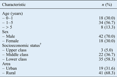
A total of 60 patients were included in the study group (42 boys and 18 girls), with an age range of 1 month to 8 years (mean age 2.01 years). Table I shows the demographic profile of these patients. Most patients (56.7 per cent) were aged one to five years. Vegetable foreign body aspiration predominantly occurred in males (male:female 2.3:1) and was more common in patients of low socioeconomic status (58.3 per cent) and rural origin (68.3 per cent).Reference Prasad6
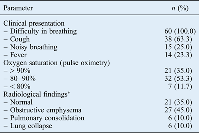
In our case series, 40 per cent of patients presented at the hospital within 24 hours of inhaling a foreign body, while 31.7 per cent presented after five or more days. Only 16.7 per cent of patients presented within 6 hours. The maximum time lag for presentation to hospital was 5 months in the case of a one-year-old boy with a foreign body (decomposed vegetable material) in the right main bronchus. There was a definite history of foreign body aspiration in 36 patients (60 per cent) and none in 24 (40 per cent). All patients had varying degrees of difficulty in breathing at presentation; a cough was the second most common presenting symptom (66.3 per cent), followed by noisy breathing (25 per cent) and fever (23.3 per cent). Oxygen saturation in room air was reduced to 80–90 per cent at presentation (as measured by pulse oximetry) in 32 patients (53.3 per cent) and to less than 80 per cent in 7 patients (11.7 per cent). Chest X-rays were normal in 21 patients (35 per cent). Obstructive emphysema was identified in 45 per cent of patients, pulmonary consolidation in 10 per cent and lung collapse in 10 per cent (Table II).
Clinical presentation, oxygen saturation at presentation and radiological findings

*Chest X-ray.
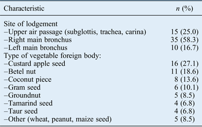
Virtual bronchoscopy identified the right main bronchus as the most common site of foreign body lodgement (58.3 per cent), followed by the left main bronchus (16.7 per cent) and upper air passage (25 per cent). In the present case series, the most common vegetable foreign body was custard apple seed (27.1 per cent), followed by betel nut (18.6 per cent; Table III).
Site of lodgement and type of vegetable foreign body
All foreign bodies were successfully removed by rigid bronchoscopy without complication, except for one case in which tracheostomy was needed to secure the airway post-operatively. Identical results (presence of a foreign body, site of lodgement, and size and shape) were identified by rigid bronchoscopy and multidetector CT virtual bronchoscopy in 59 patients (98.3 per cent). In a single case, a vegetable foreign body identified by virtual bronchoscopy was revealed by rigid bronchoscopy to be a thick mucus plug. Thus, the positive predictive value of virtual bronchoscopy was 98.3 per cent. Figure 1 shows a chest X-ray and coronal CT image of the chest and virtual bronchoscopy images for an eight-year-old boy with a vegetable foreign body (custard apple seed) in his right main bronchus. Figure 2 shows a coronal CT image of the chest and virtual bronchoscopy images of a six-year-old girl with a vegetable foreign body (coconut piece) in her left main bronchus.
Diagnostic images of an eight-year-old boy with a vegetable foreign body (custard apple seed) in the right main bronchus. (a) Chest radiography showing collapse of the right lung. (b) Coronal CT image revealing a vegetable foreign body in the right main bronchus, with the right lung showing signs of collapse. (c) Virtual bronchoscopy image showing a foreign body (arrow) in the right main bronchus. A custard apple seed was removed by bronchoscopy.
Diagnostic images of a six-year-old girl with a vegetable foreign body in the left main bronchus. (a) Coronal CT image showing a vegetable foreign body in the left main bronchus. (b) Virtual bronchoscopy image showing a foreign body (arrows) in the left main bronchus. A coconut piece was removed by bronchoscopy.
Discussion
Foreign body aspiration is an important cause of morbidity and mortality in childhood. It occurs more frequently in children aged between six months and three years.Reference Applegate, Dardinger, Lieber, Herts, Davros and Obuchowski7, Reference Metrangolo, Monetti, Meneghini, Zadra and Giusti8 Patients have varied presentations, from choking and fatal asphyxia to symptoms mimicking a chronic, recurrent chest infection. Many patients may not have a definite history of aspiration, leading to difficulties in diagnosis. In our case series, 40 per cent of patients did not have a history of foreign body aspiration. Vegetable foreign bodies are radiolucent; therefore, unlike metallic foreign bodies, they can only be diagnosed by secondary changes in chest radiography.Reference Zissin, Shapiro-Feinberg, Rozenman, Smorjik and Hertz9 Several studies have reported normal chest X-ray findings in 9–35 per cent of patients with a tracheobronchial foreign body.Reference Zissin, Shapiro-Feinberg, Rozenman, Smorjik and Hertz9–Reference Burton, Brick, Hall, Riggs and Houston12 In our case series, 35 per cent of children had a normal chest radiograph. An overlooked foreign body in the airway may cause considerable morbidity; it is therefore necessary to rule out suspected foreign body aspiration, even in the absence of radiographic changes. Rigid bronchoscopy is often performed for these reasons. However, although rigid bronchoscopy is a simple procedure, it is associated with serious complications in 6–8 per cent of children, even in skilful hands.Reference Zerella, Dimler, McGill and Pippus13, Reference Dunn, Wardrop, Lo and Cowan14
In contrast, CT virtual bronchoscopy is a non-invasive imaging technology that utilises a variety of software programs to create a 3D view of the tracheobronchial tree using spiral multidetector CT. Virtual bronchoscopy simulates an endoscopist's view of the internal surface of the airway using 3D surface rendering and volume rendering techniques: thus, the observer can interactively move through the airway.Reference Grenier, Beigelman-Aubry, Fetita, Preteux, Braener and Lenoir15 These techniques allow the accurate reproduction of major endoluminal abnormalities and provide an excellent correlation with fibre-optic bronchoscopy results regarding the location, severity and shape of airway narrowing.Reference McAdams, Palmer, Erasmus, Patz, Connolly and Goodman16 The most reliable CT virtual bronchoscopy finding for an aspirated foreign body is its identification within the lumen of the tracheobronchial tree. There may be associated secondary changes in the lung parenchyma such as hyperaeration, atelectasis, infiltration and bronchiectasis. Virtual bronchoscopy takes advantage of the natural contrast between the airway and surrounding tissues to determine the exact location, size, shape and type of foreign body. A vegetable foreign body that cannot be identified using a normal chest X-ray can be characterised by virtual bronchoscopy. Therefore, multidetector CT virtual bronchoscopy may guide the planning of tracheobronchial foreign body removal. For instance, the clinician can simulate the procedure before the actual bronchoscopy takes place. This technique may also help the surgeon to select the most appropriate type of foreign body forceps for extraction. This technique could therefore reduce operative time and subsequent morbidity and mortality. In our case series, the virtual bronchoscopic description of location, size and type of foreign body was identical to the rigid bronchoscopic findings in all cases except one. Secondly, negative virtual bronchoscopy findings could avoid the need for rigid bronchoscopy, and thus avoid complications related to rigid bronchoscopy. Thirdly, unlike rigid or flexible bronchoscopy, virtual bronchoscopy can visualise regions beyond stenosis.Reference Sodhi, Saxena, Singh, Rao and Khandelwal17 Multidetector CT technology can also enable the visualisation of segmental and sub-segmental bronchi and thus possible pathologies which a rigid bronchoscope may not be able to reach.
According to Bhat et al., many poorer patients in developing countries are unaware of the symptoms of foreign body aspiration and do not give a correct history.Reference Bhat, Hegde, Nagalotimath and Patil18 Hence, many cases present late with symptoms related to chest infection. These patients often undergo many days of medical treatment in paediatric wards for unresolved chest infections before foreign body aspiration is suspected and a request for bronchoscopy is made. In many of these cases, chest radiography is not pathognomonic for foreign body aspiration. In such cases, virtual bronchoscopy would be particularly useful to confirm the presence of a foreign body. In addition, the risks associated with rigid bronchoscopy and general anaesthesia are higher in these patients because pulmonary function has already been compromised by infection.
Multidetector CT virtual bronchoscopy is a highly sensitive and specific method of vegetable foreign body detection. Kosucu et al. concluded that multidetector CT virtual bronchoscopy may be useful for identifying the exact location of a foreign body before bronchoscopy and for ruling out foreign body aspiration in patients with a low level of suspicion and normal or non-specific chest radiography findings.Reference Kosucu, Ahmetoglu, Koramaz, Orhan, Ozdemir and Dinc19 They found the sensitivity and specificity of multidetector CT virtual bronchoscopy to be 100 per cent. Haliloglu et al. reported similar findings.Reference Haliloglu, Ciftci, Oto, Gumus, Tanyel and Senocak20 Kocaoglu et al. found multidetector CT to be 88.9 per cent sensitive, with a specificity of 91.7 per cent and accuracy of 90.5 per cent.Reference Kocaoglu, Bulakbasi, Soylu, Demirbag, Tayfun and Somuncu21 According to Bhat et al., where there is a positive clinical diagnosis and negative chest radiography, CT virtual bronchoscopy should be considered in all cases of tracheobronchial foreign body aspiration to avoid needless rigid bronchoscopy.Reference Bhat, Hegde, Nagalotimath and Patil18 Computed tomography virtual bronchoscopy is particularly useful for screening cases of occult foreign body aspiration because it has high levels of sensitivity (92.3 per cent), specificity (85.7 per cent) and validity (90 per cent). In our case series, virtual bronchoscopy identified all but one cases of vegetable foreign body aspiration; thus, it had a positive predictive value of 98.3 per cent. In a single case, a vegetable foreign body identified by virtual bronchoscopy was revealed by rigid bronchoscopy to be a thick mucus plug. This type of artefact could be minimised in the hands of an experienced radiologist. Another disadvantage of virtual bronchoscopy compared with rigid bronchoscopy is that it cannot reveal the colour, morphology and vascularity of the mucosa; it is solely a diagnostic tool.Reference Summers, Shaw and Shelhamer22 Multidetector CT also exposes patients to radiation.
In the present study, vegetable foreign body aspiration was more common in boys than in girls (2.3:1). Other authors have reported a similar male-to-female incidence: Mahyar and TarlanReference Mahyar and Tarlan23 (1.8:1) and Kamaljit et al. Reference Kaur, Sonkhya and Bapna2 (1.5:1). Vegetable foreign body aspiration was more common in the one to five years age group in our study. This could be caused by the habit of children of this age of putting everything in their mouths. In addition, the eruption of their molars and consequent gum tenderness prevents these patients from chewing hard food, and so they try to swallow it whole. Shouting, crying and playing during eating and a lack of parental supervision also contributes to the high incidence of foreign body aspiration. In our study, the most common type of vegetable foreign body was a custard apple seed, which is the most common fruit eaten in central India. However, the type of vegetable foreign body mainly depends on dietary and eating habits. Owing to their religious beliefs, parents often provide a small piece of coconut as a ‘Prasad’ (or offering from God) to their children and this can be accidentally aspirated into the airway. We found the right main bronchus to be the most common site of foreign body lodgement compared with the left (3.5:1). Kaur et al. reported the ratio of foreign body aspiration into the right and left bronchi to be 1.55:1.Reference Kaur, Sonkhya and Bapna2 Such bias may occur because the right main bronchus is shorter and wider than the left bronchus, is straight and forms a direct continuation of the trachea. Vegetable foreign body aspiration is also more common in lower socioeconomic groups (58.33 per cent). This may be attributed to a lack of awareness, lower social or education levels, and poor living conditions.
• Multidetector computed tomography (CT) virtual bronchoscopy can identify radiolucent vegetable foreign bodies in the tracheobronchial tree
• It is sensitive and specific
• It can provide a pre-operative road map for rigid bronchoscopy
• Patients with low index of suspicion of a foreign body or chronic unexplained respiratory symptoms should undergo multidetector CT virtual bronchoscopy to rule out a vegetable foreign body
With the widespread availability of helical multidetector CT scan machines in which scanning can be performed in as little as 4–6 seconds, virtual bronchoscopy relies on the radiologist generating 3D images using appropriate software. Virtual bronchoscopy provides a clear endoluminal view of airway pathology which cannot be found in conventional CT images. It may also provide a useful pre-operative road map for rigid bronchoscopy. Where there is a low index of suspicion of an airway foreign body or chronic unexplained respiratory symptoms, patients should undergo multidetector CT virtual bronchoscopy to rule out the presence of a vegetable foreign body in the tracheobronchial tree and to avoid unnecessary general anaesthesia and invasive rigid bronchoscopy.