INTRODUCTION
As already emphasized for other invertebrates, the distribution of calcareous sponges seems to be directly linked with the presence of specialized taxonomists in this group (e.g. Wörheide & Hooper, Reference Wörheide and Hooper2003; Klautau & Valentine, Reference Klautau and Valentine2003; Klautau et al., Reference Klautau, Monteiro and Borojevic2004; Rapp, Reference Rapp2006; Lanna et al., Reference Lanna, Rossi, Cavalcanti, Hajdu and Klautau2007). In Italy, despite the presence of a well-known group of sponge taxonomists, there is a low number of papers focused on this sponge class, thus, a low number of such records are reported (40 out of a total of 509 sponge species) (Pansini & Longo, Reference Pansini and Longo2008). Moreover, the last paper containing the description of a new calcareous sponge species refers to Sarà & Gaino (Reference Sarà and Gaino1971).
With the aim to improve the knowledge of this little studied group along the Italian coast, in this paper is described a new Leucosolenia (Calcarea: Calcaronea) species collected from a pond included in the ‘Laghetti di Marinello’ Natural Reserve (southern Tyrrhenian Sea) (38°08′31.30″N 15°03′10.61″E).
The genus Leucosolenia, erected by Bowerbank, 1864 comprises ‘Leucosoleniidae in which the skeleton can consist of diactines, triactines and/or tetractines, without a reinforced external layer on the tubes’ (Borojevic et al., Reference Borojevic, Boury-Esnault and Vacelet2000; Reference Borojevic, Boury-Esnault, Manuel, Vacelet, Hooper and van Soest2002). It comprises less than 50 living species distributed worldwide (van Soest et al., Reference van Soest, Boury-Esnault, Hooper, Rützler, de Voogd, Alvarez, Hajdu, Pisera, Vacelet, Manconi, Schoenberg, Janussen, Tabachnick and Klautau2008). All need thorough revision to resolve the cosmopolitan cryptic species complexes (Borojevic et al., Reference Borojevic, Boury-Esnault, Manuel, Vacelet, Hooper and van Soest2002).
Five species of Leucosolenia, L. botryoides (Ellis & Solander, 1786), L. complicata (Montagu, 1818), L. cyathus Haeckel, 1870, L. parthenopea Sarà, Reference Sarà1953 and L. variabilis Haeckel, 1870, have been reported for the Mediterranean Sea (Breitfuss, Reference Breitfuss1935; Burton, Reference Burton1963; van Soest et al., Reference van Soest, Boury-Esnault, Hooper, Rützler, de Voogd, Alvarez, Hajdu, Pisera, Vacelet, Manconi, Schoenberg, Janussen, Tabachnick and Klautau2008). Four species (L. botryoides, L. complicata, L. parthenopea and L. variabilis), are reported from Italian seas (Ligurian Sea, Southern Tyrrhenian Sea, Ionian Sea and northern Adriatic Sea) (Sarà, Reference Sarà1953, Reference Sarà1958, Reference Sarà1960, Reference Sarà1964a,b; Sarà & Siribelli, Reference Sarà and Siribelli1962; Rützler, Reference Rützler1967; Scalera-Liaci et al., Reference Scalera-Liaci, Sciscioli and Fiordiponti1976; Pronzato & Belloni, Reference Pronzato and Belloni1980–1981; Pansini & Pronzato, Reference Pansini and Pronzato1981, Reference Pansini and Pronzato1982; Reference Pansini and Pronzato1985; Longo et al., Reference Longo, Scalera Liaci and Corriero2003) three of which signalled in the southern Tyrrhenian Sea (L. botryoides, L. parthenopea and L. variabilis) (Sarà, Reference Sarà1953, Reference Sarà1958, Reference Sarà1960, Reference Sarà1964b; Pronzato & Belloni, 1980–1981; Pansini & Pronzato, Reference Pansini and Pronzato1982; Reference Pansini and Pronzato1985).
MATERIALS AND METHODS
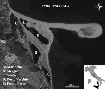
The specimens were collected by SCUBA divers in 2004 from the Porto Vecchio pond of the Marinello Marine Reserve located along the Tyrrhenian coast of Sicily (southern Tyrrhenian Sea, Messina, Italy) (Figure 1).
Map of the Tindari ponds (Messina, Italy).

The specimens were fixed with 5% formaldehyde in seawater and preserved in 70% ethanol.
Spicule slides were prepared according to the method of Manconi & Pronzato (Reference Manconi and Pronzato2000), substituting the mounting medium with DPX Fluka. Scanning electron microscope (SEM) observations were made on stubs of dissociated spicule preparations. For skeletal study, sponge fragments were dehydrated in ethanol, treated with pure acetone and embedded in Araldite® TAAB. The skeletal sections were mounted with the same resin and observed with an optical microscope.
Measurements of at least 50 spicules of each type were taken using an optical microscope with a micrometric eyepiece. The length from the tip to the base and the thickness at the base of each actine were measured. Numbers are referred to the range of measurement variations of each spicule type, with the mean values and standard deviations in parentheses.
Photographs were taken with a Nikon Eclipse 80i optical microscope equipped with a digital camera and with a SEM belonging to the Animal Health and Well-being Department, Veterinary Medicine Faculty, Bari University.
In order to evaluate differences between the length of the unpaired and paired actines of each type of triactine, analysis of variance (one-way ANOVA and Tukey's post hoc tests, P = 0.05) was performed by using SPSS software v. 16.0 for Windows.
The type material and slide preparations are deposited at the Zoology Museum of Bari University (MUZAC).
TYPE MATERIAL
Holotype: MUZAC 1244—Porto Vecchio pond, Marinello Marine Reserve, Messina, northern Sicily, southern Tyrrhenian Sea, collected by G. Corriero & C. Longo, 19 September 2004, 3 m depth.
Paratype: MUZAC 1245—Porto Vecchio pond, Marinello Marine Reserve, Messina, northern Sicily, southern Tyrrhenian Sea, collected by G. Corriero & C. Longo, 19 September 2004, 3 m depth.
TYPE LOCALITY
Porto Vecchio pond, Marinello Marine Reserve, Messina, Italy.
DESCRIPTION
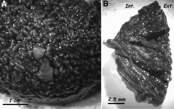
The holotype is spherical, with a diameter of 5 cm, and is formed by a dense and complex reticulation of ascon tubes, from which arise numerous tubes ending at the top with oscules of 0.5–1.5 mm in diameter (Figure 2). The surface is minutely hispid and the consistency is soft and fragile. The colour in life and in alcohol ranges from white-cream to light-brown. The small tubes may grow together with red algae (such as Polisiphonia sp. and Laurencia sp.) or to form no sessile sub-spherical formations which are scattered over the soft bottom. The sponge body includes a large amount of silt and foreign materials.
Holotype of Leucosolenia microspinata sp. nov. (A) Surface view; (B) a section showing the internal organization.

SKELETAL ARRANGEMENT
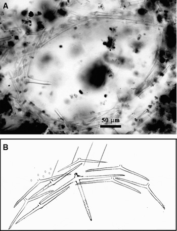
The skeleton consists of diactines, sagittal triactines of four different types and rare peculiar tetractines. The diactines are peripherally arranged, tangentially and longitudinally the larger ones and perpendicularly the smaller ones, along the external region of the tubes giving the hispid aspect of the surface. Triactines, usually with paired actines thicker than unpaired ones and sometimes deformed, are irregularly arranged in the sponge body. The tetractines have the apical actine projected towards the atrium. This actine has very thin spines at the distal end (Figure 3 A, B).
Leucosolenia microspinata sp. nov.: transversal section (A) and schematic drawing (not in scale) of the skeletal architecture (B).

SPICULES
Diactines
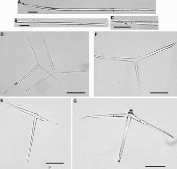
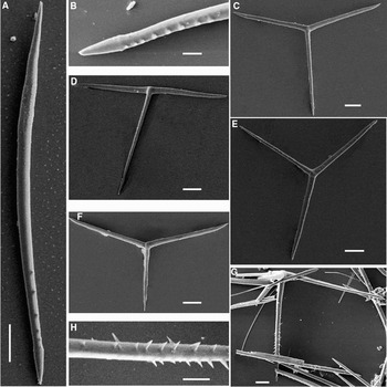
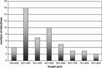
The most abundant diactines are slightly curved, often with one end slightly lanceolated and the adjacent shaft gently serrated only on the concave side. The other end is asymmetrical and its proximal shaft smooth. They have a wide size-range not separable into classes: 84–890 × 6–9 (338±167.5 × 6.9±1.4) µm (some of the longer ones are broken) (Figures 4A, 5A & B).
Spicules of Leucosolenia microspinata sp. nov. (A) Diactine slightly curved; (B) diactine straight; (C) detail of its finely spined end; (D) triactine with unpaired actine longer than paired ones and triactine with asymmetrical paired actines; (E) triactine ‘T’ shaped; (F) triactine with unpaired actines shorter than paired ones; (G) tetractine with finely spined apical actine. Scale bar: 50 µm.

Rare straight diactines, with one end fusiform or sometimes slightly lanceolated and the other extremity finely spined, are also present. They measure 216–468 × 9 (299.5±88.6) µm (Figure 4B & C).
Triactines
Four types, with actines straight or undulated and paired actines usually thicker than unpaired ones. Their ends are generally fusiform or asymmetrical (Figures 4 & 5). The paired actines are slightly curved near the tip. Some deformations are present.
SEM spicules of Leucosolenia microspinata sp. nov. (A) Serrated diactine; (B) detail of its serrated end; (C) triactine with unpaired actine longer than paired ones; (D) triactine ‘T’ shaped; (E) triactine with asymmetrical paired actines; (F) triactine with unpaired actines shorter than paired ones; (G) tetractine with apical actine finely spiny; (H) detail of its spines. Scale bar: 25 µm A, C, D, E, F, G; 10 µm B, H.

Type 1 sagittal triactine with unpaired actine longer than paired ones (Figures 4D & 5C):
Unpaired actine: 84–216 × 6 (130±20.8) µm; paired actines 60–132 × 6–9 (95±14.9 × 8.5±1.1) µm.
Type 2 ‘T’ shaped triactine (Figures 4E & 5D): unpaired actine 60–156 × 6 (105.8±25.6) µm;
Paired actines 48–108 × 9 (79.9±16.9) µm.
Type 3 sagittal triactine with asymmetrical paired actines (Figures 4D & 5E):
Longer paired actines 60–156 × 6-9 (99.1±21.9 × 7.3±1.5) µm; shorter paired actines 36–108 × 6–9 (59.8 ±20.1 × 7.5±1.5) µm; unpaired actine 84–168 × 6–9 (115.7±20.2 × 7.6±1.5) µm.
Type 4 sagittal triactine with unpaired actines shorter than paired ones (Figures 4F & 5F):
Unpaired actine 36–108 × 6–9 (55.9±15.6 × 6.6±1.2) µm; paired actines 60–132 × 6–9 (99.4±18.3 × 8.16±1.4) µm.
Tetractines
Rare, with straight and fusiform actines. The apical actine is finely spined at the distal end (Figures 4G, 5G & H): finely spined apical actine 60–130 × 6–9 (93±18.8 × 6.6±1.2) µm, triangular spines (4 µm) distally pointed; unpaired actine (often broken) 60–156 × 6–9 (108±29.4 × 8.4±1.3) µm.
Paired actines 96–144 × 9 (119.5±9.7) µm.
The length of the unpaired actines shows significant differences between the four types of triactines (F 3,196 = 163.7; P = 0.000).
The same results were obtained comparing the length of the paired actines among the four types of triactines (F4,245 = 50.6; P = 0.000). This significant difference is mainly due to the contribution of the triactines of types 2 and 4 (‘T’ shaped and sagittal with short unpaired actine, respectively) (Tukey's test, P = 0.001).
ECOLOGY
The species inhabits the Porto Vecchio pond, included in the ‘Laghetti di Marinello’ Natural Reserve established in 1998. This latter is a small littoral area located along the Tyrrhenian coast of Sicily, at the base of the Tindari Cliff (rising to 290 m above sea level) (Messina, Italy). This area is characterized by a marked geomorphological evolution able to cause rapid changes in a few years in the number, shape and dimensions of the ponds (Giacobbe & Leonardi, Reference Giacobbe, Leonardi and Miarelli1986; Leonardi et al., Reference Leonardi, Azzaro, Azzaro, Decembrini and Monticelli2000; Reference Leonardi, Azzaro, Giacobbe, Grasso and Muscolino2006). The Porto Vecchio pond is characterized by a maximum water depth of 3.40 m (mean value 1.50 m), volume less than 30.000 m3, and a salinity ranging between 29.39 and 39.26‰ during the year (Crisafi et al., Reference Crisafi, Giacobbe and Leonardi1981; Caruso et al., Reference Caruso, Monticelli, Azzaro, Azzaro, Decembrini, La Ferla, Leonardi and Zaccone2005). According to Crisafi et al. (Reference Crisafi, Giacobbe and Leonardi1981) its origin goes back to 1978. This pond is normally oligotrophic with the exception of seasonal nutrient increases (Azzaro et al., Reference Azzaro, Decembrini and Leonardi2001; Caruso et al., Reference Caruso, Monticelli, Azzaro, Azzaro, Decembrini, La Ferla, Leonardi and Zaccone2005). The bottom consists of incoherent sediment covered by a dense seagrass meadow of Cymodocea nodosa with patches occupied by a tanatocoenosis of Mytilaster sp. and Cerastoderma sp.
ETYMOLOGY
The presence of microspines on the tetractines and diactines suggested the name.
DISCUSSION
Leucosolenia microspinata is characterized by the presence of microspined tetractines and spined or serrated diactines. Among the less than fifty species of Leucosolenia distributed worldwide and considered valid by the world Porifera database (van Soest et al., Reference van Soest, Boury-Esnault, Hooper, Rützler, de Voogd, Alvarez, Hajdu, Pisera, Vacelet, Manconi, Schoenberg, Janussen, Tabachnick and Klautau2008), five species have been recorded in the Mediterranean Sea: L. botryoides, L. complicata, L. cyathus, L. parthenopea and L. variabilis. Four of them (L. botryoides, L. complicata, L. parthenopea and L. variabilis) could be considered similar to L. microspinata.
Leucosolenia microspinata shows a quite peculiar shape and spiculation in comparison with the Mediterranean Leucosolenia species. Indeed, at a first external analysis L. botryoides, L. complicata and L. variabilis can be easily recognizable thanks to their characteristic shape representing the main diagnostic character (van Soest et al., Reference van Soest, de Kluijver, van Bragt, Faasse, Nijland, Beglinger, de Weerdt and de Voogd2007). The shape of L. microspinata is not similar to other Mediterranean Leucosolenia morphotypes, which are generally arborescent with asconoid tubes creeping or rarely branched (see pl. 18 Haeckel, Reference Haeckel1872; Borojevic et al., Reference Borojevic, Boury-Esnault and Vacelet2000).
The spiculation pattern of L. microspinata represents a further element supporting its description as a new species. Leucosolenia botryoides differs from L. microspinata in the shape of the specimens and in the size and shape of the spicules. In fact, the former shows a characteristic shape with short ascon tubes arranged in grapes. Moreover, it is provided with spicules generally shorter but thicker than those of L. microspinata (Table 1). In particular, the triactines of L. botryoides have straight unpaired actines which are usually as long as, or sometimes shorter than, the paired ones (Table 1) (Arndt, Reference Arndt, Grimpe and Wagler1935; van Soest et al., Reference van Soest, Picton and Morrow2000). Leucosolenia complicata is characterized by bushy or arborescent specimens with tubes covered with short diverticula and smaller spicules than those of L. microspinata. Moreover, it has different types of diactines separable into two size-classes (Arndt, Reference Arndt, Grimpe and Wagler1935; van Soest et al., Reference van Soest, Picton and Morrow2000) (Table 1), while for the abundant diactines of L. microspinata this is not feasible (Figure 6). Leucosolenia parthenopea, whose Italian records were previously included in L. botryoides (Pansini & Longo, Reference Pansini and Longo2008), differs from L. microspinata in that the thickness of the spicules is always larger in the former. In particular, the diactines of L. parthenopea are similar in length to those of L. microspinata but their thickness may reach 29 µm (Table 1). Moreover, L. parthenopea has only one type of diactine (Sarà, Reference Sarà1953). Also the shape of the specimens is quite different (Sarà, Reference Sarà1953). Indeed, differently from L. microspinata, L. parthenopea shows two different shapes: one consisting of large and flattened tubes overlapping in layers, anastomosed and usually modified into lacunae determining a compact shape; the second has an encrusting form with marginal oscular tubes (Sarà, Reference Sarà1953). Leucosolenia microspinata can be easily distinguishable from L. variabilis, considered in the past as a variety of L. botryoides (Topsent, Reference Topsent1936; Sarà, Reference Sarà1953), because the latter has long undivided tubes with few diverticula; moreover, shorter and different types of diactines than those of L. microspinata (Table 1). Indeed, according to van Soest et al. (Reference van Soest, de Kluijver, van Bragt, Faasse, Nijland, Beglinger, de Weerdt and de Voogd2007), in L. variabilis there are three different types of diactines: large with distinct lance-shaped endings at one or both ends (190–376 by 3.5–6 µm), intermediate indistinct and overlapping with the large one (75–211 by 3–6 µm), small spined (30–126 by 0.5–2 µm) (Table 1).
Leucosolenia microspinata sp. nov.: size-class distribution of diactines.

Spicule dimensions of Leucosolenia botryoides, L. complicata, L. parthenopea, L. variabilis and L. microspinata sp. nov. Measurements are reported in μm.

Among the 40 calcareous sponge species reported along the Italian coast (Pansini & Longo, Reference Pansini and Longo2008) 32.5% are Mediterranean endemics. Considering the high number of Demospongiae endemics in the Mediterranean Sea (48.2%) (Pansini & Longo, Reference Pansini and Longo2003) it is presumable to hypothesize an underestimation of this sponge group.
This record represents a contribution to the knowledge of the calcareous sponge supporting the theory that the Mediterranean Sea could be considered a hotspot of sponge biodiversity (Pronzato, Reference Pronzato2003). According to Lanna et al. (Reference Lanna, Rossi, Cavalcanti, Hajdu and Klautau2007) the low presence of specialists, seems to be one of the probable causes of the apparent low diversity of this sponge group.
KEY TO THE MEDITERRANEAN SPECIES OF LEUCOSOLENIA
1. Arborescent or bushy… … … … … … … … … … …2
– Massive sub-spherical… … … … … … … … … …4
– Incrustant; from one to several overlapping layers made of numerous flattened tubes; one type of diactine with sub-terminal narrowing close to the lanceolate end… … … … … … … … … … …L. parthenopea
2. Divided ascon tubes… … … … … … … … … … …3
– Undivided ascon tubes, relatively long; small spiny diactines, together with two size-categories of diactines with one or both lanceolate endings ………L. variabilis
3. Tubes densely provided with diverticula; two types of diactines: larger with and smaller without lanceolate endings… … … … … … … … … … … L. complicata
– Tubes terminally swollen and arranged in grapes; diactines with lanceolate endings… … … …L. botryoides
4. Non-stipitate; two types of diactines: slightly curved with one slightly lanceolate and serrated end, straight with one spiny end .… … … … … … … … … .L. microspinata
– Stipitate; one size of diactines … … … … . L. cyathus
ACKNOWLEDGEMENTS
I am indebted to Professor Salvatore Desantis of the Animal Health and Well-being Department, Faculty of Veterinary Medicine of Bari University, for his support with SEM and to Dr Mariella Mercurio for her help with optical microscope photographs. I am grateful to the referees for their valuable suggestions, which considerably improved the content of the paper. This work was financially supported by Italian funds of Ministero dell'Economia e delle Finanze, Ministero dell'Istruzione, Ministero dell'Università e della Ricerca, Ministero dell'Ambiente e della Tutela del Territorio and Ministero delle Politiche Agricole e Forestali (Program MICENA).