Introduction
Nanotechnology has demonstrated great potential to improve cancer treatments by affecting the ways in which cancer is diagnosed, prevented, and treated in the clinic. Reference Peer, Karp, Hong, Farokhzad, Margalit and Langer1–Reference Chauhan and Jain4 In particular, targeted drug delivery using nanoscale carriers holds great promise to substantially improve therapeutic indices (the ratio between the toxic dose and the therapeutic dose of a drug) of incorporated drug molecules; in fact, a number of them are currently under advanced-stage clinical trials or are being used in clinical settings. Reference van der Meel, Vehmeijer, Kok, Storm and van Gaal5,Reference Sengupta and Kulkarni6 Although formidable progress has been achieved in this field, problems related to variable pharmacokinetics, nanoparticle (NP) instability, insufficiently selective tumor accumulation, and premature drug release continue to plague their fast clinical translation. Reference van der Meel, Vehmeijer, Kok, Storm and van Gaal5,Reference Davis7,Reference Hubbell and Langer8 One of the major drawbacks of most, if not all, NP systems can be linked to their inability to maintain their designed biological functions in various physiological and pathological conditions. Reference Kim, Shi, Kim, Park and Cheng9,Reference Chrastina, Massey and Schnitzer10 Therefore, to design an effective NP system, it is necessary to develop a comprehensive understanding of the interactions that occur between the nanomaterials and biological systems (i.e., nano-bio interactions) at each stage of the drug delivery process.
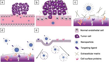
Figure 1 depicts the important in vivo barriers that NPs encounter during their journey from injection to reaching targets in the human body. To achieve a successful therapeutic result, NPs must survive blood circulation, accumulate at the tumor site, diffuse through the extracellular matrix (ECM), interact with the cell membrane, internalize into the target cell, and reach the subcellular target. At each stage, the surrounding microenvironment plays a significant, yet differing role in determining the fate of the NP. Reference Nel, Mädler, Velegol, Xia, Hoek, Somasundaran, Klaessig, Castranova and Thompson11–Reference Doane and Burda13 The considerable differences between the important interactions at each step necessitate the development of NPs that are optimally engineered to function in various physiological settings.
Various barriers associated with nanoparticle (NP) delivery to tumor cells. To effectively achieve a therapeutic response, the NP must (a) survive blood circulation, (b) accumulate at the tumor site by extravasation through leaky tumor vasculature, (c) diffuse through the extracellular matrix to reach the target cell surface, (d) interact with the target cell surface, preferably via specific ligand-receptor interaction, and (e) internalize and bypass the degradation pathways associated with endocytosis to reach the subcellular target.

This article summarizes recent advances in the understanding of nano-bio interactions occurring at those multiple stages by dividing it into sections that describe interactions of NPs with the tumor microenvironment, membranes and receptors, and intracellular compartments. Within each section, we discuss the chemical and structural compositions of each biological environment, challenges associated with NP delivery to that environment, and various approaches implemented to overcome the challenges to enable the formation of desired nano-bio interactions. It is our aim to provide a succinct overview of these important interactions and suggest key design rationales for NPs to be considered to help design better performing nanomaterials for the ultimate use as anti-cancer therapies.
Nanoparticle-tumor microenvironment interactions
Tumor microenvironments
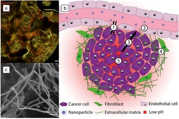
The microenvironment of solid tumors is highly complex and heterogeneous, often characterized by vascular abnormalities and a unique pathology. Unlike healthy tissue, tumor vasculature is characterized by asymmetric blood vessel distributions, dilated vascular structures, and high levels of disorganization ( Figure 2 a). Reference Jain14,Reference Vakoc, Lanning, Tyrrell, Padera, Bartlett, Stylianopoulos, Munn, Tearney, Fukumura, Jain and Bouma149 These vascular abnormalities, in combination with vessel compression from rapid tumor growth, can affect blood flow and impair tumor perfusion. Reference Kamoun, Chae, Lacorre, Tyrrell, Mitre, Gillissen, Fukumura, Jain and Munn15,Reference Padera, Stoll, Tooredman, Capen, Tomaso and Jain16 To maintain the rapid growth of the tumor, cancer cells continuously induce the formation of new vasculature through the process of angiogenesis. Reference Kerbel17 Angiogenesis in tumors is typically dysregulated, or physiologically impaired, due to the overabundance of angiogenesis activators, including vascular endothelial growth factor (VEGF), basic fibroblast growth factor (bFGF), and tumor necrosis factor alpha (TNF-α) that result in the formation of defective endothelial gap junctions (200–800 nm in general). Reference Torchilin18 This causes many solid tumors to exhibit leaky vasculature and promotes metabolic physiological changes. Reference Hobbs, Monsky, Yuan, Roberts, Griffith, Torchilin and Jain19,Reference Hashizume, Baluk, Morikawa, McLean, Thurston, Roberge, Jain and McDonald20
Characteristics of the tumor microenvironment. (a) Optical frequency-domain image of chaotic vasculature of the tumor environment. The color encodes depth: yellow (superficial) to red (deep) (scale bar: 500 μm). Reprinted with permission from Reference 149. © 2009 Macmillan Publishers Ltd. (b) Various interactions occurring within the tumor microenvironment: (1) diffusion and convection through the blood vessel; (2) interactions with fenestrations (perforations) and leaky endothelial connections; (3) high interstitial pressure, producing unfavorable mass transport away from the tumor; (4) fibroblasts and the secreted extracellular matrix (ECM) protein network; and (5) low core extracellular pH, increasing outward toward the periphery. (c) Scanning electron microscope image of a HN12 tumor ECM showing a dense collagen network (scale bar: 10 μm). Reproduced with permission from IOP Publishing. All rights reserved. Reference Anna, Gary, Stella and Yeudall51

Tumors exhibit unique pH, nutrient, oxygen, and redox gradients that differ dramatically from normal tissues due to abnormal physiology and metabolism, as shown in Figure 2b. The pH values at the core of the tumor are typically lower compared to its periphery mostly due to an insufficient supply of nutrients and oxygen to the fast-expanding tumor cells. Metabolic dysregulation, including up-regulated glycolysis, increased production of lactic acid, and hydrolysis of adenosine triphosphate (ATP) under nutrient deficient/hypoxic conditions is known to induce the acidification of the tumor microenvironment. Reference Tannock and Rotin21 Specifically, the pH changes are a result of the primary activation of membrane-based ion exchangers such as the Na+/H+ exchanger NHE1 and the H+/lactate cotransporter. Reference Cardone, Casavola and Reshkin22 Hydration of CO2 produced by cells under hypoxic conditions into H+ and HCO3 – by the cell surface enzyme carbonic anhydrase IX also contributes to the acidification of the tumor microenvironment. Reference Swietach, Vaughan-Jones and Harris23,Reference Švastová, Hulíková, Rafajová, Zat’ovičová, Gibadulinová, Casini, Cecchi, Scozzafava, Supuran, Pastorek and Pastoreková24 Redox states within the tumor are heterogeneous compared to normal tissues due to differences in oxygen levels, growth states of tumor cells, and cellular composition that alter the ability of cells to handle free radicals and reactive oxygen species (ROS) through disruption of the redox buffer network. Reference Cook, Gius, Wink, Krishna, Russo and Mitchell25 The imbalance with respect to ROS within the tumor microenvironment coupled with dysregulated ROS homeostasis can result in abnormal induction of signaling networks that affect intracellular processes from cell proliferation and activation to growth inhibition and cell death, which causes genomic instability that potentially leads to the formation of tumors. Reference Acharya, Das, Chandhok and Saha26
In addition to physiological dysregulation at the tumor site due to abnormal vasculature and varied chemical and metabolic gradients, the physical structure of the tumor, largely provided by the ECM, is also highly disorganized. An overproduction of ECM components such as proteins, glycoproteins, proteoglycans, and polysaccharides have also been linked to tumor formation. Reference Lu, Weaver and Werb27 The collagen-rich and fibrotic tumor-associated ECM can enhance growth factor signaling, potentiating the development of a phenotype that is typically associated with malignancies and ultimately resulting in the formation of an abnormally dense protein network. Reference Lu, Weaver and Werb27,Reference Ramanujan, Pluen, McKee, Brown, Boucher and Jain28 While the distinct physiology of the tumor microenvironment presents significant barriers to efficient NP drug delivery, it also helps in developing unique design strategies that can be used to enhance the deposition of NPs into the tumor.
Challenges in NP delivery to the tumor microenvironment
As noted previously, the tumor microenvironment presents three major challenges for the effective delivery of NPs: (1) limited accessibility and perfusion; (2) abnormal tumor-associated chemical and metabolic gradients; and (3) a dense protein-rich ECM.
The reduced blood flow and perfusion of the solid tumor presents a significant barrier for NPs to access the tumor from circulation. Reference Jain and Stylianopoulos29 Due to a lack of ordered vessel branching and disconnection from normal physiology, it is difficult to attain homogenous distributions of NPs throughout the tumor. Moreover, these structural changes alter the local blood flow, increasing both viscous and geometric resistance within the vessels, which further limits the accumulation of NPs at the tumor site. Reference Jain30 When combined with vessel compression from rapidly proliferating tumor cells, these forces can act against efficient NP access and distribution to the tumor. Reference Padera, Stoll, Tooredman, Capen, Tomaso and Jain16 A high interstitial fluid pressure (IFP) is typically associated with the core of solid tumors and can also impair permeation of NPs into the tumor. Reference Jain31 Additionally, both increased vessel permeability and compression of lymphatic vessels block lymphatic drainage, contributing to increased tumor IFP. These physiological changes also promote differences in metabolic and chemical gradients within the tumor relative to that of normal physiology.
The unique chemical gradients associated with the tumor microenvironment, in addition to normal physiological conditions, necessitate that the macromolecular structure of NPs be stable in a variety of conditions. Particularly in the case of polymeric NPs, acidic conditions may facilitate degradation of the NP structure due to acid-catalyzed hydrolysis mechanisms. Reference Raemdonck, Braeckmans, Demeester and De Smedt32 Components sensitive to changes in redox states can also induce degradation of NPs by the tumor microenvironment. Reference Policastro, Ibañez, Notcovich, Duran and Podhajcer33–Reference Ganta, Devalapally, Shahiwala and Amiji35 These premature degradation paths for the NPs can result in sub-optimal drug release, thereby decreasing its therapeutic efficacy. Additionally, the high concentration of proteins in the tumor microenvironment may cause self-assembled NPs to destabilize by surface adsorption of proteins that alter the hydrophilic-lipophilic balance. Reference Azagarsamy, Yesilyurt and Thayumanavan36
The tumor-associated ECM also presents a significant barrier to the transport and penetration of NPs through solid tumors. The increase in collagen and high density of the protein network secreted from stromal cells and fibroblasts hinder the effective diffusion and homogeneous distributions of NPs throughout solid tumors. Reference Jain and Stylianopoulos29
Approaches to overcoming the challenges
To overcome the challenges of NP delivery to the tumor microenvironment mentioned previously, numerous design strategies have been investigated to improve NP interactions with the tumor microenvironment. Many of these design strategies have been incorporated into several nanocarriers that have reached advanced stages of clinical translation, including Doxil, Caelyx, SGT-53, SGT-94, BIND-014, and CALAA-01. Reference van der Meel, Vehmeijer, Kok, Storm and van Gaal5,Reference Torchilin18
Passive targeting through size control of NPs
While the altered vascular environment of tumors presents challenges to efficient NP access, the increased permeability has been utilized as a commonly used tumor targeting strategy. As described in the previous section, tumor sites typically exhibit leaky vasculature and impaired lymphatic drainage, which is termed enhanced permeability and retention (EPR) effect. Reference Danhier, Feron and Préat37,Reference Maeda, Sawa and Konno38 The EPR effect forms the basis for passive targeting, allowing for appropriately sized NPs to selectively accumulate within the tumor. It is generally accepted that NPs with sizes between 20–200 nm can exploit this phenomenon. Reference Peer, Karp, Hong, Farokhzad, Margalit and Langer1,Reference van der Meel, Vehmeijer, Kok, Storm and van Gaal5,Reference Torchilin18,Reference Danhier, Feron and Préat37–Reference Cho, Wang, Nie, Chen and Shin39 However, it is noteworthy that a controversy exists with regard to the mechanisms by which NPs can utilize passive targeting. This is because diffusive and convective forces have been cited as major contributors, accounting for the passive accumulation of NPs at the tumor site; however, recent studies have demonstrated that these forces are minimal compared to bulk flow in blood vessels and increased pressure within tumors. Reference Kratz and Warnecke40–Reference Yu and Tannock44
Stimuli-responsive NPs
Metabolic and chemical differences within the tumor microenvironment offer unique opportunities for NPs to be designed with stimuli-sensitive properties, including pH, redox potential, and temperature, to facilitate drug release or NP degradation. Several reviews have extensively described the use of stimuli-responsive materials to achieve enhanced delivery of therapeutic payloads. Reference Lee and Nguyen45–Reference Cabane, Zhang, Langowska, Palivan and Meier47 Specifically, conjugation of drug molecules (e.g., doxorubicin) to the surface of NPs using pH stimuli-responsive linkers such as hydrazone can increase the stability and selectivity of the drug to target cells in the acidic tumor microenvironment. Reference Patil, Portilla-Arias, Ding, Konda, Rekechenetskiy, Inoue, Black, Holler and Ljubimova48 Also, NPs that employ disulfide linkages or polymers such as poly(N-isopropylacrylamide) (PNIPAM) can modulate their molecular conformations in response to differences in redox states or temperature, respectively. Reference Lee and Nguyen45
Strategies for efficient tumor penetration of NPs
ECM components such as collagen and other structural proteins typically act as physical barriers to the diffusion of NPs (Figure 2c). Reference Lu, Weaver and Werb27,Reference Frantz, Stewart and Weaver49–Reference Anna, Gary, Stella and Yeudall51 Effective penetration of NPs through the ECM and tumor is determined by the particle size, shape, and surface charge. Reference Popovic, Liu, Chauhan, Lee, Wong, Greytak, Insin, Nocera, Fukumura, Jain and Bawendi52–Reference Kim, Han, Toley, Kim, Rotello and Forbes54 In general, smaller, flexible, and charge neutral NPs tend to penetrate better than larger, more rigid, and charged NPs. Reference Chauhan, Popovic, Chen, Cui, Fukumura, Bawendi and Jain53,Reference De Jong and Borm55
Modulation of the size of NPs is known to effectively control their tumor penetration. Huang et al. showed that tumor penetration is size-dependent, where Au NPs with 2 nm and 6 nm diameters could penetrate deeply into multicellular tumor spheroids (MCTS), and 15 nm NPs could not. Reference Huang, Ma, Liu, Huo, Kumar, Wei, Zhang, Jin, Gan, Wang, He, Zhang and Liang56 The size-dependent tumor penetration was further supported by a live animal (intravital) imaging study conducted using mixed polymeric micelles (30, 50, 70, and 100 nm in diameter), consisting of 1,2-diaminocyclohexane-platinum(II) (DACHPt)-encapsulated PEG-b-poly(glutamic acid) and poly(glutamic acid). It was shown that 30 and 70 nm micelles exhibited similar penetration depths measured up to 100 μm in hypervascularized tumors that have an abnormally large number of blood vessels attached to them. In contrast, in hypovascularized tumors, only the 30 nm micelles were able to penetrate deep into the tumor (∼80 μm), whereas the 70 nm micelles localized at the perivascular regions after 24 h. Reference Cabral, Matsumoto, Mizuno, Chen, Murakami, Kimura, Terada, Kano, Miyazono, Uesaka, Nishiyama and Kataoka57 Additionally, Sunoqrot et al. also reported that polyamidoamine (PAMAM) dendrimers (tree-like polymers with spherical morphology) with sizes of ∼5 nm were able to penetrate more effectively through MCTS, compared to ∼100 nm poly(lactide-co-glycolide)-b-poly(ethylene glycol) (PLGA-PEG) NPs. Reference Sunoqrot, Liu, Kim and Hong58
The shape, conformational flexibility, and surface charge of the NPs are additional parameters that affect the penetration of NPs into tumors. For instance, fluorescent quantum dot-based nanorods penetrated more rapidly into tumors than nanospheres due to improved transport through tumor pores. Reference Chauhan, Popovic, Chen, Cui, Fukumura, Bawendi and Jain53 Pluen et al. compared proteins, dextrans, polymer beads, and DNA with the same hydrodynamic radii for their ability to diffuse in agarose gels. Reference Pluen, Netti, Jain and Berk59 It was found that the diffusion coefficients of the flexible macromolecules were greater than those of rigid or spherical macromolecules. This study suggests that conformational flexibility of a macromolecule to allow reptation through the ECM network is important to penetrate into the core of the tumor mass. Besides shape and flexibility, the surface charge of the NP can also determine its penetration efficiency. In general, charge neutral particles are reported to be more efficient in tumor penetration than charged counterparts, leading to more homogeneous tumor distributions. Reference Waite and Roth60 While cationic molecules may have efficient initial accumulation, charged particles can non-specifically interact with components of the tumor environment, such as the positively charged collagen or negatively charged hyaluronan, which hinders their efficient penetration. Reference Waite and Roth60,Reference Stylianopoulos, Poh, Insin, Bawendi, Fukumura, Munn and Jain61
In addition, one method that may be useful for overcoming the dense collagen network associated with tumors is to coat NPs with collagenase that actively degrades collagen. Using MCTS, Goodman et al. observed a fourfold enhancement in the number of collagenase-coated polystyrene NPs (100 nm in diameter) delivered to the spheroid core compared to the same-sized, non-coated NPs. Reference Goodman, Olive and Pun62,Reference Goodman, Chen, Matveev and Pun63 Other recent approaches to enhancing NP penetration within tumors include normalizing tumor vasculature and anti-angiogenesis therapies that can restore NP accessibility to a tumor by increasing flow and restoring normal pressure gradients across the vessel wall. Reference Jain and Stylianopoulos29,Reference Jain64,Reference Fukumura and Jain65
As described previously, interactions of nanomaterials with the tumor microenvironment can play a significant role in governing the intratumoral distribution of the materials and their payloads. The ability of NPs to overcome the challenges presented by the tumor microenvironment enables it to proceed to the next important biological interaction.
Nanoparticle-membrane interactions
Cell membranes and surface receptors
The cell membrane comprises the boundary of living cells and functions to separate the intracellular compartments from the surrounding extracellular microenvironment. The cell membrane consists of multiple components, especially phospholipids and cholesterol that are arranged into a flexible, elastic, and highly deformable bilayer structure. The exterior surface of the membrane is decorated with a variety of peripheral and integral membrane proteins and receptors that function as key mediators of cellular signaling, binding, and internalization. Reference Spector and Yorek66 Particularly, surface-bound receptors are important for the transport of ions and macromolecules across the membrane that are otherwise membrane impermeable since the tightly controlled composition of the cell membrane typically allows passive diffusion of small and nonpolar molecules only. Reference Verma and Stellacci67
Receptors are integral membrane proteins that respond to specific chemical signals and function as signal transducers. Their structure typically consists of extracellular, transmembrane, and intracellular domains. The surface of most mammalian cells is covered with thousands of receptors that mediate various cell surface interactions, ranging from adhesion/binding phenomena to cell-cell communication. Reference Albelda and Buck68,Reference Dale69 Importantly, cell surface receptors are differentially expressed between normal and malignant cell types, which can be used as a method to identify cancerous cells. For example, receptors such as folic acid (FA) receptor, integrins, prostate-specific membrane antigen, CD44 (a cell-surface glycoprotein), human epidermal growth factor receptor 2, vascular endothelial growth factor (VEGF), vascular cell adhesion protein 1, and epidermal growth factor receptor have all been demonstrated to be overexpressed on the surface of particular cancer cells. Reference Slamon, Godolphin, Jones, Holt, Wong, Keith, Levin, Stuart, Udove and Ullrich70–Reference Al-Hajj, Wicha, Benito-Hernandez, Morrison and Clarke73 More extensive lists of the receptors overexpressed by cancer cells can be found elsewhere. Reference Li, Huang and Peng74–Reference Hynes and Lane76
Challenges in NP interactions with cell membranes
Among the many challenges that the cell membrane presents to the effective targeted delivery of NPs, this section will focus on: (1) formation of strong, specific NP-cell surface interactions; and (2) accessibility of NP targeting ligands to their membrane target receptors. We describe the issues caused by these challenges that the membrane poses to the delivery of NPs.
Engineering NPs to achieve strong, specific interactions with cell membranes is critical to achieve high targeting efficacy of the NPs. The NP surfaces can be decorated with a ligand that specifically binds to a target receptor on the cancer cell membrane, improving targeted delivery of various therapeutic agents such as small molecules, proteins, and nucleic acids. However, cell surfaces are typically covered with charged moieties and hydrophobic patches that often cause non-specific interactions of NPs. Additionally, the weak binding affinity of some cancer-specific ligand-receptor pairs such as the binding of the arginine-glycine-aspartic acid (RGD) peptide ligand, glycine-arginine-glycine-aspartic acid-serine (GRGDS), to αvβ3 integrin (half maximal inhibitory concentration (IC 50) = 1 μM) may hinder the effective binding and uptake of NPs. Reference Simnick, Valencia, Liu and Chilkoti77,Reference Koivunen, Gay and Ruoslahti78
Another challenge is associated with the ligand accessibility to the receptor, which, when hindered, reduces the cellular interactions of targeted NPs. The surface accessibility of targeting ligands conjugated to the surface of NPs can be hindered by two factors: the formation of a protein corona Reference Mirshafiee, Mahmoudi, Lou, Cheng and Kraft79,Reference Salvati, Pitek, Monopoli, Prapainop, Bombelli, Hristov, Kelly, Aberg, Mahon and Dawson80 and poor presentation of the ligand at the surface of the NP due to its poor water solubility. Reference Valencia, Hanewich-Hollatz, Gao, Karim, Langer, Karnik and Farokhzad81 The formation of the protein corona on the surface of the NP occurs immediately after injection into the bloodstream to effectively lower the free energy associated with the NP. It has been suggested that the tight, but not irreversible, binding of the protein corona to the NP surface decreases the surface presentation of targeting, hindering desired ligand-receptor interactions. Reference Monopoli, Aberg, Salvati and Dawson82 Additionally, conjugation of hydrophobic targeting ligands to the surface of NPs often decreases their availability to bind with cell surface receptors due to differences in solubility that affect the percentage of the ligand present on the NP surface. The challenges presented by the cell membrane and surface receptors are some of the most difficult challenges to overcome when designing NPs for drug delivery.
Approaches to overcoming the challenges
To overcome the challenges presented to NPs by the cell membrane and receptors, a variety of strategies have been utilized to develop effective, targeted drug delivery systems. The strong, specific binding formation between NPs and cell membranes with minimal non-specific uptake can be achieved through the appropriate choice of physicochemical composition of the NP. For NPs to form specific cellular interactions, they should offer modularity in their surface functionality. Modification of the surfaces of NPs with reactive handles such as amino and carboxyl groups enables employing a variety of conjugation chemistries to covalently attach functional moieties such as targeting, imaging, and therapeutic agents. Reference Hermanson83 Ligand accessibility can be improved by minimizing the formation of the protein corona and controlling hydrophobicity of the NPs.
Surface charge effect on NP-membrane interactions
To ultimately increase the specificity of NPs to their target cell surface receptors, the surface groups of the NPs should be carefully chosen. Surface-charge-based cellular interactions have been studied using a variety of nanocarriers, including inorganic NPs, polymeric NPs, dendrimers, liposomes, and micelles. Reference Kim, Shi, Kim, Park and Cheng9,Reference Verma and Stellacci84–Reference Miller, Bondurant, McLean, McGovern and O′Brien86 In general, positively charged surface groups are known to induce the formation of non-specific electrostatic interactions with negatively charged cell membranes. For example, positively charged, amine-terminated PAMAM dendrimers exhibited strong non-specific interactions with cell membranes, whereas negatively charged or charge neutral PAMAM dendrimers did not. Reference Hong, Rattan, Majoros, Mullen, Peters, Shi, Bielinska, Blanco, Orr, Baker and Holl87,Reference Hong, Bielinska, Mecke, Keszler, Beals, Shi, Balogh, Orr, Baker and Holl88 In contrast, our recent study revealed that positively charged dendron micelles showed minimal non-specific interactions due to the high density PEG layers, low surface-group-to-molecular-weight ratio, and sequestration of amine termini into the PEG backbone. Reference Pearson, Patra, Hsu, Uddin, Kral and Hong89 Nonetheless, the results of these studies generally support that the use of NPs with surface-exposed positively charged groups increases non-specific interactions with the cell surfaces and often results in cytotoxicity, which is typically not desired for targeted nanocarriers.
Active targeting by ligand incorporation into NPs
To achieve target-specific binding with a low level of non-specific cellular interaction, the NP surface is preferred to be negatively charged or charge neutral and conjugated with targeting ligands. This delivery strategy exploiting specific ligand-receptor interactions enables NPs to actively target tumor cells. Given that active targeting represents one of the two most commonly used targeting strategies available, a number of NP systems using this approach have been reported, as reviewed elsewhere. Reference Danhier, Feron and Préat37,Reference Lammers, Hennink and Storm90,Reference Byrne, Betancourt and Brannon-Peppas91 Among many promising systems, Kiziltepe et al. developed very late antigen-4 (VLA4)-targeted micellar NPs to target multiple myeloma (MM) cells with cell-adhesion-mediated drug resistance. In a MM xenograft model, it was found that the targeted NPs achieved ∼10-fold higher drug accumulation than non-targeted NPs, resulting in significant tumor growth inhibition. Reference Kiziltepe, Ashley, Stefanick, Qi, Alves, Handlogten, Suckow, Navari and Bilgicer92,Reference Acharya, Dilnawaz and Sahoo93 Acharya et al. prepared rapamycin-encapsulated EGF-targeted PLGA NPs and evaluated their cell uptake and cytotoxicity against malignant MCF-7 cells. Reference Acharya, Dilnawaz and Sahoo93 The results demonstrated an enhanced therapeutic index of rapamycin that was attributed to increased cell uptake over non-targeted PLGA NPs and low burst release (only 18% in the first 24 h). Enhanced cellular uptake in combination with stable drug encapsulation offers an opportunity to design effective targeted-NPs for clinical applications in the future.
Multivalent binding
One of the most promising approaches to promote specific ligand-receptor interactions is to take advantage of multivalent interactions. Multivalent binding is defined as the simultaneous binding of multiple ligands to multiple receptors, which can lead to a substantial increases in binding constants through specific arrangements of lower affinity ligands. Reference Mammen, Choi and Whitesides94 This advantage of “avidity” has led to the development of numerous ligand-targeted drug delivery systems to treat a variety of diseases. Reference Davis, Chen and Shin95 Of the NPs that utilize multivalent binding, dendrimers have been considered to be one of the ideal nanomaterials to facilitate multivalent binding interactions. Reference Lee, MacKay, Frechet and Szoka96,Reference Pearson, Bae and Hong97 The binding kinetics of PAMAM dendrimers functionalized with FA or anti-epithelial-cell-adhesion-molecule (aEpCAM) to folate binding protein or EpCAM, respectively, was evaluated using surface plasmon resonance. It was found that dendrimers effectively mediated multivalent binding, resulting in substantially enhanced dissociation constants (K D) (up to 150,000- and 1 million-fold enhancement compared to free FA and aEpCAM, respectively). Reference Hong, Leroueil, Majoros, Orr, Baker and Banaszak Holl98,Reference Myung, Gajjar, Saric, Eddington and Hong99 Using linear-dendritic block copolymer micelles, a multivalent binding effect was also used to maximize their targeting efficacy. Reference Poon, Chen, Engler, Lee, Atas, von Maltzahn, Bhatia and Hammond100 Bae et al. also demonstrated that the cell uptake of polymeric micelles composed of folate-PEG-poly(aspartate hydrazone adriamycin) was significantly enhanced due to multivalent binding interactions by approximately 10-fold. Reference Bae, Jang, Nishiyama, Fukushima and Kataoka101
Additional considerations: Protein corona and ligand solubility
The ability of ligands at the surfaces of NPs to interact with their receptor counterparts significantly affects the outcome of specific binding interactions. The first major challenge for NPs to overcome to form specific interactions with the cell surface is the protein corona. Immediately upon entering a biological fluid, biomolecules (especially proteins) adsorb onto the NP surface forming a protein corona. Reference Monopoli, Aberg, Salvati and Dawson82 The protein corona was characterized using polystyrene and silica NPs of various sizes and surface functionalities. Reference Tenzer, Docter, Kuharev, Musyanovych, Fetz, Hecht, Schlenk, Fischer, Kiouptsi, Reinhardt, Landfester, Schild, Maskos, Knauer and Stauber102 Over 300 proteins were found to adhere to the surface of the NPs in less than 30 seconds, and the amount of proteins on the NPs decreased over time. The relative amounts of proteins adsorbed on the NPs at different time points were found to affect biological properties such as hemolysis (rupturing of red blood cells), thrombocyte (platelet) activation (promotes coagulation), uptake, and endothelial cell death. Reference Tenzer, Docter, Kuharev, Musyanovych, Fetz, Hecht, Schlenk, Fischer, Kiouptsi, Reinhardt, Landfester, Schild, Maskos, Knauer and Stauber102 Recently, research has focused on determining the effects of the protein corona on NPs and how it affects their biological interactions. Salvati et al. synthesized 50 nm fluorescent silica NPs and conjugated transferrin to their surface to determine the effect of the protein corona on targeted cellular interactions. Reference Salvati, Pitek, Monopoli, Prapainop, Bombelli, Hristov, Kelly, Aberg, Mahon and Dawson80 They revealed that the specificity of the targeted NPs was lost because the protein corona screened the specific interaction of transferrin-NPs with the transferrin receptor. The results of these studies suggest that the targeting ability of NPs should be evaluated as close to physiological conditions as possible to obtain the most accurate results in terms of their in vivo behaviors.
In addition to the formation of the protein corona, ligand solubility can have an impact on its presentation at the surface of NPs, potentially altering its targeting ability. Using NPs composed of PLGA-PEG, it was found that nearly 100% of a hydrophilic targeting peptide RGD was present on the NP surface, whereas only 20% of the total ligand content in the NP was present on the surface when hydrophobic FA was used. Reference Valencia, Hanewich-Hollatz, Gao, Karim, Langer, Karnik and Farokhzad81 One of the methods to overcome this issue is to use PEG linkers that extend the targeting ligands at its termini from the surface of NPs, allowing greater accessibility of the ligand to the receptor. Reference Stefanick, Ashley, Kiziltepe and Bilgicer103
Nanoparticle-intracellular interactions
Cell entry of NPs and intracellular environment
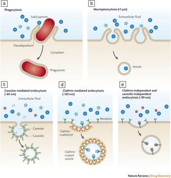
The intracellular compartments of a cell are uniquely different in composition from the extracellular environment. Within a eukaryotic cell, organelles are separated from the cytoplasm by lipid membranes, and thus form many small isolated compartments. Unlike passive transport mechanisms that enable the passage of small, non-charged molecules across the cell membrane, NPs are usually internalized into cells through one or a combination of endocytosis mechanisms ( Figure 3 ). Reference Petros and DeSimone104 Depending on the type of non-specific or specific interactions that NPs have with the cell surface, energy-dependent (more frequent) and energy-independent (less frequent) mechanisms exist to mediate their entry into the cell. The details related to each of the mentioned internalization pathways have been extensively reviewed in the literature. Reference Leroueil, Hong, Mecke, Baker, Orr and Holl85,Reference Khalil, Kogure, Akita and Harashima105,Reference Hong, Leroueil, Janus, Peters, Kober, Islam, Orr, Baker and Banaszak Holl106
Representative endocytosis mechanisms of nanoparticles. (a) Large particles are internalized by phagocytosis. (b) Internalization of smaller particles can occur through non-specific macropinocytosis (>1 μm). Other multiple internalization pathways are available to facilitate the uptake of nanoparticles such as (c) caveolar-mediated endocytosis (∼60 nm), (d) clathrin-mediated endocytosis (∼120 nm), and (e) clathrin-independent and caveolin-independent endocytosis (∼90 nm). Reprinted with permission from Reference 104. ©2010 Macmillan Publishers Ltd.

Endocytosis, or energy-dependent cell uptake, can be classified into two broad categories: phagocytosis and pinocytosis. In general, phagocytosis is receptor-mediated and functions with the purpose of removing pathogens from the body as part of the innate immune system. Reference Hillaireau and Couvreur107–Reference Champion, Walker and Mitragotri109 It can be performed only by specialized cells such as neutrophils, macrophages, dendritic cells, monocytes, and mast cells (Figure 3a). Reference Galli, Maurer and Lantz110,Reference Savina and Amigorena111 In contrast to phagocytosis, pinocytosis occurs in almost all cell types and is the major energy-dependent mechanism by which molecules are internalized into the cell. Four basic pinocytic mechanisms are currently known: macropinocytosis (Figure 3b), caveolae-mediated endocytosis (Figure 3c), clathrin-mediated endocytosis (Figure 3d), and mechanisms independent of clathrin or caveolin (Figure 3e). Reference Petros and DeSimone104,Reference Conner and Schmid112 Each of these mechanisms is distinctly different from one another. Specifically, they differ with regard to the composition of the coated pits, size of the vesicle, and fate of the internalized molecules. Reference Khalil, Kogure, Akita and Harashima105
Macropinocytosis internalizes particles non-specifically and traffics them through the endolysosomal route for degradation. Caveolae-mediated endocytosis internalizes molecules using vesicles coated with caveolin-1 or caveolae. Specific ligands (e.g., FA) or certain pathogens such as Simian Virus 40 (SV40) can induce internalization via a caveolae-mediated endocytic mechanism. Reference Hayer, Stoeber, Ritz, Engel, Meyer and Helenius113,Reference Pelkmans and Helenius114 The exact intracellular trafficking route of caveolae is still under debate, but it is generally accepted that this pathway is non-acidifying and digestion free, although lysosomal routes cannot be completely disregarded. Reference Hayer, Stoeber, Ritz, Engel, Meyer and Helenius113,Reference Parton and Simons115 In contrast, clathrin-mediated endocytosis internalizes molecules by forming clathrin-coated vesicles that consequently enter the endolysomal trafficking route for degradation. Uptake through this pathway can be induced by specific ligands (e.g., low density lipoprotein [LDL], transferrin, EGF, and insulin). Reference Sigismund, Argenzio, Tosoni, Cavallaro, Polo and Di Fiore116,Reference Goldstein, Brown, Anderson, Russell and Schneider117 Other clathrin- and caveolin-independent endocytosis mechanisms exist, but further research is necessary to fully understand them. Reference Mayor and Pagano118
Among the organelles, the nucleus is one of the most important intracellular compartments since it is the site of DNA replication and transcription. The nucleus is surrounded by a double-layered nuclear envelope that isolates it from the cytosol, the intracellular fluid. While molecules smaller than the size of the nuclear pore complex (9 nm), such as DNA, may diffuse freely into the nucleus, larger molecules require active transport mediated by nuclear import receptors. Reference Mann, Thakur, Shukla and Ganguli119 Several protein sequences that act as nuclear localization signals (NLSs) have been discovered to activate protein receptors on the nucleus membrane and cause nuclear internalization. Reference Duvshani-Eshet, Keren, Oz, Radzishevsky, Mor and Machluf120,Reference Lange, Mills, Lange, Stewart, Devine and Corbett121 The classical NLSs that include SV40 large T-antigen NLSs and nucleoplasm NLSs, consist of clusters with four or more repeats of basic amino acids at the N-terminus, which can be discriminated by import receptors.
Mitochondria occupy a considerable volume of the cytosol within eukaryotic cells and function mainly as energy generators. Mitochondria have also been implicated in playing roles in cellular processes such as signaling, cellular differentiation, and cell death. A variety of disorders, including neurodegenerative disease, cardiovascular disease, diabetes, and cancer, have been associated with mitochondria dysfunction, making it a promising therapeutic target. Reference Suomalainen122
Challenges in NP-intracellular interactions
The intracellular compartment presents significant challenges to the delivery of NPs, including (1) endosomal escape and (2) subcellular targeting efficiency of NPs.
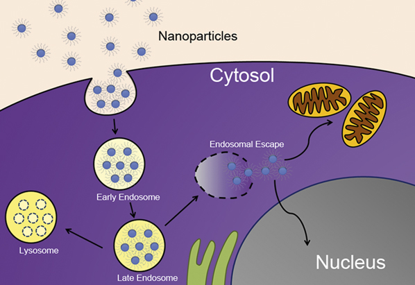
Depending on the internalization mechanism, NPs can be trafficked via the endolysosomal pathway. In this case, NPs and their payload will be degraded by the harsh conditions present in the lysosomal compartment such as low pH and various enzymes, which will dramatically deteriorate therapeutic effects. For example, the transfection efficiency (TE) of plasmid DNA/glycosylated polylysine complexes to HepG2 cells was 10-fold lower than complexes prepared with fusogenic peptides that allow efficient endosomal escape. Reference Midoux, Mendes, Legrand, Raimond, Mayer, Monsigny and Roche123 Similarly, the TE of PEGylated cationic liposome/DNA complexes was significantly diminished when acid-resistant PEG lipids were used due to the inability of the complex to efficiently escape the endosome via membrane fusion. Reference Chan, Majzoub, Shirazi, Ewert, Chen, Liang and Safinya124 Therefore, it is important for the NP to escape the endosome before it is subjected to lysosome-mediated digestion ( Figure 4 ). Reference Dominska and Dykxhoorn125,Reference Varkouhi, Scholte, Storm and Haisma126
Desired intracellular trafficking of nanoparticles (NPs) through endosomal escape that enables NP accumulation in the cytosol and allows subcellular targeting. Appropriate design of NPs using materials can act as proton sponges or incorporation of fusiogenic peptides can alleviate NP degradation in the lysosome (shown as dotted NPs). After escaping the endosome, NPs accumulate in the cytosol and release their therapeutic cargo or navigate to more specific subcellular compartments such as the mitochondria or the nucleus to achieve more targeted effects.
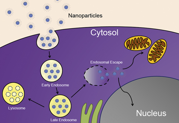
Depending on the cargo being delivered, subcellular targeting may be necessary. Exogenously introduced plasmid DNA, for instance, has to reach the nucleus to cause protein expression. In this case, the NPs need to travel through the cytoplasm, find their targeted organelles, and induce translocation of their cargo across the nuclear envelope. While this delivery strategy may further enhance the efficiency of therapeutics, it also further complicates the design considerations of NPs. Fortunately, strategies have been developed to overcome the challenges associated with the intracellular compartments.
Approaches to overcoming the challenges
Strategies for efficient endosomal escape of NPs
To overcome the challenges presented to NPs by the intracellular environment, a number of strategies have been developed. Since NPs are internalized into cells mainly via endocytosis, the release from endolysosomal trafficking is essential to remain therapeutically effective. The proton sponge effect is the most commonly used strategy to enhance endosomal escape. Basic polymers such as polyethyleneimine (PEI) and PAMAM dendrimers have the capacity to buffer the influx of hydrogen ions pumped into the endosome due to the presence of many secondary and tertiary amine groups in their chemical structure. Consequently, this increases osmotic pressure, resulting in an influx of water. Reference Behr127 The osmotic swelling of the vesicle eventually causes the endocytic vesicle to rupture, thus releasing the entrapped NPs. Amphiphilic pH-sensitive fusiogenic (facilitating fusion) compounds have also been shown to enhance the escape of NPs from endosomes by membrane fusion and subsequent destabilization of the endosomal membrane. Reference Horth, Lambrecht, Khim, Bex, Thiriart, Ruysschaert, Burny and Brasseur128,Reference Marsh and Helenius129 Both of these mechanisms result in partially degraded NPs being released into the cytosol of the cell, which, in many cases, is enough for the encapsulated active therapeutic agents to be released from the NP and achieve the desired results.
Several viruses and bacteria exploit the caveolae-mediated pathway to avoid lysosomal degradation. Reference Carver and Schnitzer130 Therefore, utilizing caveolae-mediated endocytosis is an alternative strategy for NPs to bypass lysosomal degradation. Reference Rejman, Conese and Hoekstra131 Some of the targeting ligands, such as FA, that preferentially direct NP uptake toward this pathway have been used as another strategy to overcome the payload degradation. Reference Sahay, Kim, Kabanov and Bronich132 However, achieving complete control over the internalization pathway utilized by NPs still remains a challenge. Reference Sahay, Kim, Kabanov and Bronich132 Furthermore, understanding of the caveolae-mediated pathway is relatively limited compared to the clathrin-mediated pathway, requiring further investigation.
Intracellular targeting
Although intracellular targeting is not necessary for all types of NP-based drug delivery, it substantially increases the therapeutic efficacy of NPs carrying cargos specific to intracellular targets. Reference Branden, Mohamed and Smith133 Specific material properties of NPs can be engineered to mediate the delivery of the payloads to subcellular compartments. Reference Rajendran, Knolker and Simons134 As mentioned in the previous section, the coupling of NLSs can be used to facilitate the passage of large molecules across the nuclear envelope to enhance their TE. Reference Duvshani-Eshet, Keren, Oz, Radzishevsky, Mor and Machluf120,Reference Lange, Mills, Lange, Stewart, Devine and Corbett121,Reference Fernandez, Baumhover, Anderson and Rice135,Reference Zhang, Mitin and Vinogradov136 Another emerging approach is to employ protein transduction domains (PTDs), Reference Gupta, Levchenko and Torchilin137 which contain non-classical NLSs and effectively condense the DNA or RNA using their cationic domains. Commonly used PTDs include TAT (GRKKRRQRRRPQ) peptides derived from transactivator of transcription, antennapedia, and VP22 protein that have been shown to efficiently deliver DNA into the nuclei. Reference Min, Kim, Kim, Ge, Lee, Park, Park, Hwang, Cho and Yeom138,Reference Ziegler and Seelig140
NPs bearing positively charged molecules on their surfaces are known to usually accumulate on mitochondria, Reference Callahan and Kopecek141–Reference Weissig, Lasch, Erdos, Meyer, Rowe and Hughes143 because the outer membrane potential of mitochondria is approximately –130 mV to –150 mV, which is much lower than other organelles. Reference Chen144 However, it might not be practical to use positively charged NPs for mitochondria targeting due to their short circulation half-life, non-specific binding, and potential cytotoxicity as discussed in the previous sections. In contrast, decorating the surface of NPs with mitochondria-targeting peptides is a promising alternative. Reference Wallace145–Reference Rozenzhak, Kadakia, Caserta, Westbrook, Stone and Naik148 While the intracellular compartments present challenges that can interfere with the successful delivery of therapeutic payloads, the strategies mentioned offer useful solutions to achieve the desired biological responses.
Summary
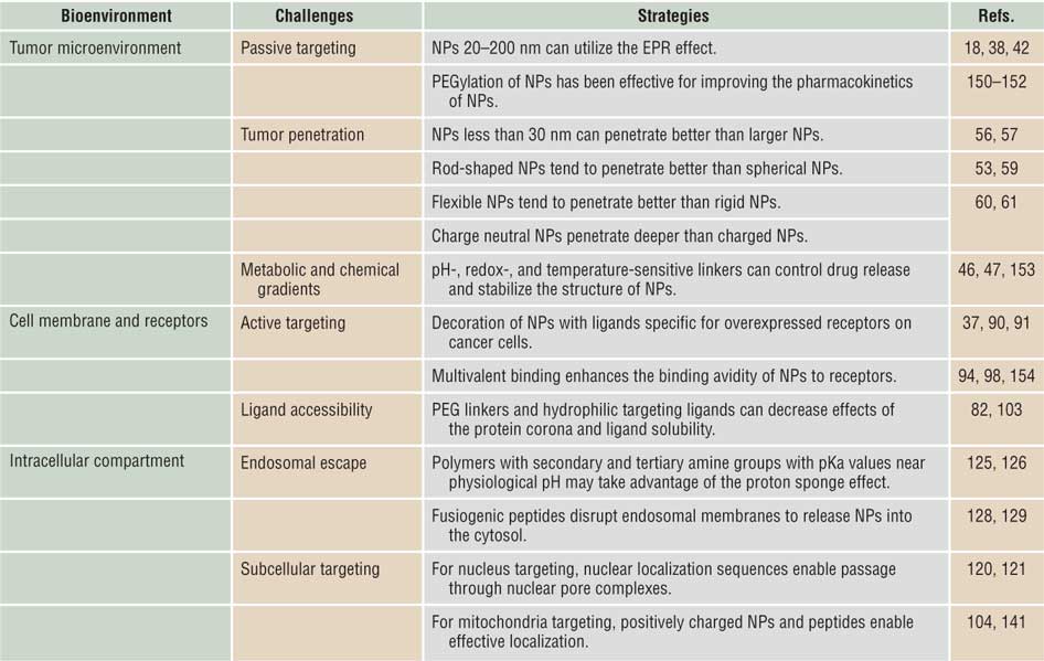
It is clear that nanoparticles (NPs) face a number of difficult challenges throughout their journey from injection into the blood stream to ultimately reaching their target site and provoking desired biological responses. In order to develop an effective NP, it will be critical to engineer NPs that efficiently surpass each of the biological barriers. Through systematic design of NPs to preferentially controlling the nano-bio interactions, many trial-and-error processes can be avoided, and the time needed for in vitro to in vivo translation can be dramatically decreased. In Table I , we summarize the design features for NPs described in this article that have been proven to lead to positive results. For effective tumor-targeted delivery of NPs, the size of the NP must be large enough to accumulate at the tumor site by the enhanced permeability and retention effect, and yet small enough to penetrate deep within the tumor site. The NP must also be able to form a strong, specific interaction with the cell membrane, transport across the cell membrane, be released from vesicular trafficking, and reach its subcellular target. However, all the strategies discussed herein simply cannot be incorporated into a single nanocarrier system. Rather, design features of NPs should be incorporated with the consideration of specific applications in order to achieve a desired biological or clinical outcome. As a number of NPs are currently going into clinical trials, we will likely obtain clinically relevant knowledge regarding the ways in which NPs interact with their surrounding biological environments, which will ultimately lead to transforming NPs to a true solution to many diseases.
Important features of nanoparticles (NPs) to enhance drug delivery.
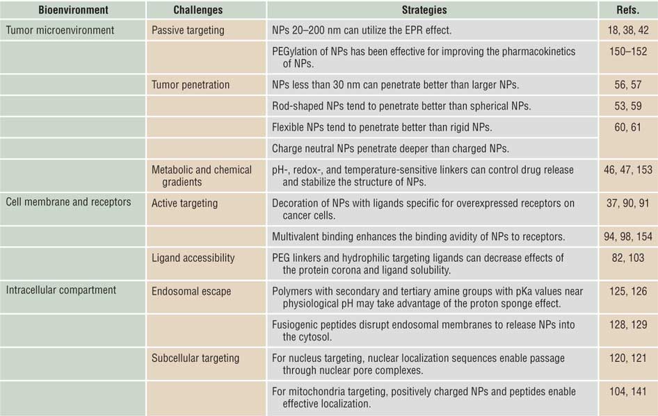
Note: EPR, enhanced permeability and retention; PEG, poly(ethylene glycol); pKa, acid dissociation constant at logarithmic scale.
Acknowledgments
This work was supported by the Susan G. Komen Foundation under Grant No. KG100713, Alex’s Lemonade Stand Foundation for Childhood Cancer, and Korean Ministry of Small & Medium Business Administration. R.M.P. acknowledges support from UIC in the form of the Dean’s Scholarship.