Introduction
The human larynx is classically described as receiving its nerve supply principally from two vagal branches: the superior laryngeal nerve and the recurrent laryngeal nerve, contributing both motor and sensory innervation.Reference Wyke, Kirchner, Hinchcliff and Harrison1
Early neurophysiological studies demonstrated that most fibres in the superior laryngeal nerve are sensory, innervating the supraglottic mucosa through the internal branch, with a motor supply to the cricothyroid via the external branch.Reference Onodi2, Reference Lemere3 These internal branch fibres are predominantly smaller diameter afferent myelin fibres, and those in the external branch are medium diameter efferent fibres.Reference Tomasch and Britton4, Reference Domeij, Carlsoo, Dahlqvist, Hellstrom and Kortopoulos5
By contrast, the recurrent laryngeal nerve contains both afferent and efferent myelin fibres. It innervates all the intrinsic muscles of the larynx except for the cricothyroid, and supplies sensation to all the subglottic mucosa.Reference Lemere3, Reference Suzuki and Kirchner6
In the last century, the so-called plexus theory proposed that the laryngeal nerves were derived from a single plexus within the vagus; thus, a small number of fibres in the superior laryngeal nerve would be associated with a corresponding high number in the recurrent laryngeal nerve.Reference Scheur7
Numerous anatomical studies have also identified internal laryngeal anastomoses between the superior laryngeal nerve and recurrent laryngeal nerve. For example, Steiberg et al. Reference Steinberg, Khane and Fernandes8 confirmed that the posterior branch of the laryngeal nerve (one of the extralaryngeal divisions of the recurrent nerve) supplied sensory innervation to the mucosa inferior to the vocal fold and then anastomosed with the internal laryngeal nerve, forming the loop of Galen. This arrangement has also been described in other studies.Reference Nguyen, Junien-Lavillauroy and Faure9 Such anastomoses may account for the delayed reinnervation of contralateral intrinsic laryngeal muscles following ipsilateral nerve transection. They may also explain the different final position of a paralysed vocal fold (i.e. median, paramedian or lateralised), depending on the degree of synkinesis in different intrinsic laryngeal muscle groups.
The proportions of sensory and motor fibres in the laryngeal nerves are not clearly defined. Few human studies exist on the neuroanatomy of these nerves, and there has been little investigation of the effects of injury upon axonal populations of both large myelinated fibres and smaller unmyelinated fibres. However, a recent study has demonstrated rapid unmyelinated fibre degeneration following acute injury in the central nervous system of rats, following traumatic brain injury.Reference Reeves, Phillips and Povlishock10 Therefore, the degree of myelination of the laryngeal nerves could have important prognostic implications for the recovery of laryngeal function and sensation following injury.
Our study aimed to compare the numbers of myelinated versus unmyelinated fibres in the human laryngeal nerves.
Materials and methods
Cadaveric dissection was performed on five specimens (four fixed, one fresh frozen), by three dissectors of varying otolaryngological experience (one consultant, two registrars).
The superior laryngeal nerve was identified 3 cm from the thyrohyoid membrane, in line with previous studies.Reference Tiago, Pontes and Brasil Ode11 The recurrent laryngeal nerve was sampled 4 cm from the lower border of the cricoid cartilage. This harvesting involved two cross-sectional cuts perpendicular to the long axis of the nerve, to obtain a 1 cm specimen. Five nerve samples were lost to analysis: four because previous cadaveric dissections prevented nerve identification, and one because of mistaken vessel identification and harvesting.
Following standard blocking and cutting of the remaining 15 nerve samples, 7 µm thick sections were produced and stained using standard Solochrome R techniques.Reference Clark12 The first slide from each of these specimens was examined under a Leica photomicroscope system (Leica Microsystems; Wetzlar, Germany), comprising a DM5000B microscope with DFC300FX camera.
Any specimens subsequently found to be non-neurological in nature were discarded.
No further dissection was performed.
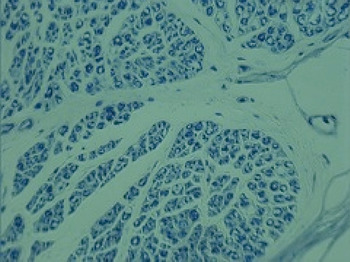
Image analysis equipment (Leica Analysis Suite) was then utilised to electronically count both myelinated and unmyelinated nerve fibres in the whole cross-section, under ×40 magnification. An example of such an image is shown in Figure 1. All slides were counted, using analysis software, by two independent, non-blinded counters. The mean of the counts was taken as the final value. If the two researchers’ counts differed by more than 10 per cent of the lower total, both parties performed a re-count.
Photomicrograph of typical cross-section of a right superior laryngeal nerve. (Solochrome R; ×40)

Analysis of results was performed using the Statistical Package for the Social Sciences version 17.0 for Windows software program (SPSS Inc, Chicago, Illinois, USA). The (non-parametric) Wilcoxon signed rank test was utilised for statistical analysis. A p value of 0.05 or less was considered significant.
Results and analysis
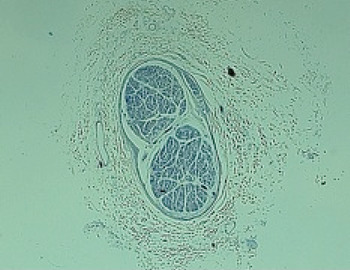
Post-mortem cadaveric dissections were performed on five patients aged between 60 and 96 years (mean age 76.8 years, standard deviation (SD) 13.1 years). There was no known history of significant chronic neurological disease or laryngeal disease affecting any of the cadavers. Figures 1 and 2 show typical photomicrographs obtained.
Photomicrograph of typical cross-section of a right superior laryngeal nerve. (Solochrome R; ×2.5)

Tables I and II show descriptive data for the subdivided and total nerve counts. The number of fascicles in each nerve section ranged from one to 13 (median three, SD 3.03).
Right laryngeal nerve data

Data represent numbers of nerve fibres. RLN = recurrent laryngeal nerve; SLN = superior laryngeal nerve; NRLN = non-recurrent laryngeal nerve
Left laryngeal nerve data

Data represent numbers of nerve fibres. SLN = superior laryngeal nerve; RLN = recurrent laryngeal nerve
Combining results for both nerves, there was a significant difference in the total number of myelinated versus unmyelinated fibres (p = 0.001). Furthermore, subset analyses demonstrated a significantly larger proportion of myelinated fibres, compared with unmyelinated fibres, for both the recurrent laryngeal nerve (p = 0.018) and the superior laryngeal nerve (p = 0.012).
Comparison between the two nerves identified a significantly greater number of myelinated fibres in the superior laryngeal nerve compared with the recurrent laryngeal nerve (p = 0.028). However, there was no significant difference between the numbers of unmyelinated fibres, comparing the two nerves (p = 0.116).
Discussion
Our results demonstrate significantly greater total fibre counts than many previous anatomical studies.
Murtagh and CampbellReference Murtagh and Campbell13 reported fibre counts in four human recurrent laryngeal nerves, but did not provide information on the level of the sections. The number of myelinated fibres ranged from 1598 to 2891.
ScheurReference Scheur7 specified the level of sectioning for both internal and recurrent laryngeal nerves in three hemilarynges, finding total fibre counts of 2012, 3646 and 4668, and 1493, 788 and 687, respectively.
A 1986 French studyReference Germain, Trotoux and Bruneau14 of 100 fresh larynxes demonstrated that the number of fibres per nerve ranged from 511 to 2244, with the number of fascicles ranging from three to 14.
Our findings are more in keeping with those of Tiago and colleagues’ recent studies.Reference Tiago, Pontes and Brasil Ode11, Reference Tiago, Pontes and do Brasil15 This group described the superior laryngeal nerve as having over double the number of myelinated fibres of the recurrent laryngeal nerve. Our results also concur with this and other studiesReference Germain, Trotoux and Bruneau14 showing a significantly greater number of myelinated nerves in the superior laryngeal nerve compared with the recurrent laryngeal nerve. However, there is no evidence of a difference between the number of unmyelinated fibres in the two nerves.
This is further evidence against the historical plexus theory, which assumed that high counts in one superior laryngeal nerve would be compensated by low counts in the ipsilateral recurrent laryngeal nerve.Reference Scheur7 Alternatively, in the words of Dilworth, ‘these nerves were a plexus [, with the] vagus represented by a continuous nerve joining the internal and recurrent nerves and […] separation from this strand form[ing] the individual nerves of the larynx’.Reference Dilworth16
Interestingly, the greatest number of fibres found in our study was from the cadaver with the youngest age of death (60 years); this was especially so for the superior laryngeal nerve. We accept that this finding represents at most a notable point of interest; however, it does correspond with findings from Tiago and colleagues’ studies,Reference Tiago, Pontes and Brasil Ode11, Reference Tiago, Pontes and do Brasil15 which clearly demonstrated a significant decrease in the total number of myelin fibres in elderly patients. These authors proposed that this reduction in fibre numbers may be a factor in the impairment of protective reflexes seen in the elderly, increasing the risk of aspiration and resultant pneumonia.
Recent neurophysiological studies have demonstrated a differing response to traumatic injury, with preferential vulnerability of small, unmyelinated axons, which show more dramatic and persistent electrophysiological changes.Reference Reeves, Phillips and Povlishock10 This implies that this axonal subpopulation is uniquely susceptible to injury; furthermore, the changes observed may influence the potential for recovery.
This is of obvious significance in cases of traumatic injury, and when considering laryngeal nerves and the risk of injury during neck and thyroid surgery. The significantly greater proportion of myelinated fibres in the laryngeal nerves suggests that they may have greater recovery potential, compared with other nerves with a greater proportion of unmyelinated fibres. More interestingly, in our day-to-day clinical practice we are presented with vocal fold motor deficits much more commonly than laryngeal sensory deficits (although historically this may have been partly a diagnostic problem). This is despite the fact that, during thyroidectomy, the frequency of iatrogenic nerve injury is estimated at between <1 to 8 per cent for the recurrent laryngeal nerve, but 14 to 68 per cent for the external branch of the superior laryngeal nerve.Reference Kierner, Aigner and Burian17
A combination of electromyography and judicious frequency range testing may hold promise as a more reliable form of diagnosis, compared with laryngoscopic findings. However, until the natural history, treatment and overall significance of superior laryngeal nerve dysfunction is clarified, the degree to which we should be investigating and treating disorders relating to abnormal laryngeal sensory innervations (e.g. chronic cough) is unclear.
Although not examined in this study, the results of the previously cited studies suggest that the greater proportion of myelinated fibres in the superior laryngeal nerve, and the resulting reduced susceptibility to injury, compared with the recurrent laryngeal nerve, may be a possible explanation for the more common presentation of vocal fold paralysis to the otolaryngologist.
We believe that neurophysiological studies on the susceptibility of human laryngeal nerves to injury, and their potential for recovery, are vital in order to elucidate the natural history of laryngeal nerve disease.
Characterisation of the degree of human laryngeal nerve myelination takes on increased importance when discussing options for laryngeal reinnervation surgery. When considering nerve anastomosis techniques, it would seem prudent to use a transfer nerve with similar morphometric and neuroanatomical qualities, especially as there is considerable evidence that reinnervated muscle takes on the characteristics of the donor nerve.Reference Hengerer and Tucher18 The ansa cervicalis is a commonly used donor nerve for laryngeal reinnervation techniques. Although the anatomical variations in this nerve have been extensively investigated, its degree of myelination has not undergone the same level of study. This could have important implications for the success of these surgical techniques.
Published studies of human laryngeal nerve morphology, dating back to the 1950s, have suffered from numerous well documented methodological flaws. As well as the inevitable limitation of the small sample sizes involved in cadaveric studies, a broad range of analytical techniques have been used, ranging from manual counting with printed photographs to automated image analysis. Two notable criticisms of many previous studies are (1) their failure to report the level of nerve sectioning, and (2) their attempt to compare results from studies with differing data measurement techniques.Reference Harrison19 In the present study, we took a similar approach to Tiago and colleaguesReference Tiago, Pontes and Brasil Ode11 regarding definition of nerve sectioning level, based on previous anatomical studies.
More recently, nerve morphometry studies have had the benefit of image analysers and adjunctive software to minimise error. However, there has still been a tendency to use well recognised sampling techniquesReference Mayhew and Sharma20 to estimate fibre frequency. Although steps to reduce error (e.g. margin effect) have often been taken,Reference Tiago, Pontes and do Brasil15 we believe that the use of such new techniques as image-intensified photographic magnification to calculate a complete nerve count, whilst time-consuming, would ultimately enable more accurate results.
We acknowledge a number of methodological problems in the present study, which need to be addressed in future studies.
• The plexus theory states that high nerve fibre counts in one superior laryngeal nerve are compensated for by low counts in the ipsilateral recurrent laryngeal nerve
• Nerve fibre counts range from 511 to 4668 per laryngeal nerve
• There are more myelinated fibres than unmyelinated fibres in both the recurrent and superior laryngeal nerves
• There are more myelinated fibres in the superior laryngeal nerve than the recurrent laryngeal nerve
• This evidence argues against the plexus theory
Firstly, the problem of limited sample size, which has affected previous, similar studies, was unfortunately still evident in this study. However, the study method of avoiding sampling by utilising individual fibre counts means that any larger study with a greater sample size would involve significantly more time-consuming data collection.
Secondly, a number of nerve samples were lost in the present study. Whilst the majority were due to previous cadaveric dissection, there was one error of mistaken vessel harvesting. In future, the provision of a light microscope close to the dissecting room, to confirm correct specimen collection, would help prevent this mistake.
Conclusion
This anatomical study of human laryngeal nerves demonstrated that both the recurrent laryngeal nerve and the superior laryngeal nerve have a significantly greater number of myelinated fibres than unmyelinated fibres. This result adds to the body of evidence arguing against the historical plexus theory of laryngeal nerve morphology.
In addition, this study demonstrated a significantly greater proportion of myelinated fibres in the superior laryngeal nerve, compared with the recurrent laryngeal nerve. The clinical significance of this finding, in relation to the susceptibility to and recovery from nerve injury, is yet to be determined.
Acknowledgement
This research was funded by the Voice Research Fund, Royal Sussex County Hospital.