Introduction
Myositis ossificans (MO) or heterotopic ossification (HO) is a benign disease characterised by lamellar bone formation in the extra-skeletal tissues involving muscles, tendons and ligaments. The lower extremities are more commonly affected than the upper, followed by head and neck, axilla or paraspinal muscles. Leading triggers include trauma, neurogenic, chronic inflammation, genetic susceptibility and burns. Risk factors for MO include hypercalcaemia, prolonged immobilisation or hypoperfusion driven by venous thromboembolism or arteriovenous shunting in the affected tissues that can alter haemodynamic and metabolic processes. Reference Wong, Mychasiuk and O’Brien1 It presents as a painful, firm, mobile soft tissue mass over the affected site. On X-ray, a typical intramuscular eggshell calcification with radiolucent zones is seen. Surgical excision is the mainstay of treatment, although a few cases are not amenable to resection owing to proximity to critical neurovascular bundle. Radiation therapy (RT) can be an effective therapeutic option for MO to alleviate the symptoms as we describe in our case report. Reference Montero Luis, Hernanz de Lucas and Hervás Morón2–Reference Huang, Chen and Kuo4
Case Report
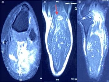
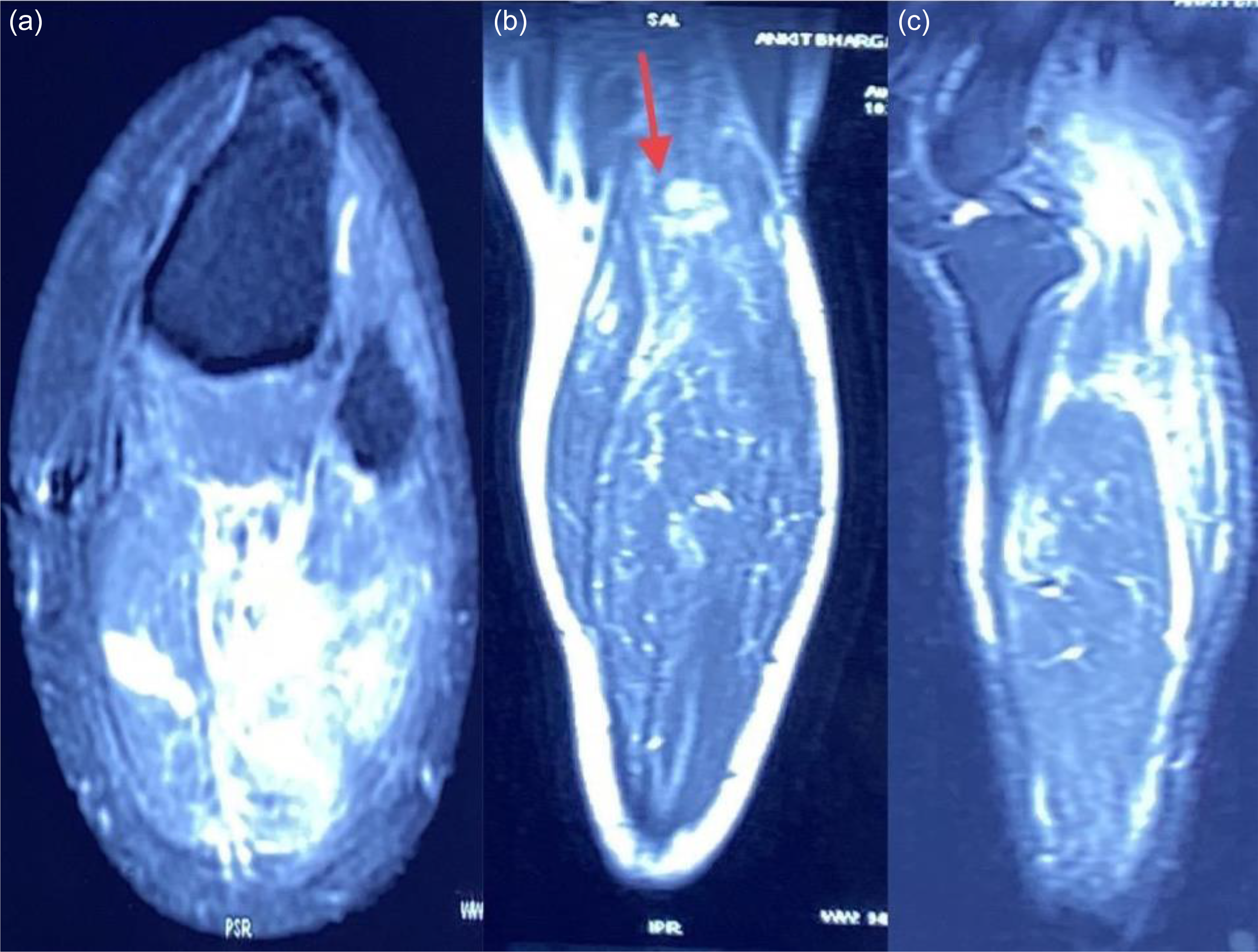
A 36-year-old gentleman presented with progressive radiating pain in the left lower limb for 6 weeks, impaired mobility for 3 weeks, unresponsive to medications. On examination, a localised, swelling in the left popliteal fossa extending along the back of the left lower limb with numbness and paresthesia was noted. There was no history of trauma, infection or similar complaints in the family. A magnetic resonance imaging (MRI) showed a focal abnormal signal intensity lesion measuring 16 × 12 × 23 mm showing patchy calcification in region of popliteal vein with focal aneurysmal dilatation and thrombosis (Figure 1). A nerve conduction velocity test detected left sural sensory axonal neuropathy. Local excision of calcified mass arising from fascia of left popliteal fossa engulfing both sciatic nerve branches was done; on histopathological examination multiple foci of well-defined bony trabeculae surrounded by fibroblasts/myofibroblasts were consistent with MO features. On the post-operative computed axial tomograph (CT) scan, a 2.6 × 1.9 cm residual lesion was detected abutting both heads of the left gastrocnemius muscle and the popliteal artery. The patient reported persistent pain and movement restrictions with swelling of the left knee.
MRI of left leg: (a) axial section shows marked surrounding inflammatory changes around the lesion with small fluid collection in popliteal region; (b) on coronal section a focal abnormal signal intensity lesion measuring 16 × 12 × 23 mm (red arrow) showing patchy calcification in region of popliteal vein; (c) sagittal section depicts popliteal vein focal aneurysmal dilatation with thrombosis. MRI, magnetic resonance imaging; mm, millimetre.
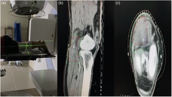
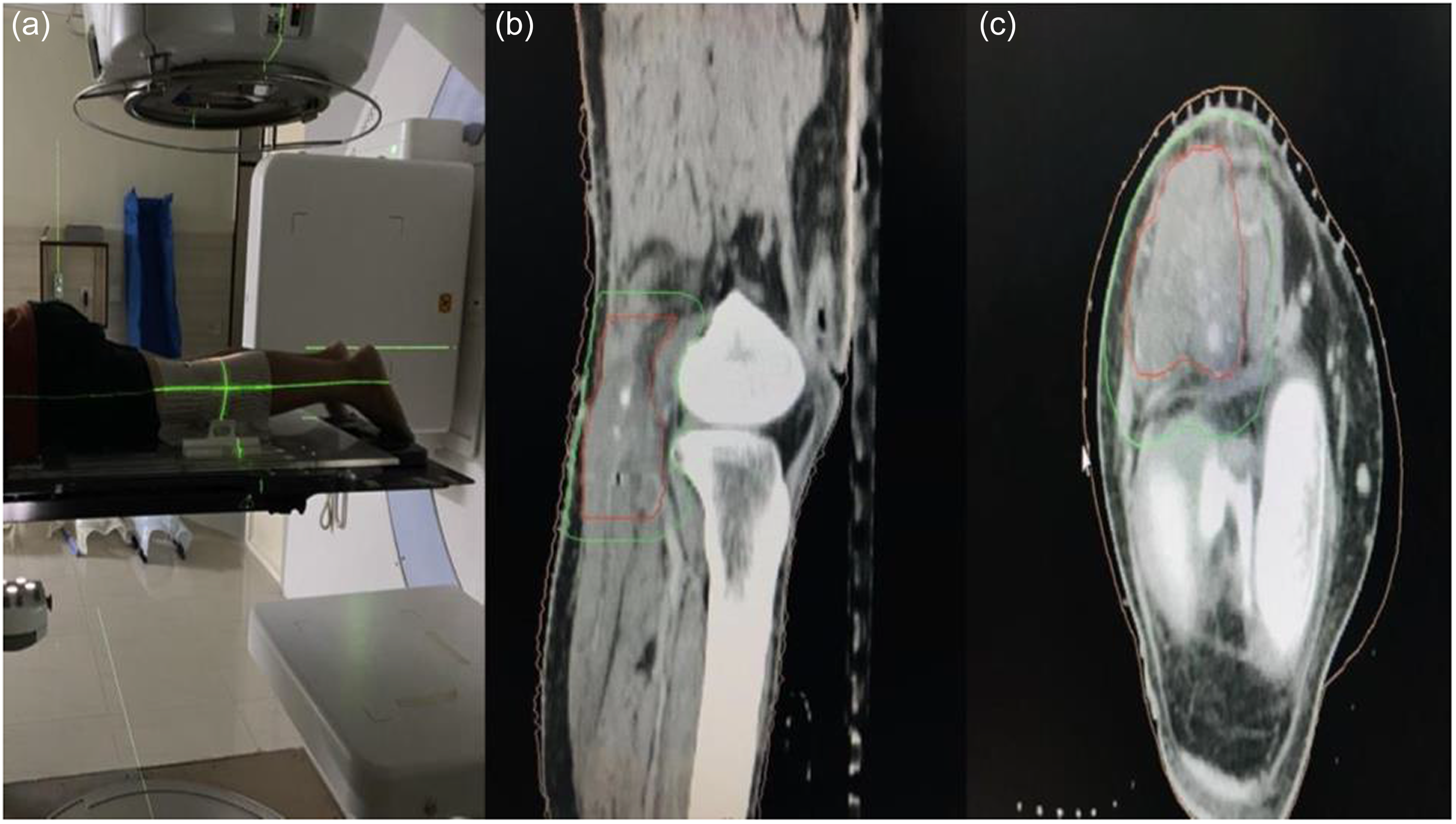
Despite multiple lines of analgesics and physiotherapy, the patient’s symptoms were not alleviated. Thus, RT was planned for him. A written informed consent was taken from the patient after explaining the proposed line of therapy and potential risk of complications. Patient was treated with adjuvant external beam radiation therapy (EBRT) after 2 weeks with a low dose of 10 Gy (Gray) in 2 fractions on 2 consecutive days in a prone position with thermoplastic immobilisation by three-dimensional conformal radiotherapy (3D-CRT) using 6 MV (mega voltage) photon beam on linear accelerator (Figure 2). Patient was pain-free after radiotherapy (visual analogue scale score reduced from 10 to 0), without any related acute toxicities. Patient is on a 3-month follow-up and reports no pain or restricted mobility after 2 years.
(a) Radiation delivery of left popliteal region in prone position using 6-MV photon energy on linear accelerator; (b) sagittal section of left leg with contouring of residual lesion in left popliteal fossa; (c) axial section of post-operative site showing CTV (red) and PTV (green). CTV, clinical target volume; PTV, planning target volume.
Discussion
MO lying in close proximity to a neuromuscular bundle poses a unique surgical challenge. It is diagnosed by CT or MRI with angiography that aids in delineating neurovasculature. Complete excision may not be possible without permanent disability, leading to high chances of residual or recurrence. Meta-analysis have shown that a single fraction of RT of 7–8 Gy given within 3–4 days post-operatively was as effective as a fractionated course. Reference Pellegrini, Konski, Gastel, Rubin and Evarts5 For benign lesions, Esenwein et al reported fractionated irradiation to be effective at inhibiting the differentiation of pluripotent mesenchymal cells into osteoblasts, which improves the quality of life by reducing pain and tissue inflammation. Reference Esenwein, Sell and Herr6 Coventry and Scanlon reported 42 patients of total hip arthroplasty who received adjuvant RT to prevent ectopic bone formation. Reference Coventry and Scanlon7,Reference Chong, Kneebone and Kirsh8 The role of EBRT in earlier reported series is well established for prophylaxis following surgery. Ko et al from Taiwan reported treating MO causing venous compression, mimicking Deep vein thrombosis in a patient with spinal cord injury Reference Ko, Weng, Liu and Chen9 . In our case, EBRT was given for relief of persistent symptoms following incomplete resection as the disease was in close proximity to the sciatic nerve.
Conclusion
EBRT is an effective modality for post-operative MO patients with residual disease or persistent symptoms, especially if the disease is in the vicinity of neurovascular structures and complete resection is not feasible.
Acknowledgements
None.
Financial support
None.
Competing interests
None.