Anomalous drainage of the right superior caval vein into the left atrium is a very rare congenital anomaly, especially in the absence of other congenital lesions.Reference De Leval, Ritter, McGoon and Danielson 1 Diagnosis is usually made in young, asymptomatic adults with hypoxaemia or cyanosis who are referred for further investigation. Among the most serious complications of this anomaly are brain abscess and paradoxical emboli,Reference Park, Summerer, Preuss, Armstrong, Mahomed and Hamilton 2 and the only definite treatment is surgical correction.
Case
We present the case of a 72-year-old female patient with unrepaired anomalous right superior caval vein drainage to the left atrium. She was initially diagnosed in the 1960s, and subsequently followed-up at our hospital. Over this time, the patient was largely asymptomatic, complaining only of infrequent short-lived palpitations. Surgical repair was repeatedly offered to the patient; however, she was not happy to proceed with an operation as she had a successful pregnancy in the past and her exercise capacity or symptoms remained unchanged through all these years. Her follow-up cardiac investigations revealed normal biventricular size and function, her repeated cardiopulmonary exercise tests were reported as normal, and her blood test results remained unchanged. In 2015, the patient presented with a myocardial infarction, and coronary angiography after the event showed unobstructed coronary arteries. On her last clinical examination, she was cyanosed without clubbing. Her saturation in air was 89%. Her blood pressure was 110/70 mmHg. Cardiac auscultation revealed normal heart sounds with no additional murmurs. She had no clinical signs of heart failure, and the respiratory examination was unremarkable. Contrast echocardiography demonstrated the anomalous drainage of the right superior caval vein into the left atrium. Biventricular size was within normal limits with preserved systolic function (Supplementary Figure 1).
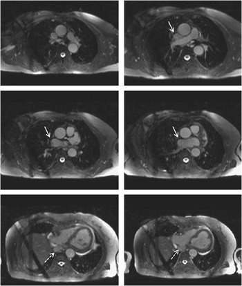
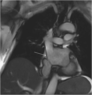
Her cardiac magnetic resonance study, after the myocardial infarction, demonstrated the right superior caval vein drainage into the left atrium, excluding the presence of a persistent left superior caval vein. There was no evidence of other congenital anomalies apart from a small right upper pulmonary vein draining into the superior caval vein and to the left atrium (Figs 1 and 2). Phase-contrast cardiac magnetic resonance of pulmonary arterial and aortic flow showed a significant right-to-left shunt with a Qp:Qs of 0.6. Her biventricular size and function were normal, with normal-sized atria. There were no regional wall motion abnormalities of the left and right ventricles.
Transaxial plane, steady free precession cardiac magnetic resonance, demonstrating in descending-ordered consecutive frames the right superior caval vein (white arrow) flow into the left atrium and the inferior caval vein (dashed line) draining normally to the right atrium.

Steady free precession cine CMR in an oblique view demonstrating the right superior caval vein (white arrow) flow into the left atrium.

Discussion
Isolated anomalous superior caval vein connection to the left atrium was first described in 1956 by Wood.Reference Wood 3 Park et alReference Park, Smith and Silberstein 4 were the first to report an adult case of isolated anomalous drainage of the right superior caval vein to the left atrium in 1973, diagnosed by radionuclide studies. In 1975, De Leval et al reported only 28 cases of anomalous drainage of the superior caval vein to the left atrium among 5127 consecutive congenital cardiac patients. Of those, only 19 were right superior caval vein drainage to the left atrium, mostly in children.Reference De Leval, Ritter, McGoon and Danielson 1 Most of the previously reported cases were in children presenting with mild hypoxaemia or cyanosis. A few cases of patients with cyanosis or chronic hypoxaemia who were diagnosed in adulthood have been previously published.Reference Park, Smith and Silberstein 4 , Reference Ezekowitz, Alderson and Bulkley 5 To the best of our knowledge, ours is the oldest reported case of unrepaired anomalous drainage of the right superior caval vein to the left atrium.
Although the right superior caval vein-to-left atrium connection is a very rare congenital anomaly, it should be included in the differential diagnoses of hypoxaemia and cyanosis along with other congenital anomalies causing intracardiac right-to-left shunt, pulmonary arteriovenous collaterals, primary lung disorders, or haemopoietic defects.Reference Aminololama-Shakeri, Wootton-Gorges, Pretzlaff, Reyes and Moore 6
In our case, apart from the right superior caval vein-to-left atrium connection, anomalous drainage of a small upper pulmonary vein to the superior caval vein was also present; however, other congenital cardiac anomalies have been associated with this condition, such as ventricular septal defect, atrial septal defect, univentricular hearts or Eisenmenger syndrome, tetralogy of Fallot, and transposition of the great arteries. Other extracardiac defects such as coarctation of aorta, patent ductus arteriosus, and pulmonary arteriovenous collaterals may also be present and should be excluded.
Cardiac magnetic resonance was used even in 1989 to confirm the diagnosis of a superior caval vein-to-left the atrium connection in cases diagnosed by cardiac catheterisation.Reference Rosenkranz, Stäblein, Deutsch, Verhoeven and Erdmann 7 Today, cardiac magnetic resonance may be the investigation of choice in cases with hypoxaemia or cyanosis as it can demonstrate the anatomy, and thus establish or exclude the diagnosis. In addition, it permits accurate quantification of right-to-left shunt, the evaluation of the size and function of cardiac chambers and valves, and exclusion of other cardiac or extracardiac anomalies.
Formation of brain abscess and paradoxical embolisation are the most important risk factors in patients with this cardiac anomaly. Our patient experienced a myocardial infarction, which was attributed to paradoxical embolisation as no coronary artery disease was documented.Reference Aminololama-Shakeri, Wootton-Gorges, Pretzlaff, Reyes and Moore 6 , Reference Shapiro, Al-Sadir, Campbell, Thilenius, Anagnostopoulos and Hays 8 Brain abscess occurred in 20% of the reported cases with anomalous connection of the right superior caval vein to the left atrium;Reference Aminololama-Shakeri, Wootton-Gorges, Pretzlaff, Reyes and Moore 6 , Reference Shapiro, Al-Sadir, Campbell, Thilenius, Anagnostopoulos and Hays 8 thus, intravenous infusion via upper body veins should be avoided in these patients.Reference Ezekowitz, Alderson and Bulkley 5 – Reference Aminololama-Shakeri, Wootton-Gorges, Pretzlaff, Reyes and Moore 6
Surgical therapy with anatomical correction of the anomalous connections is the only definitive treatment for this condition, and patients should be advised to undergo surgical correction once the diagnosis is established. At the time of diagnosis in our patient, the risk of surgical repair was considered high; thus, operation was deferred as the patient was asymptomatic.
Acknowledgements
The authors thank Dr Philip Kilner and George Mathew for their contribution towards image acquisition and reporting of the cardiac magnetic resonance scan.
Financial Support
This research received no specific grant from any funding agency or commercial or non-for-profit sectors.
Conflicts of Interest
None.
Supplementary material
To view supplementary material for this article, please visit https://doi.org/10.1017/S1047951116002808