Introduction
Citrus leprosis (CL) is a viral disease transmitted by mites in the genus Brevipalpus (Rodrigues et al., Reference Rodrigues, Kitajima, Childers and Chagas2003). It has been reported in several countries in America, from Argentina to Mexico (Hartung and León, Reference Hartung and León2020). CL has been associated with at least five viruses within the genera Cilevirus and Dichorhavirus (Ramos-González et al., Reference Ramos-González, Chabi-Jesus, Arena, Tassi, Kitajima and Freitas-Astúa2018). Furthermore, orchid fleck virus (OFV, Dichorhavirus orchidaceae) is transmitted by B. californicus to both orchids and citrus (Kondo et al., Reference Kondo, Maeda and Tamada2003; Roy et al., Reference Roy, Stone, Shao, Otero-Colina, Wei, Choudhary, Achor, Levy, Nakhla, Hartung, Schneider and Brlansky2015) and causes CL disease in Mexico, Colombia, South Africa, and Hawaii, USA (Roy et al., Reference Roy, León, Stone, Hartung and Brlansky2014; Cook et al., Reference Cook, Kirkman, Clase, Steyn, Basson, Fourie, Moore, Carstens and Hattingh2019; Olmedo-Velarde et al., Reference Olmedo-Velarde, Roy, Padmanabhan, Nunziata, Nakhla and Melzer2021; Cruz-Jaramillo et al., Reference Cruz-Jaramillo, Ruiz-Medrano, Rojas-Morales, López-Buenfil, Morales-Galván, Chavarín-Palacio, Ramírez-Pool and Xoconostle-Cázares2014). In Brazil, OFV has only been recorded in orchids and not citrus, which is likely due to the absence of B. californicus in citrus orchards (Chabi-Jesus et al., Reference Chabi-Jesus, Ramos-González, Tassi, Rossetto Pereira, Bastianel, Lau, Canale, Harakava, Novelli, Kitajima and Freitas-Astúa2023). However, the citrus leprosis virus N (CiLV-N, Dichorhavirus leprosis), transmitted by B. phoenicis sensu stricto (s.s.), and citrus chlorotic spot virus (CiCSV, Dichorhavirus citri), associated with B. yothersi cause CL in Brazil; and citrus bright spot virus (CiBSV, Dichorhavirus australis) has recently been detected infecting orange orchards and has been linked to B. azores (Chabi-Jesus et al., Reference Chabi-Jesus, Ramos-González, Tassi, Rossetto Pereira, Bastianel, Lau, Canale, Harakava, Novelli, Kitajima and Freitas-Astúa2023). Commercial orchards in Mexico and Brazil are almost exclusively affected by citrus leprosis virus C (CiLV-C, Cilevirus leprosis) which has been associated with B. yothersi (Sánchez-Velázquez et al., Reference Sánchez-Velázquez, Santillán-Galicia, Novelli, Nunes, Mora-Aguilera and Valdez-Carrasco2015; Beltrán-Beltrán et al., Reference Beltrán-Beltrán, Santillán-Galicia, Guzmán-Franco, Teliz-Ortiz, Gutiérrez-Espinoza, Romero-Rosales and Robles-García2020; Nunes et al., Reference Nunes, Novelli, da Cunha, Soares, de Mineiro, Freitas-Astúa and Bastianel2020). The relationship between mites and viral transmission for cileviruses has been described as persistent circulative (Tassi et al., Reference Tassi, Garita-Salazar, Amorim, Novelli, Freitas-Astúa, Childers and Kitajima2017). This disease causes localised chlorotic or necrotic spots, which are circular or oval in shape and have yellow, green, or brown halos on the leaves, branches, and fruits, usually near the feeding sites of the mite. (Locali-Fabris et al., Reference Locali-Fabris, Freitas-Astúa, Souza, Takita, Astúa-Monge, Antonioli-Luizon, Rodrigues, Targon and Machado2006; Bastianel et al., Reference Bastianel, Noveli, Bassanezi, Kitajima, Machado, Kubo and Freitas-Astúa2010).
Brevipalpus spp. has a worldwide distribution but mostly in tropical and subtropical climates (Childers et al., Reference Childers, French and Rodrigues2003). In México, Brevipalpus californicus, B. yothersi, and B. papayensis have been recorded (Sánchez-Velázquez et al., Reference Sánchez-Velázquez, Santillán-Galicia, Novelli, Nunes, Mora-Aguilera and Valdez-Carrasco2015; Salinas-Vargas et al., Reference Salinas-Vargas, Santillán-Galicia, Guzmán-Franco, Hernández-López, Ortega-Arenas and Mora-Aguilera2016; Beltrán-Beltrán et al., Reference Beltrán-Beltrán, Santillán-Galicia, Guzmán-Franco, Teliz-Ortiz, Gutiérrez-Espinoza, Romero-Rosales and Robles-García2020; Domínguez-Gabriel et al., Reference Domínguez-Gabriel, Guillén-Navarro, Otero-Colina, Valle-Mora and González-Gómez2021). B. yothersi has been associated with transmission of the CiLV-C, and it is the most widely distributed Brevipalpus species in Mexico (Sánchez-Velázquez et al., Reference Sánchez-Velázquez, Santillán-Galicia, Novelli, Nunes, Mora-Aguilera and Valdez-Carrasco2015; Salinas-Vargas et al., Reference Salinas-Vargas, Santillán-Galicia, Guzmán-Franco, Hernández-López, Ortega-Arenas and Mora-Aguilera2016; Beltrán-Beltrán et al., Reference Beltrán-Beltrán, Santillán-Galicia, Guzmán-Franco, Teliz-Ortiz, Gutiérrez-Espinoza, Romero-Rosales and Robles-García2020). Although transmission of CiLV-C is widely attributed to B. yothersi, the fact that B. yothersi and B. californicus can be found in the same citrus orchards, suggests the latter species may also be involved in virus transmission. A recent publication reported similar proportions of field-collected specimens of B. yothersi and B. californicus were positive for CiLV-C and OFV-citrus (Beltrán-Beltrán et al., Reference Beltrán-Beltrán, Santillán-Galicia, Guzmán-Franco, Teliz-Ortiz, Gutiérrez-Espinoza, Romero-Rosales and Robles-García2020), although their ability to transmit the virus was not evaluated. However, García-Escamilla et al. (Reference García-Escamilla, Duran-Trujillo, Otero-Colina, Valdovinos-Ponce, Santillán-Galicia, Ortiz-García and Sánchez-Soto2018) reported that B. californicus only transmitted the nuclear type of the CL virus (OFV-citrus). It has been reported that virus dispersion may occur mainly due to viruliferous mite movement caused by their feeding behaviour (Rodrigues et al., Reference Rodrigues, Kitajima, Childers and Chagas2003). However, recent studies failed to find a relationship between mite presence and CiLV-C infected leaf distribution in citrus orchards (Andrade et al., Reference Andrade, Lorençon, Siqueira, Novelli and Bassanezi2018; Gómez-Mercado et al., Reference Gómez-Mercado, Santillán-Galicia, Guzmán-Franco, Valdovinos-Ponce, Becerril-Román and Robles-García2019).
It is clear that interactions between Brevipalpus spp., CiLV-C, and CL are complex and could be multifactorial. Interactions among biological, biochemical, and molecular factors occurring in the virus, vector, and plant may affect acquisition and transmission a virus by its vector (Zhou et al., Reference Zhou, Drucker and Ng2018), although higher virus incidences and severities were associated with higher densities of a vector (Bua et al., Reference Bua, Mawa, Ayiga and Ocwa2024). Here we investigated the effect of mite density on acquisition and transmission of CiLV-C; and whether this relationship was consistent regardless of Brevipalpus species such as B. yothersi and B. californicus.
Materials and methods
Brevipalpus colonies
For B. yothersi, we used mites from a colony that had been initiated with mites collected from orange trees in Sinaloa state, Mexico and maintained at the Acarology laboratory since 2015 (Salinas-Vargas et al., Reference Salinas-Vargas, Santillán-Galicia, Guzmán-Franco, Hernández-López, Ortega-Arenas and Mora-Aguilera2016). The B. californicus colony was established with mites collected from orange trees located at the Colegio de Postgraduados, Texcoco, Mexico. The identity of the mites in each colony was confirmed when the colonies were stablished. For B. yothersi, we used the morphological attributes as reported by Sánchez-Velázquez et al. (Reference Sánchez-Velázquez, Santillán-Galicia, Novelli, Nunes, Mora-Aguilera and Valdez-Carrasco2015). For B. californicus, the morphological traits reported by Salinas-Vargas et al. (Reference Salinas-Vargas, Santillán-Galicia, Valdez-Carrasco, Mora-Aguilera, Atanacio-Serrano and Romero-Pescador2013) and the morphology of the spermatheca described by Beard et al. (Reference Beard, Ochoa, Braswell and Bauchan2015) were used. For both species, the identity was further confirmed using molecular methods described by Salinas-Vargas et al. (Reference Salinas-Vargas, Santillán-Galicia, Guzmán-Franco, Hernández-López, Ortega-Arenas and Mora-Aguilera2016). Subsequent identity confirmation was conducted every 3 months, based solely on morphological traits. Colonies of both species were maintained using the same method described by Salinas-Vargas et al. (Reference Salinas-Vargas, Santillán-Galicia, Guzmán-Franco, Hernández-López, Ortega-Arenas and Mora-Aguilera2016); briefly, mites were maintained on orange fruits contained in transparent plastic boxes (12.5 × 12.5 × 7.5 cm) at 25 ± 3°C, 60 ± 10% relative humidity, and a 12:12 light:dark regime.
Virus source and virus detection
To source the virus, orange fruits with CL symptoms were collected from an orchard in the locality of Cardenas, Tabasco state, Mexico. Control oranges (without virus) were collected from orange trees within the Colegio de Postgraduados grounds. Before experiments, the presence of CiLV-C in all orange fruits with leprosis symptoms was confirmed by RT-PCR following the methods of Gómez-Mercado et al. (Reference Gómez-Mercado, Santillán-Galicia, Guzmán-Franco, Valdovinos-Ponce, Becerril-Román and Robles-García2019). Briefly, 10 mg of orange tissue from oranges with or without leprosis symptoms were used for RNA extraction using the kit Quick-RNA Plant Miniprep (Zymo Research, Irvine, CA, USA) following the manufacturer’s instructions. Reverse transcriptase reactions were done using the RNA extracted to obtain cDNA using the kit RevertAid™ First Strand cDNA Synthesis (Thermo Fisher Scientific Inc., Waltham, MA, USA) according to the manufacturer’s instructions. The presence of virus in samples was confirmed by conventional PCR using primers developed by Locali et al. (Reference Locali, Freitas-Astua, De Souza, Takita, Astua-Monge, Antonioli, Kitajima and Machado2003) and the cDNA previously obtained as a template. PCRs were done in a final reaction volume of 25 µL as described by Gómez-Mercado et al. (Reference Gómez-Mercado, Santillán-Galicia, Guzmán-Franco, Valdovinos-Ponce, Becerril-Román and Robles-García2019). PCR products were visualised on a 1.5% agarose gel, stained with ethidium bromide (10 mg mL−1) and photographed.
Effect of mite density on acquisition of CILV-C
Experimental setup
To determine the effect of mite density on virus acquisition, three densities of B. yothersi – 5, 10, and 15 mites – were each exposed to a virus source in a 2.5 cm diameter designated area. For B. californicus, only the 15-mite density was evaluated. These mite densities were selected based on natural population densities previously reported, which ranged from 1 to 15 mites per orange tree (Gómez-Mercado et al., Reference Gómez-Mercado, Santillán-Galicia, Guzmán-Franco, Valdovinos-Ponce, Becerril-Román and Robles-García2019). The methods were the same for both mite species. Orange fruits with CL symptoms (the presence of the virus had been confirmed using specific primers (Locali et al., Reference Locali, Freitas-Astua, De Souza, Takita, Astua-Monge, Antonioli, Kitajima and Machado2003) as described previously) were used as a virus source. Oranges with CL were prepared as described by Salinas-Vargas et al. (Reference Salinas-Vargas, Santillán-Galicia, Guzmán-Franco, Hernández-López, Ortega-Arenas and Mora-Aguilera2016). Briefly, a 2.5 cm diameter area containing the infected tissue was covered with a mixture of flour, plaster, and sand (1:1:1) which served as a refuge. Mites remained within the designated area during the acquisition period. Each fruit was maintained separately in a plastic box (12.5 × 12.5 × 7.5 cm) as described above. Control oranges (the lack of infection was confirmed as described previously) were prepared as described above.
Virus acquisition
Prior to experimentation, adult females of each Brevipalpus species were transferred to 60 mm diameter Petri dishes (50 adult females per dish) and maintained without access to food for 4 hours. Then, groups of 5, 10, and 15 females of B. yothersi, and 15 females of B. californicus, were allocated to virus-infected oranges using a size 20/0 fine brush and incubated for 7 days to allow feeding on the infected area of the oranges (Ferreira et al., Reference Ferreira, Nunes, Sinico, Soares and Novelli2020) contained within a 2.5 cm diameter circular area. For B. californicus, only the 15-female density was tested, based on preliminary trials indicating no virus acquisition; however, this experiment was conducted formally to confirm those results. For control treatments, the same densities for both mite species were used except that all mites were allowed to feed within the same area (2.5 cm diameter area) on non-infected oranges. After the 7 days of virus acquisition, all mites were collected and deposited in 0.2 mL tubes, with all mites from a specific treatment deposited in a separate tube.
Detection and quantification of CILV-C on mites
The proportion of mites positive for the virus in each treatment was determined using RT-PCR. For this, RNA from each individual mite was extracted using the kit ZR Tissue & Insect RNA MicroPrep (Zymo Research, Irvine, CA) following the manufacturer’s instructions. cDNA was obtained using Sensiscript (QIAGEN, Hilden, Germany) following the manufacturer’s instructions and conventional PCRs to detect the virus were done using the primers of Locali et al. (Reference Locali, Freitas-Astua, De Souza, Takita, Astua-Monge, Antonioli, Kitajima and Machado2003) according to Rodríguez-Ramírez et al. (Reference Rodríguez-Ramírez, Santillán-Galicia, Guzmán-Franco, Ortega-Arenas, Teliz-Ortiz, Sánchez-Soto and Robles-García2019).
For virus quantification in mites from each treatment, TaqMan probe and one set of primers were designed using the movement protein gene region of the complete and partial RNA2 sequences of CiLV-C (GenBank Accessions: Supplemental Table 1). The primers (F: TGA CAC CGA ACG ATA TGA TTT CTC, R: ATA TCT GCC TAA AAG ACT CCA CAG) and the TaqMan probe (CCC CAG CGT CTT CAG CTT TAC GGT) were designed using the software AlleleID 6 (Premier Biosoft, Palo Alto, CA, USA) and synthesized by Oligo T4 (Irapuato, Guanajuato, México). FAM and BHQ1 were used to label the probe at the 5′ and 3′ end, respectively.
A standard curve was obtained using a sample containing copies obtained by cloning a region within the CiLV-C putative protein movement gene kindly provided by the ‘Centro Nacional de Referencia Fitosanitaria’ (CNRF). The original sample (100 ng µL−1) was diluted to 1 ng µL−1 followed by a series of logarithmic dilutions from 1 × 10⁻² to 1 × 10⁻⁷, which were used as a template for qPCR assays. To quantify the amount of virus contained within mites from each treatment, we combined the RNA from three mites per replicate and treatment. Prior to this, RNA concentration and purity were assessed using a NanoDrop spectrophotometer (Thermo Fisher Scientific, Wilmington, DE, USA), then we selected mite samples from each replicate with an RNA concentration of 6 ng µL−1, samples with less than this concentration were not used and samples with greater concentrations were adjusted by dilution to the selected RNA concentration. For qPCR assays, we used a one-step process that included RT and PCR using the kit AgPath-ID One-Step RT-PCR (Applied Biosystems, Massachusetts, USA). The reaction contained 12.5 μl of RT-PCR buffer (2X), 0.4 µl of each primer (10 µM), 0.5 µl of the TaqMan probe (6 µM), 1 µl of RT-PCR Enzyme Mix (25X), 1 µl of the RNA, and sufficient nuclease free water to achieve a final volume of 25 μl. RT-qPCR was done in 0.2 µL eight-strip tubes with flat caps (Bio-Rad Laboratories Inc., Hercules, CA). Thermal conditions were one cycle of 10 min at 45°C, one cycle of 10 min at 95°C, followed by 40 cycles of 10 s at 95°C and 30 s at 55°C. All RT-qPCR were done in triplicate including the control tubes which contained nuclease free water instead of RNA. The C t value for each reaction was automatically calculated and analysed by the Bio-Rad CFX Manager 3.1 software. This software transformed the C t values automatically into ng of CiLV-C in each sample.
Experimental design and data analyses
The experiment was conducted using a completely randomised design with three replicates per treatment. For B. yothersi, the entire experiment was repeated three times on separate occasions, whereas for B. californicus, it was conducted only once with three replicates. Because some mites did not reach the required RNA concentration of 6 ng µL−1, the number of mites was adjusted; therefore, for B. yothersi we used 215 out of 270 processed mites, and for B. californicus we used 43 out of 45 processed mites. The proportion of mites positive for the virus were analysed using a generalized linear model (GLM) with binomial distribution and logit link function (logistic regression), assuming data followed a binomial distribution with sample sizes equal to the number of mites analysed. The quantity of virus per sample analysed was compared using analysis of variance (ANOVA) with hierarchical contrasts where the 5-mite treatment was compared to the combined effect of 10- and 15-mite treatments, followed by the comparison between 10- and 15-mite treatments. The analysis was done with a nested blocking structure representing repetitions on different occasions and repetitions within each occasion. Data did not require prior transformation for the analysis as no evidence of skewness was detected. All analyses were done using GenStat v 24 (VSN International, 2022).
Transmission of CILV-C
For this experiment, only B. yothersi was used. Mites were exposed to a virus source as described previously using the same densities (5, 10, and 15 mites), each with their respective control treatment (plants infested with mites not exposed to the virus). Then, after the 7 days of feeding, all mites were transferred to 7-day-old bean plants (Phaseoulus vulgaris var. Jamapa). For this, one leaf was randomly selected and petroleum jelly was applied to the petiole to prevent mites from escaping and moving to other leaves. On the abaxial surface of the leaf, a 3 × 1 cm area was covered with a mixture of sand, plaster, and flour to provide a refuge for the mites. Mite-infested plants were maintained at 25 ± 3°C, 60 ± 10% HR, and a 12/12 light:dark regime. Seven days after mite infestation of bean plants, the presence of symptoms was assessed and all mites were retrieved from the leaves with a size 20/0 fine brush, deposited in 0.2 mL sterile tubes, and maintained at −20°C until required. The presence of the virus in all mites was confirmed using the methods previously described. The proportion of mites positive for the virus at the end of the experiments was compared using logistic regression.
Virus quantities in the bean leaves were estimated as describes previously but with the following modifications. RNA was extracted from 10 mg of leaf tissue using the kit Quick-RNA Plant Miniprep (Zymo Research, Irvine, CA, USA) following the manufacturer’s instructions. Complementary DNA (cDNA) was obtained through reverse transcriptase reactions using the RevertAid™ First Strand cDNA Synthesis (Thermo Fisher Scientific Inc., Waltham, MA, USA) following the manufacturer’s instructions and according to Gómez-Mercado et al. (Reference Gómez-Mercado, Santillán-Galicia, Guzmán-Franco, Valdovinos-Ponce, Becerril-Román and Robles-García2019). The presence of the virus was first confirmed using the primers developed by Locali et al. (Reference Locali, Freitas-Astua, De Souza, Takita, Astua-Monge, Antonioli, Kitajima and Machado2003) as described previously. For RT-qPCR assays of virus in infected bean leaves, we first quantified the RNA concentration as described previously, and samples from each replicate with 10 ng µL−1 were selected; samples with lower concentrations were not included in analysis; and samples with higher concentrations were diluted to the selected RNA concentration. Quantitative assays were done in 96 well plates (Bio-Rad Laboratories Inc., Hercules, CA) with 5 µl AllTaq Master Mix (QIAGEN, Hilden, Germany), 0.4 µl of each primer (10 µM), 0.5 µl of the TaqMan probe (6 µM), 1 µl of cDNA, and enough nuclease free water for a final reaction volume of 25 μL. Thermal conditions were one cycle of 10 min at 95°C followed by 35 cycles 95°C for 10 s and 55°C for 30 s. All reactions were done in triplicate and three control reactions were included containing only nuclease-free distilled water. The C t value for each reaction was automatically calculated and analysed by the Bio-Rad CFX Manager 3.1 software. This software transformed the C t values automatically into ng of CiLV-C in each sample. The quantity of virus per bean leaf was compared among treatments by ANOVA as described previously. Prior to analysis, data were transformed using the square-root method to fulfil the normality requirements of the analysis.
Relationship between number of mites and leaf area infected by CILV-C
From the transmission experiment, all bean leaves exposed to mites positive for the virus were digitalised using a scanner (HP Deskjet 1500) with a resolution of 600 DPI. All images obtained were analysed using the software EBImage: pliman and the commands image_import, image_combine, and measure_disease (Olivoto et al., Reference Olivoto, Andrade and M Del Ponte2022). Calculation of the severity percentage was based on RGB colour information extracted from each of the three colour palettes representing different subareas of the images: solid leaf background (B), symptomatic areas (S), and healthy areas (H) of the leaf. The background colour was white and contrasted with the colours of the leaf and disease symptoms. The reference colour palettes were manually produced by sampling small representative areas from the three subareas of the image to create a composite image. According to Olivoto et al. (Reference Olivoto, Andrade and M Del Ponte2022), this methodology uses a generalised linear model (binomial family, automatically generated by R) fitted to RGB data to segment the leaf from the background. This allows for calculation of severity using images of leaves with a contrasting background. In addition, the number of spots on each leaf and across treatments was counted. The data obtained were analysed using ANOVA. Data did not require transformation, as there was no evidence of skewness.
Results
Effect of mite density on acquisition of CILV-C
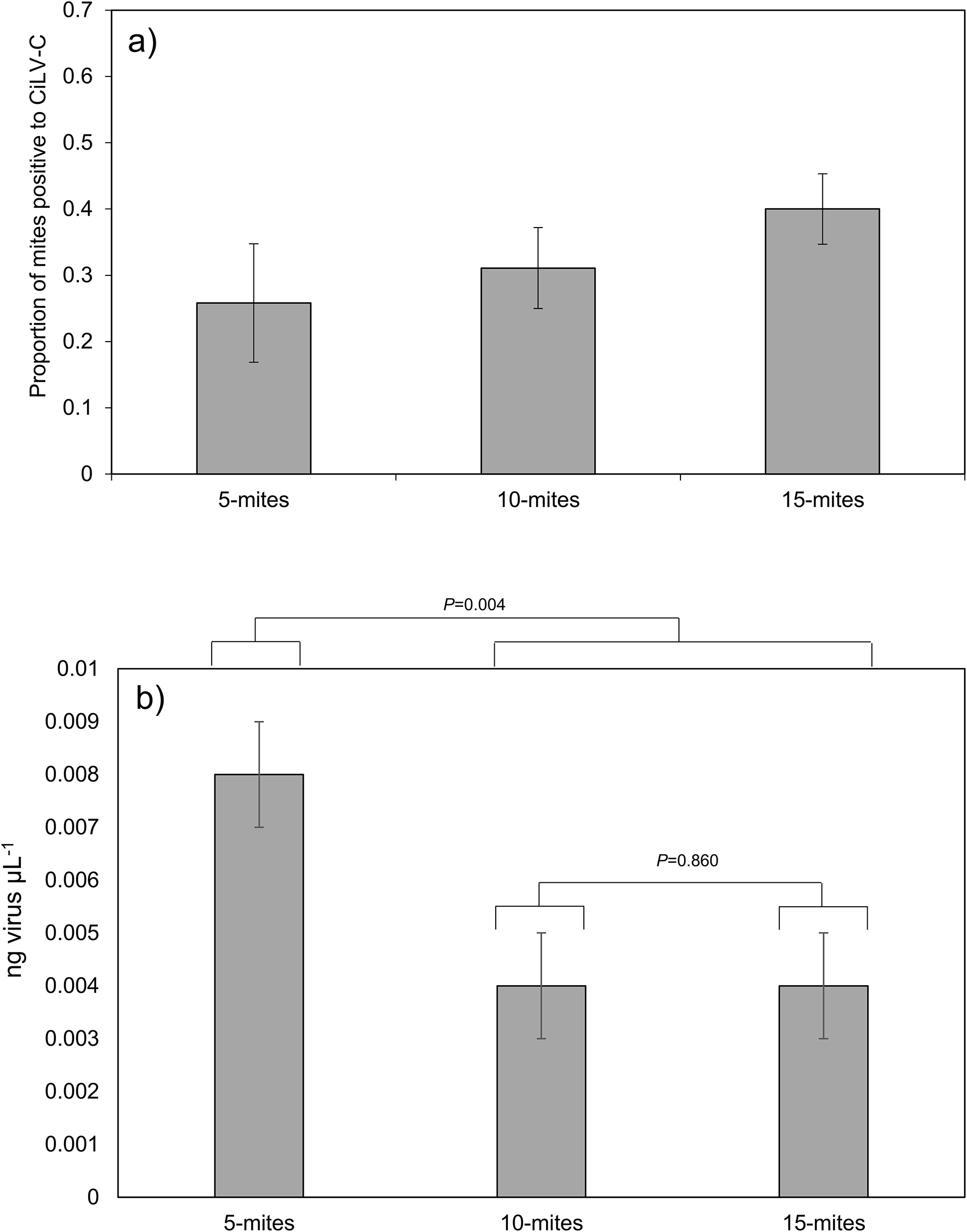
For B. yothersi, the proportion of mites positive to CiLV-C were similar regardless of mite density (F 2, 212 = 1.12, P = 0.329). The average proportion of mites positive for the virus was 0.32 (approx. 32%) (fig. 1a). All B. californicus mites exposed to a virus source were negative for the virus (n = 43).
(a) Proportion of B. yothersi mites positive for CiLV-C among the three mite densities evaluated. Error bars represent 1 × SE (df = 212). (b) Mean virus quantity estimated for mites positive for virus in the three mite densities evaluated. Error bars represent 1 × SE (df = 22).

When the quantity of virus in B. yothersi mites from the three density treatments were compared a greater amount of virus was found in the 5-mite treatment compared to the 10- and 15-mite treatments combined (F 1, 22 = 10.20, P = 0.004), with no differences between the 10- and 15-mite treatments (F 1, 22 = 0.03, P = 0.860) (fig. 1b).
Effect of mite density on transmission of CILV-C
All bean leaves exposed to 5-, 10-, or 15-mite density populations of B. yothersi were infected with CiLV-C, as all presented symptoms of CL (fig. 2). The proportion of B. yothersi mites positive for the virus was similar to the previous experiment; no significant differences were detected among the three densities of mites (F 2, 212 = 1.12, P = 0.329) and the average proportion of mites positive for virus was 0.45 (approx. 45%) (fig. 3a).
Necrotic spots caused by CiLV-C infection in bean leaves infested with (a) 5 mites, (b) 10 mites, and (c) 15 mites.

(a) Proportion of B. yothersi mites positive for CiLV-C among the three mite densities evaluated and used for the transmission experiment. Error bars represent 1 × SE (df = 212). (b) Mean virus quantity estimated in infected bean leaves exposed to three mite densities. Error bars represent 1 × SE (df = 70). (c) Mean infected area in bean leaves exposed to the three mite densities. Error bars represent 1 × SE (df = 21).

When the quantity of virus in leaves exposed to the different B. yothersi mites’ densities were compared, no differences was obtained (F 2, 70 = 1.20, P = 0.307). The mean quantity of virus in bean leaves was 0.01 ng µL−1 (fig. 3b).
Relationship between number of mites and leaf area infected by CILV-C
No significant differences were found when the infected area of bean leaves was compared among the three densities of B. yothersi mites (F 2, 21 = 0.74, P = 0.488). The mean percentage area of bean leaves infected was 3% (fig. 3c).
Discussion
Our study provides new insights into the interaction between Brevipalpus mites and CiLV-C, particularly regarding the effect of vector density on virus acquisition and transmission. Our results showed that B. californicus was unable to acquire the virus, confirming its limited role as a vector in citrus orchards. In contrast, B. yothersi efficiently acquired and transmitted CiLV-C at all densities tested. The average CiLV-C acquisition efficiency with B. yothersi was about 40% (figs 1a and 3a), which is in line with previous reports on C. sinensis leaves infected with CiLV-C2 (Leon et al., Reference Leon, Guillermo, Choudhary and Brlansky2017). It has been suggested that aggregation may reduce each individual’s acquisition efficiency when vector density is high, thus the acquisition rate parameter would not be constant but rather inversely related to vector density. However, plants have a finite number of favourable feeding sites and, as vector density increases, the likelihood of a vector occupying a suitable location on an infected plant decreases (Zhang et al., Reference Zhang, Holt and Colvin2000). Tassi et al. (Reference Tassi, Garita-Salazar, Amorim, Novelli, Freitas-Astúa, Childers and Kitajima2017) mentioned several reasons why a certain number of mites exposed to an infected source may not become viruliferous: e.g. a particular mite might not have access to infected tissues, or a mite could feed only in areas free from lesions. We believe this partially explains our data, as we did not find any statistically significant differences in acquisition efficiency across the three B. yothersi mite densities evaluated (figs 1a and 3a). Although conducting experiments with higher mite densities to confirm the relationship between mite density and virus acquisition would be valuable, we consider the densities we tested to be appropriate because they were chosen based on previous field studies. Brevipalpus yothersi populations in the field are typically low. In a study carried out over 1 year in 100 orange trees where 20 leaves per tree were sampled, most trees harboured between 1 and 3 mites (Gómez-Mercado et al., Reference Gómez-Mercado, Santillán-Galicia, Guzmán-Franco, Valdovinos-Ponce, Becerril-Román and Robles-García2019).
The number of mites capable of acquiring and transmitting CiLV-C after accessing a virus source provides important data for epidemiological studies. Gómez-Mercado et al. (Reference Gómez-Mercado, Santillán-Galicia, Guzmán-Franco, Valdovinos-Ponce, Becerril-Román and Robles-García2019) found no correlation between disease symptoms and the presence of the vector mite in a sweet orange orchard when studying the spatial relationship between CiLV-C and its vector B. yothersi. Our findings on approximately 40% of mites acquiring the virus, partially explains the findings of Gómez-Mercado et al. (Reference Gómez-Mercado, Santillán-Galicia, Guzmán-Franco, Valdovinos-Ponce, Becerril-Román and Robles-García2019), as only a portion of the population in the field was viruliferous. Therefore, the presence of the mite does not necessarily explain the presence of the virus.
The virus concentration in B. yothersi determined using RT-qPCR showed statistically significant differences between the mite populations evaluated, with the treatment involving five mites showing the highest virus concentration (fig. 1b). To our knowledge, there are no reports on the interaction between virus acquisition efficiency and vector density. However, we hypothesise that as the number of mites increases, the efficiency of virus acquisition may decrease. This could be due to the competing for feeding sites, or it could be that the feeding period is longer because of a lack of competition for food. However, further experimental data are needed to confirm this.
In the acquisition experiment, no B. californicus individuals tested positive for CiLV-C. Interestingly, Beltrán-Beltrán et al. (Reference Beltrán-Beltrán, Santillán-Galicia, Guzmán-Franco, Teliz-Ortiz, Gutiérrez-Espinoza, Romero-Rosales and Robles-García2020) reported field-collected B. californicus individuals that tested positive for CiLV-C and OFV-citrus viruses. The reason why no B. californicus individuals acquired the virus in our experiment remains unclear. It is possible that this species only acquires the virus under specific conditions that we did not consider in the experiment, such as longer periods of exposure to a virus source. In addition, helper components (HCs), which are non-structural proteins encoded by the virus, have been shown to be necessary for successful transmission (Di Mattia et al., Reference Di Mattia, Zeddam, Uzest and Blanc2023). Therefore, it could be possible that OFV-citrus produces proteins functioning as HCs that assist in acquisition of CiLV-C by B. californicus when these viruses are found simultaneously in the same mite as reported by Beltrán-Beltrán et al. (Reference Beltrán-Beltrán, Santillán-Galicia, Guzmán-Franco, Teliz-Ortiz, Gutiérrez-Espinoza, Romero-Rosales and Robles-García2020). However, experimental data are required to support this hypothesis. Nevertheless, the lack of virus acquisition by B. californicus corroborates the findings of García-Escamilla et al. (Reference García-Escamilla, Duran-Trujillo, Otero-Colina, Valdovinos-Ponce, Santillán-Galicia, Ortiz-García and Sánchez-Soto2018) and Ávalos-Cerdas et al. (Reference Ávalos-Cerdas, Otero-Colina, Ochoa-Martínez, Villegas-Monter and Suárez-Espinosa2023), who reported that B. californicus could not acquire and transmit CiLV-C when fed with sour orange leaves positive for CilVC.
Brevilapus yothersi were able to inoculate CiLV-C into bean plants, confirming previous data obtained by Garita et al. (Reference Garita, Tassi, Calegario, Kitajima, Carbonell and Freitas-Astúa2013, Reference Garita, Tassi, Calegario, Freitas-Astúa, Salaroli, Romão and Kitajima2014) and Ferreira et al. (Reference Ferreira, Nunes, Sinico, Soares and Novelli2020). Tassi et al. (Reference Tassi, Garita-Salazar, Amorim, Novelli, Freitas-Astúa, Childers and Kitajima2017) reported that B. yothersi’s transmission rate could be from 20 to 75% in P. vulgaris plants. Ferreira et al. (Reference Ferreira, Nunes, Sinico, Soares and Novelli2020) reported that B. yothersi could achieve transmission efficiencies of 83% in sweet orange and 87.5% in common bean, which is in line with our results as we found 100% of bean plants with CL symptoms tested positive for CiLV-C. We expected the greatest quantity of virus to be in bean leaf tissue exposed to 15 mites compared with the other mite densities. However, no significant differences were observed between the evaluated population densities. These results are not entirely surprising, as only a mean of 40% of B. yothersi mites acquired the virus (figs 1a and 3a), regardless of the mite density exposed to the virus source. Therefore, it would be expected that a similar quantity of virus would be inoculated into bean plants, regardless of the mite density, producing a similar quantity of virus at the end of the experiment (fig. 3b). In addition, even when acquiring the virus via ingestion, due to individual variation, the virus may fail to cross the midgut and/or salivary gland barriers of the mite, thus preventing inoculation. Moreover, experimental stress imposed on the mites during handling may affect their ability to transmit the virus.
When we evaluated signs of virus infection on bean leaves, we found necrotic spots near the veins where the mites had fed, which is in line with what has been reported previously (Garita et al., Reference Garita, Tassi, Calegario, Kitajima, Carbonell and Freitas-Astúa2013; Ferreira et al., Reference Ferreira, Nunes, Sinico, Soares and Novelli2020). Tassi et al. (Reference Tassi, Garita-Salazar, Amorim, Novelli, Freitas-Astúa, Childers and Kitajima2017) observed necrotic spots within 5 days on P. vulgaris plants inoculated with B. yothersi positive for CiLV-C. In the analysis of bean leaves infested with B. yothersi, no significant differences were observed in the percentage of leaf area damaged among the treatments. Tassi et al. (Reference Tassi, Garita-Salazar, Amorim, Novelli, Freitas-Astúa, Childers and Kitajima2017) evaluated CiLV-C transmission by placing individual B. yothersi mites (fed on fruit with symptoms) on bean leaves, observing that each mite could cause an average of eleven lesions per leaf. However, under the conditions of our experiment, the damage per mite was four, three, and two spots in the different groups evaluated (data not shown).
In conclusion, mite density did not affect the proportion of B. yothersi mites acquiring CiLV-C but affected the quantity of virus acquired. We obtained 100% infection of bean leaves but no differences in the quantity of virus in the bean leaves. B. californicus did not acquire CiLV-C. Our findings provided new insights into the effect of mite-virus interactions on CiLV transmission.
Supplementary material
The supplementary material for this article can be found at https://doi.org/10.1017/S0007485326100832.
Acknowledgements
The authors are grateful to CONAHCYT for the scholarship provided to RGM for his PhD; the Colegio de Postgraduados for all the facilities and resources to carry out this research; the ‘Comité Estatal de Sanidad Vegetal de Tabasco’ staff for their valuable help providing CL-infected oranges throughout the entire research.
Author contributions
All authors contributed to the study conception and design. Material preparation, data collection, and analysis were performed by Rene Gómez-Mercado, Ma. Teresa Santillán-Galicia, Ariel. W. Guzmán-Franco, and Marcelino Martínez-Nuñez. Rene Gómez-Mercado and Ma.Teresa Santillán-Galicia wrote the first draft of the manuscript and all authors commented on previous versions of the manuscript. All authors read and approved the final manuscript.
Competing interests
The authors declare none.
Data availability
The datasets generated during and/or analysed during the current study are available from the corresponding author on reasonable request.