There is little doubt that chronic and intense exposure to stress early in life increases risk for psychological (e.g., Gunnar, Fisher, & The Early Experience, Stress, and Prevention Network, Reference Gunnar and Fisher2006) and physical disorders (e.g., Cohen & Hamrick, Reference Cohen and Hamrick2003). Evidence for this well-established relationship has come from diverse fields, including investigations of the chronic stress of childhood poverty (Evans & English, Reference Evans and English2002; Wadsworth et al., Reference Wadsworth, Raviv, Reinhard, Wolff, Santiago and Schachter2008), child abuse, and neglect (Manly, Kim, Rogosch, & Cicchetti, Reference Manly, Kim, Rogosch and Cicchetti2001), and animal models of the effects of early deprivation (Sanchez, Ladd, & Plotsky, Reference Sanchez, Ladd and Plotsky2001). With this knowledge, prevention researchers have focused increasingly on identifying and targeting mediators of the association between stress and health problems, which often may be more amenable to change than stress itself.
One mediating pathway in the link between stress exposure and deleterious outcomes that has received increasing attention is dysregulated autonomic reactivity (e.g., Repetti, Taylor, & Seeman, Reference Repetti, Taylor and Seeman2002), particularly heightened reactivity of the sympathetic nervous system (SNS). Experiencing patterns of heightened SNS stress reactivity is central to the development of stress-related psychopathology and physical illness (e.g., Cohen, Janicki-Deverts, & Miller, Reference Cohen, Janicki-Deverts and Miller2007; Uchino, Cacioppo, & Kiecolt-Glaser, Reference Uchino, Cacioppo and Kiecolt-Glaser1996). Among children, SNS stress reactivity has negative effects on a range of outcomes, including adjustment problems, reduced classroom effort, increased anxiety, greater reactive aggression, and impaired immune functioning (Bakker, Tijsen, van der Meer, Koelman, & Boer, Reference Bakker, Tijsen, van der Meer, Koelman and Boer2009; Carapetian, Siedlarz, Jackson, & Perlmuter, Reference Carapetian, Siedlarz, Jackson and Perlmuter2008; El-Sheikh, Erath, Buckhalt, Granger, & Mize, Reference El-Sheikh, Erath, Buckhalt, Granger and Mize2008; Hubbard et al., Reference Hubbard, Smithmyer, Ramsden, Parker, Flanagan and Dearing2002; Kiecolt-Glaser & Glaser, Reference Kiecolt-Glaser and Glaser1995).
Given the clearly detrimental impact of SNS stress reactivity on children's health, research seeking to reveal physiological and environmental predictors of SNS stress reactivity is vital to preventing stress-related pathology. However, few studies have investigated the joint influence of physiology and environment on SNS stress reactivity, especially among young children. To address this gap in the literature, the present study examined the interactive influence of one physiological factor, vagal regulatory capacity (VRC), and one environmental factor, social support, on young children's SNS stress reactivity. As discussed in more detail below, VRC is defined as an individual's typical vagal response to stressors, relative to baseline vagal activity. We assessed individual differences in children's VRC and examined whether such differences interacted with experimentally manipulated social support levels to predict SNS stress reactivity. More specifically, drawing upon tenets of the polyvagal perspective (Porges, Reference Porges2007) discussed below, we hypothesized that high VRC levels would predict lower SNS stress reactivity in socially supportive environments for young children.
Sympathetic Stress Reactivity and Health
The research is clear on the detrimental impact of heightened SNS stress reactivity on a range of mental and physical health outcomes (e.g., Cohen et al., Reference Cohen, Janicki-Deverts and Miller2007; Uchino et al., Reference Uchino, Cacioppo and Kiecolt-Glaser1996). Greater electrodermal reactivity, one index of SNS stress reactivity, robustly predicts both internalizing and externalizing symptoms among children (El-Sheikh, Reference El-Sheikh2005). Further, SNS-induced releases of norepinephrine are associated with childhood and adolescent anxiety disorders (Bakker et al., Reference Bakker, Tijsen, van der Meer, Koelman and Boer2009; Gerra et al., Reference Gerra, Zaimovic, Zambelli, Timpano, Reali and Bernasconi2000). SNS stress reactivity is also elevated among low-income children with more cumulative psychosocial risk and greater learned helplessness (Evans, Reference Evans2003), risk factors for depressive disorders. With regard to externalizing symptoms, Hubbard and colleagues (2002) found exaggerated electrodermal reactivity predicts greater reactive aggression among children in response to experimentally induced anger. Finally, extensive research documents the negative impact of SNS stress reactivity on physical health. SNS stress reactivity has been implicated as the primary mediator of the relationship between stress and impaired immune functioning (Kiecolt-Glaser & Glaser, Reference Kiecolt-Glaser and Glaser1995; Manuck, Cohen, Rabin, Muldoon, & Bachen, Reference Manuck, Cohen, Rabin, Muldoon and Bachen1991; Uchino, Cacioppo, Malarkey, & Glaser, Reference Uchino, Cacioppo, Malarkey and Glaser1995), contributing to a range of cardiovascular, pulmonary, and infectious diseases (e.g., Cohen et al., Reference Cohen, Janicki-Deverts and Miller2007; Lundberg, Reference Lundberg2006). These findings, and others, raise the crucial question of what factors predict exaggerated SNS stress reactivity among children. Understanding what factors lead to variations in SNS stress reactivity across children and contexts would inform efforts to prevent excessive SNS reactivity and its negative health consequences.
VRC and Sympathetic Stress Reactivity
The polyvagal perspective (Porges, Reference Porges2007) provides a useful conceptual framework to generate hypotheses about predictors of SNS stress reactivity. According to this framework, stress response systems developed earlier in evolutionary history are activated only when more contemporary stress response systems fail. The polyvagal perspective suggests that prior to the evolutionary development of the ventral vagal complex, through which the parasympathetic nervous system (PNS) operates on the heart, humans responded to stress primarily with the SNS, a system that elicits fight or flight reactions by increasing heart rate and mobilizing metabolic resources. Through the ventral vagal complex, the PNS stays steadily active under nonstressful conditions, inhibiting cardiac output by applying a “vagal brake” on the heart (Porges, Reference Porges2007). During stressful situations, this vagal brake can be withdrawn rapidly to increase cardiac output without requiring engagement of the more metabolically taxing SNS (Porges, Reference Porges2007). Vagal withdrawal allows for more incremental, focused, and transient adjustments of heart rate in response to stressors, as well as other environmental demands requiring attentional and cognitive processes (Beauchaine, Reference Beauchaine2001; Doussard-Roosevelt & Porges, Reference Doussard-Roosevelt, Porges, Lewis and Ramsey1999; Gottman, Reference Gottman1994). As such, vagal withdrawal has been considered to be a more adaptive, first line of response to stress, which may prevent excessive SNS stress reactivity.
Greater vagal flexibility, reflected by an individual's capacity to reliably withdraw the vagal brake in response to stress (Kashdan & Rottenberg, Reference Kashdan and Rottenberg2010; Rottenberg, Clift, Bolden, & Salomon, Reference Rottenberg, Clift, Bolden and Salomon2007), has been proposed as a key physiological mechanism that prevents excessive SNS reactivity (Porges, Reference Porges2007; Whitson & El-Sheikh, Reference Whitson and El-Sheikh2003). In the present study, we conceptualized vagal flexibility as a stable, traitlike measure on which individuals vary, and we have termed this construct VRC. VRC was operationalized as the average change from baseline in children's vagal responses to four different types of stressors (i.e., social, cognitive, physical, and emotional) using a standardized stress induction protocol. Higher VRC reflects a relatively large decrease in vagal responses from baseline to the stressor tasks (i.e., more vagal withdrawal). Although there is considerable evidence linking VRC to improved emotional and behavioral functioning among children (for an extensive review, see Beauchaine, Reference Beauchaine2001), the hypothesis that VRC can prevent excessive SNS stress reactivity has not yet been tested. The present study not only is among the first to investigate this hypothesis, but, drawing from multiple theoretical frameworks, goes one step further to propose that the mitigating influence of VRC on SNS stress reactivity depends on the availability of social support in the environment.
VRC and Social Support
According to the polyvagal perspective (Porges, Reference Porges2007), there is a close and complex relationship between VRC and social support. First, VRC is thought to elicit behaviors that facilitate social support utilization, such as eye contact and listening, via neuroanatomical connections between the ventral vagal complex and nerves involved in social–emotional processes (Doussard-Roosevelt, Montgomery, & Porges, Reference Doussard-Roosevelt, Montgomery and Porges2003; Porges, Reference Porges2001; Stifter, Fox, & Porges, Reference Stifter, Fox and Porges1989). In addition, the effects of VRC and these behaviors are thought to be moderated by social support availability, because VRC facilitates an individual's assessment of social support levels in the environment (Porges, Reference Porges2007). Thus, when an individual with heightened VRC is in a socially supportive context, one would expect inhibition of SNS stress reactivity. The present study examined this second, moderating relationship between VRC and social support. To our knowledge, no prior studies have explicitly tested this prediction. The present study sought to investigate physiological and contextual influences on SNS stress reactivity by testing the interaction between individual differences in children's VRC and experimental manipulations of contextual social support levels on SNS stress reactivity.
The context dependence of the influence of VRC on SNS stress reactivity is also supported by the biological sensitivity to context (BSC; Boyce & Ellis, Reference Boyce and Ellis2005) and differential susceptibility (DS; Belsky & Pluess, Reference Belsky and Pluess2009) theories. Drawing from evolutionary principles, BSC and DS theories posit that some individuals, through a combination of genetic and early environmental factors, are more susceptible than others to the influence of social support. As such, when in supportive social conditions, only susceptible individuals experience health-related benefits, both psychological and physical (e.g., Boyce et al., 1995; El-Sheikh & Whitson, Reference El-Sheikh and Whitson2006; Obradovic, Bush, Stamperdahl, Adler, & Boyce, Reference Obradovic, Bush, Stamperdahl, Adler and Boyce2010; Pluess & Belsky, Reference Pluess and Belsky2010; Quas, Bauer, & Boyce, Reference Quas, Bauer and Boyce2004). Through this lens, a pattern of strong vagal responses to stress (i.e., VRC) would signify greater susceptibility to available social support; thus, under supportive conditions, heightened VRC may exert a protective influence on health-related outcomes, such as exaggerated SNS stress reactivity. For the present study, the BSC and DS theories would predict, concurrent with our hypotheses guided by the polyvagal perspective, that high VRC is associated with lower SNS stress reactivity for children in socially supportive contexts.
BSC and DS proponents would also predict that high VRC is associated with higher SNS stress reactivity for children in negative social contexts, as heightened VRC is thought to reflect greater sensitivity to both positive and negative contextual conditions (Belsky & Pluess, Reference Belsky and Pluess2009). A recent study by Obradovic and colleagues (2010) illustrated that high VRC was associated with maladaptive behavioral outcomes only for children in a negative social context (i.e., high family adversity). However, because the present study's hypotheses focused exclusively on the interaction between VRC and a positive social context in predicting SNS stress reactivity, we only created experimental conditions of social support and the absence of social support, and did not establish a negative social context. Given the dearth of research examining how high VRC interacts with positive, supportive environments to predict health-related outcomes (see Belsky & Pluess, Reference Belsky and Pluess2009), the present study's focus on the influence of social support represents an important step in testing a key component of the polyvagal perspective as well as tenets of BSC and DS theories.
Present Study
The primary goal of the present study was to test whether higher VRC predicts lower SNS stress reactivity in socially supportive contexts. To do so, we assessed differences in children's SNS stress reactivity as a function of individual variation in VRC and experimentally manipulated contextual support levels. All participants were initially assessed for VRC patterns using a standardized stress induction protocol (Alkon et al., Reference Alkon, Goldstein, Smider, Essex, Kupfer and Boyce2003). Then they were randomly assigned to either a supportive or nonsupportive condition, administered another standardized stress induction protocol, and SNS stress reactivity was measured.
We chose to conduct this research with a sample of young children living in poverty. Though our predictions should apply to children from all segments of the population, poor children are at greater risk than other children for stress exposure and experiencing early adversity, which are associated with developing exaggerated SNS stress reactivity (Ellis, Essex, & Boyce, Reference Ellis, Essex and Boyce2005; Oosterman, De Schipper, Fisher, Dozier, & Schuengel, Reference Oosterman, De Schipper, Fisher, Dozier and Schuengel2010) and experiencing psychopathology (Evans, Reference Evans2003; Wadsworth et al., Reference Wadsworth, Raviv, Reinhard, Wolff, Santiago and Schachter2008; Wolff, Santiago, & Wadsworth, Reference Wolff, Santiago and Wadsworth2009). As such, the socioeconomic characteristics of our sample improve the ability of this research to be directly applicable to vulnerable children living in poverty, while also providing a greater opportunity to examine children at heightened risk for developing patterns of excessive SNS stress reactivity. Similarly, early childhood is a particularly vulnerable age when developmental trajectories begin to be set; thus, it is especially important to conduct research with this age group. In addition, SNS stress reactivity levels may be relatively malleable and variable among younger children (e.g., Alkon et al., Reference Alkon, Goldstein, Smider, Essex, Kupfer and Boyce2003; Bar-Haim, Marshall, & Fox, Reference Bar-Haim, Marshall and Fox2000; Borstein & Suess, Reference Borstein and Suess2000; Calkins & Keane, Reference Calkins and Keane2004; Whitson & El-Sheikh, Reference Whitson and El-Sheikh2003). Recruiting a sample with greater autonomic variability strengthens our ability to measure and characterize a full range of individual differences in the joint influence of VRC and social support on SNS stress reactivity.
The present study also implemented a number of methodological advances. First, because inconsistency in children's responses to specific stressor types may contribute to noise in assessing reactivity patterns (e.g., Calkins & Keane, Reference Calkins and Keane2004), stress inductions in the present study consisted of multiple stressors, thus providing more reliable indices of autonomic stress reactivity (Boyce et al., Reference Boyce, Quas, Alkon, Smider, Essex and Kupfer2001; Kamarck, Debski, & Manuck, Reference Kamarck, Debski and Manuck2000). Second, the present study utilized standardized and well-validated laboratory stress induction and social support manipulation procedures (Alkon et al., Reference Alkon, Goldstein, Smider, Essex, Kupfer and Boyce2003; Quas et al., Reference Quas, Bauer and Boyce2004). Finally, the physiological indices underlying SNS stress reactivity and VRC in the current study, namely, preejection period (PEP) and respiratory sinus arrhythmia (RSA), are known to be reliable, valid, and specific measures of SNS and PNS functioning, respectively (Alkon et al., Reference Alkon, Goldstein, Smider, Essex, Kupfer and Boyce2003; Cacioppo, Uchino, & Berntson, Reference Cacioppo, Uchino and Berntson1994).
Hypotheses
We predicted that children's VRC and the social support level of the experimental context would interact to predict children's SNS stress reactivity. More specifically, we hypothesized that higher VRC would predict lower SNS stress reactivity, but only in a socially supportive context. In contrast, we did not expect higher VRC in a nonsupportive context, or lower VRC in either context, to predict SNS stress reactivity.
Method
Participants
Seventy-three children between the ages of four and five participated in this study. Of these children, six did not complete the protocol due to the child refusing to participate, and six were removed from analyses due to equipment or software failure. Thus, 12 participants were excluded, yielding a final sample size of 61 participants. The excluded participants did not differ significantly from the included participants on most demographic variables, including sex, ethnicity (Latino or non-Latino), primary language spoken in the home, marital status (married or not married), educational attainment (completed high school or did not complete high school), annual income, and self-reported indicators of current family finances and changes in finances over the past 6 months. However, the age (months) of excluded participants (M = 51.3, SD = 5.3) was significantly lower than the age of included participants (M = 55.8, SD = 6.4; t = −2.54, p < .05).
Within the final sample, 60.7% (n = 37) were girls and 39.3% (n = 24) were boys. Age ranged from 48 to 71 months (M = 55.8, SD = 6.5). Further, 59.0% of participants were identified by their parents as Latino, 16.4% African American, 14.8% multiracial, 3.3% Caucasian, 1.6% American Indian, 1.6% other, and 3.3% were missing data on race/ethnicity. Over 49% of participants spoke Spanish as their primary language at home and were administered Spanish-language protocols and questionnaires by experimenters fluent in Spanish. All study materials had been translated and backtranslated by bilingual individuals. Nearly 53% of primary caregivers were currently married. With regard to educational attainment, 11.4% of primary caregivers had at least a bachelor's or associate's degree, 50.8% had a high school degree or General Educational Development (GED) equivalency, 18.0% did not complete high school, 8.2% were currently working toward a GED or college credit, and 11.5% were missing data.
Families were recruited through flyers and information sessions at Head Start early education centers in the Denver-metro area. Interested parents were contacted by phone to schedule a visit to a university research laboratory for one 60-min session. Families received transportation vouchers by mail prior to the session and $40 compensation at the completion of the session. To be eligible, the family's income had to be at or below the federal poverty threshold and the participating child had to be either 4 or 5 years of age at the time of data collection.
Procedure
Overview
Each 60-min data collection session consisted of establishing informed consent, familiarizing the child with the physiological assessment equipment, applying sensors, administering the Phase 1 (P1) stress induction protocol, giving a 7-min snack break, administering the Phase 2 (P2) stress induction protocol by a second supportive or nonsupportive experimenter, removing sensors, and debriefing the family. Children were allowed to choose a toy after all sensors were disconnected.
Prior to the session, children were randomly assigned to either the support or no-support condition for P2. All experimenters were blind to study hypotheses and trained to administer all protocols in a standardized fashion. P1 experimenters were blind to participants' P2 condition. Based on evidence that social support has a stronger effect on reducing autonomic reactivity when provided by women (Glynn, Christenfeld, & Gerin, Reference Glynn, Christenfeld and Gerin1999), all experimenters were female. While children completed the reactivity protocols, parents filled out questionnaires in an adjacent room, where they watched their child on a video monitor. Two parents opted to stay in the testing room and sit unobtrusively behind their children. Reactivity profiles for these two children were comparable to others, and results stayed the same when they were omitted from the analyses.
P1 and P2 stress induction protocols
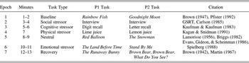
The present study included two standardized protocols (Alkon et al., Reference Alkon, Goldstein, Smider, Essex, Kupfer and Boyce2003) designed to be mildly stressful for 4- to 5-year-olds and to assess individual differences in children's VRC and SNS stress reactivity (see Table 1). Mild stressors were used not only for ethical purposes in conducting research with young children, but also as a way to assess children's responses to externally valid, day to day stressors. Each protocol consisted of seven epochs presented in a fixed order to all participants, with physiological data collected continuously. Both protocols began with a baseline epoch consisting of a calm story read aloud,Footnote 1 and then presented the child with a series of mildly challenging stressors from four domains (social, cognitive, physical, and emotional). The social stressor consisted of a structured social interview about day-to-day events and situations in children's lives (e.g., What color is your toothbrush?). The cognitive stressor consisted of the child repeating aloud increasingly difficult sets of digits at P1 and letters at P2. The physical stressor involved the child identifying unknown liquids (lime juice at P1; lemon juice at P2) placed on the tongue by pipette twice. The emotional stressor consisted of a fear-evoking video clip.
Standardized stress induction protocols

Note: Materials were commercially available in English and Spanish and provided in the language chosen by the primary caregiver. Data from the neutral and recovery epochs were not used in the present analysis. GSRT, Gesell School Readiness Test.
All participants were administered the same types of stressors at P1 and P2. However, they were randomly assigned to either a no-support or support condition at P2. The critical difference between the two conditions was the absence or presence of social support, created by manipulations in experimenter demeanor. Following an established protocol (Quas et al., Reference Quas, Bauer and Boyce2004),Footnote 2 P1 experimenters and no-support condition experimenters at P2 were trained to (a) silently sit apart from the child for 2 min upon entering the experiment room, while appearing to do paperwork, (b) sit in front of but face slightly away from the child, (c) speak in a neutral, monotone voice, (d) not smile, (e) only maintain minimal eye contact with the child to ensure his or her attention, and (f) never provide verbal encouragement. In contrast, support condition experimenters at P2 were trained to (a) build rapport with the child by playing games for 2 min upon entering the experiment room, (b) sit in front of the child and directly face him or her, (c) speak in a fluctuating, positive voice tone, (d) smile frequently, (e) maintain eye contact with the child as much as possible, and (f) provide verbal encouragement throughout the session.
Measures
RSA and PEP scores were obtained for each child at baseline and during stressor tasks at P1 and P2 in order to compute VRC and SNS stress reactivity, as described in detail below. Electrocardiograph (ECG) and impedance cardiograph (ICG) signals used to derive RSA and PEP were acquired and filtered through BioPac MP150 and MindWare Impedance Cardiograph equipment, and then digitized at 1000 Hz. Physiological signals were monitored by the project leader (B.C.W.) on a computer using the BioPac AcqKnowledge software in an adjacent control room during the data collection session. ECG signals were obtained using disposable spot electrodes placed in a standard lead II configuration and were automatically and visually inspected to remove artifacts. ICG signals were obtained using disposable spot electrodes in a standard tetrapolar configuration following standardized procedures described elsewhere (Alkon et al., Reference Alkon, Goldstein, Smider, Essex, Kupfer and Boyce2003). RSA and PEP were edited, scored and reduced using ANSLAB (Autonomic Nervous System Laboratory Software, University of Basel), a customized physiological scoring software package (Wilhelm, Grossman, & Roth, Reference Wilhelm, Grossman and Roth1999).
RSA
RSA, an index of PNS control of cardiac functioning separate from SNS influence, reflects the rhythmic fluctuation of heart rate (i.e., heart rate variability) within the high frequency band associated with respiration (Berntson, Norman, Hawkley, & Cacioppo, Reference Berntson, Norman, Hawkley and Cacioppo2008). For the young children in the present study, a frequency band of 0.15–0.80 Hz was used, as recommended by past research (Bar-Haim et al., Reference Bar-Haim, Marshall and Fox2000; Berntson, Quigley, & Lozano, Reference Berntson, Quigley, Lozano, Cacioppo and Tassinary2007). To compute RSA, heart period (i.e., R-R interbeat intervals) scores were first converted into equidistant time series data with a 4-Hz resolution using weighted averaging interpolation. The time series data were then linearly detrended and quantified with a power spectral analysis using the Welch method of spectral averaging. RSA was calculated as the natural logarithm of the power spectral density values over the high-frequency band (i.e., 0.15–0.80 Hz) with the unit of measurement in ms2. RSA has been shown in past studies to have reasonable temporal consistency (Berntson et al., Reference Berntson, Norman, Hawkley and Cacioppo2008). In the present study, RSA difference scores (i.e., stressor mean – baseline) demonstrated high test–retest reliability from P1 to P2 for the entire sample (r = .68, p < .001), and separately among participants in the no-support (r = .75, p < .001) and support (r = .62, p < .001) conditions.
PEP
PEP (rather than skin conductance or heart rate) was selected as the measure of SNS activation due to its specific dependence on cardiac SNS control and its independence from vagal control (Berntson, Cacioppo, & Quigley, Reference Berntson, Cacioppo and Quigley1991; Cacioppo et al., Reference Cacioppo, Uchino and Berntson1994; Kreibig, Reference Kreibig2010). Derived from both ECG and ICG signals, PEP reflects the time interval (milliseconds) from the start of the heart's ventricular depolarization marked by the onset of the ECG Q wave to the opening of the aortic valve and the simultaneous onset of left ventricular ejection of blood marked by the B point of the ICG dZ/dt wave (Cacioppo et al., Reference Cacioppo, Uchino and Berntson1994; Uchino et al., Reference Uchino, Cacioppo, Malarkey and Glaser1995). From the ICG signal, ensemble averages were created for all dZ/dt data collected within each minute. Minute-by-minute dZ/dt ensemble averages were then automatically and visually inspected for outliers 2 SD above and below the mean. Outliers were removed so the data would more closely approximate prototypical ICG waveforms. PEP has been shown in past studies to be a valid and reliable measure of SNS functioning in young children (Quigley & Stifter, Reference Quigley and Stifter2006). In the present study, PEP difference scores (i.e., stressor mean – baseline) demonstrated moderate test–retest reliability from P1 to P2 (r = .39, p < .05) among control participants who did not receive the support intervention.
VRC and SNS stress reactivity
Measures of baseline RSA and PEP were collected at P1, reflecting a child's initial PNS and SNS activity, respectively, after adjusting to the novel lab environment. At P1 and P2, the social, cognitive, and emotional stressor epochs were each 2 min in length, and the physical stressor was 1 min (see Table 1). For each 2-min epoch, only individual cleaned minutes with at least 30 s of usable data were averaged to create one RSA and one PEP score per stressor epoch.Footnote 3
Defined as the average change in RSA between baseline and the four P1 stressors, VRC was computed as follows: (a) the child's P1 RSA baseline score was subtracted from each P1 RSA stressor epoch score, creating four separate P1 RSA difference scores, and (b) these four P1 RSA difference scores were averaged together to calculate each child's VRC score. The Cronbach α value of the four P1 RSA difference scores was 0.85, confirming that averaging P1 RSA difference scores across tasks was appropriate.
Defined as the average change in PEP between baseline and the four P2 stressors, SNS stress reactivity was computed as follows: (a) the child's P1 PEP baseline score was subtracted from each P2 PEP stressor epoch score, creating four separate PEP difference scores; (b) these four P2 PEP difference scores were then averaged together to calculate each child's SNS stress reactivity score. The Cronbach α value of the four P2 PEP difference scores was 0.95, confirming that averaging P2 PEP difference scores across tasks was appropriate.
Difference scores are commonly employed in psychophysiological research to assess change in autonomic indices (e.g., Alkon et al., Reference Alkon, Goldstein, Smider, Essex, Kupfer and Boyce2003; Boyce et al., Reference Boyce, Quas, Alkon, Smider, Essex and Kupfer2001; Moore & Calkins, Reference Moore and Calkins2004; Propper et al., Reference Propper, Moore, Mills-Koonce, Halpern, Hill-Soderlund and Calkins2008). In the present study, more negative RSA difference scores indexed greater VRC (i.e., more vagal withdrawal), and more negative PEP difference scores indexed greater SNS stress reactivity.
Results
Preliminary and descriptive analyses
Random assignment resulted in approximately half of the participants being assigned to the no-support condition (n = 31) and half to the support condition (n = 30). We evaluated the success of randomization by comparing key demographic variables (i.e., child's sex, ethnicity, and primary language spoken at home, and caregivers' marital status, educational attainment, annual income, current family finances, and recent changes in family finances). No significant differences in these variables were found between the no-support and support conditions (p > .05).
The mean VRC score for the entire sample (M = −0.24, SD = 0.73) reflected an overall, and statistically significant, t (60) = −2.55, p < .05, decrease in RSA between baseline and the P1 stressors. Mean VRC scores for children in the no-support (M = −0.27, SD = 0.78) and support (M = −0.20, SD = 0.67) conditions, which were computed at P1 prior to randomization to the two conditions, were not significantly different from each other, t (59) = −0.35, p > .05, suggesting no preexisting group difference in VRC levels. To evaluate the stability of RSA difference scores underlying VRC, we computed bivariate Pearson product–moment correlations of P1 and P2 RSA difference scores for each of the four stressor types for the entire sample. Results were all moderate to high and statistically significant as follows: social (r = .51, p < .001), cognitive (r = .58, p < .001), physical (r = .42, p < .01), and emotional (r = .61, p < .001). These findings, in addition to the high test–retest reliability of VRC in both conditions described earlier, support the trait interpretation of VRC proposed in the present study.
The mean SNS stress reactivity score for the entire sample at P1 (M = −0.98, SD = 9.40) did not reflect a statistically significant overall change in PEP between baseline and the four P2 stressors, t (60) = 0.82, p > .05, suggesting the stressor tasks, on average, did not elicit SNS stress reactivity. This lack of overall SNS stress reactivity coincides with previously published normative data from the developers of this study's stress induction protocol (Alkon et al., Reference Alkon, Goldstein, Smider, Essex, Kupfer and Boyce2003). The present study did not predict SNS stress reactivity would occur on average, instead expecting that an interaction between children's VRC and the supportiveness of the social context would predict individual differences in SNS stress reactivity.
Bivariate Pearson product–moment correlations were conducted to assess whether there were significant associations between VRC scores and SNS stress reactivity scores at P1 and P2 for the participants in the no-support condition. VRC was not significantly correlated with P1 SNS stress reactivity (r = .14, p > .05) or P2 SNS stress reactivity (r = .03, p > .05). These results suggest that, on the whole, changes in VRC are uncoupled from changes in SNS stress reactivity. In the primary analyses, we evaluated our hypothesis that VRC, rather than show a main effect on SNS stress reactivity, would interact with experimentally manipulated social support levels to predict SNS stress reactivity.
Primary analyses
Although age was not significantly correlated with the baseline or reactivity measures in our sample, it was included as a covariate in the primary analyses for two reasons. First, based on the attrition analysis, children included in the final sample were on average older than those who were excluded. Second, increases in age among young children are associated with higher baseline PNS and SNS activation (e.g., Alkon et al., Reference Alkon, Goldstein, Smider, Essex, Kupfer and Boyce2003). Baseline RSA was also included as a covariate in the model to control for any preexisting differences in baseline vagal activity. All predictor variables and covariates, other than condition coded 0 = no support and 1 = support, were mean-centered for ease of interpretation.
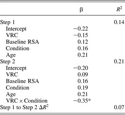
Following recent moderation analysis guidelines (Cohen, Cohen, West, & Aiken, Reference Cohen, Cohen, West and Aiken2003; Dearing & Hamilton, Reference Dearing and Hamilton2006), a linear regression analysis was conducted to examine whether VRC interacted with condition to predict P2 SNS stress reactivity. Results are presented in Table 2. P2 SNS stress reactivity was first regressed on VRC, baseline RSA, condition, and age. Then the VRC by condition interaction term was included to assess its additional contribution to the variance explained by the model. As expected, VRC did not demonstrate a main effect on SNS stress reactivity, either with or without the interaction term in the model. As shown in Table 2, the VRC by condition interaction term was a significant predictor of P2 SNS stress reactivity (β = −0.35, p < .05). Further, the interaction term contributed a significant amount of additional variance explained to the model, ΔF (1, 55) = 4.74, p < .05, with the R 2 increasing from .14 to .21. Finally, the VRC by condition interaction term remained a significant predictor of P2 SNS stress reactivity when the model was run without baseline RSA as a covariate (β = −0.32, p < .05), without age as a covariate (β = −0.37, p < .05), and with neither baseline RSA nor age as covariates (β = −0.34, p < .05).Footnote 4
Sympathetic nervous system stress reactivity, predicted by VRC, Baseline RSA, Condition, Age, and VRC × Condition interaction

Note: The betas are standardized beta coefficients. VRC, vagal regulatory capacity; RSA, respiratory sinus arrhythmia.
*p < .05.
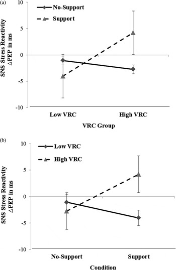
Figure 1 visually depicts the two ways to conceptualize this interaction: (a) social support condition as the moderator and VRC group as the predictor, showing P2 SNS stress reactivity scores for children in the no-support and support conditions across low VRC (1 SD < mean) and high VRC (1 SD > mean) groups; and (b) VRC group as the moderator and social support condition as the predictor, showing P2 SNS stress reactivity scores for children in the low VRC and high VRC groups across the no-support and support conditions. The plots reveal the direction of the interaction effect is consistent with the study's primary hypothesis. For the first plot, follow-up t tests were conducted to assess whether the simple slope (i.e., conditional effect) of each condition (i.e., no-support and support) across low and high VRC groups was significantly different from zero. Results indicated that the regression slope (β = −5.84) of the support condition was significant (t = −2.33, p < .05), reflecting lower P2 SNS stress reactivity for children in the high VRC group relative to children in the low VRC group in the support condition. The regression slope (β = 1.16) of the no-support condition was not significant (t = 0.52, p > .05), suggesting that P2 SNS stress reactivity did not differ across low and high VRC groups in the no-support condition.
The interaction of the vagal regulatory capacity (VRC) and social support condition predicts sympathetic nervous system (SNS) stress reactivity: (a) the social support condition as the moderator and VRC group as the predictor and (b) the VRC group as the moderator and social support condition as the predictor. Note: A lower change in preejection period scores reflects greater SNS stress reactivity.

For the second plot, follow-up t tests were conducted to assess whether the simple slope of each VRC group's regression slope across conditions was significantly different from zero. Results indicated that the regression slope (β = 7.19) of the high VRC group was significant (t = 2.21, p < .05), reflecting lower P2 SNS stress reactivity in the support condition relative to the no-support condition for children with high VRC. The regression slope (β = −2.97) of the low VRC group was not significant (t = −0.90, p > .05), suggesting that P2 SNS stress reactivity did not differ across conditions for individuals with low VRC.
The present study hypothesized that VRC levels would interact with condition to predict P2 SNS stress reactivity. As such, it was important to test the specificity of this model by ruling out key alternative models, as recommended by previous research in this area (e.g., Belsky, Bakermans-Kranenburg, & van IJzendoorn, 2007). One such alternative model would switch the susceptibility and outcome factors of the model, testing whether P1 SNS stress reactivity interacted with condition to predict P2 VRC. These two variables (i.e., P1 SNS stress reactivity and P2 VRC) were created in a parallel manner to the variables in the primary analytical model (i.e., P2 SNS stress reactivity and P1 VRC), but with PEP scores obtained during the four P1 stressors and RSA scores obtained during the four P2 stressors. To test this alternative model, the same linear regression steps were followed as before, except P1 SNS stress reactivity became the predictor variable and P2 VRC became the outcome variable. P2 VRC was first regressed on P1 SNS stress reactivity, baseline PEP, condition, and age. Then the P1 SNS stress reactivity by condition interaction term was included to assess its additional contribution to the variance explained by the model. P1 SNS stress reactivity neither had a main effect (β = 0.11, p > .05), nor an interactive effect (β = −0.04, p > .05) with social support condition, on P2 VRC.
Another alternative analysis would remove VRC from the model, instead testing the influence of baseline activation of the ventral vagal complex (i.e., vagal tone measured as baseline RSA), and its interaction with social support, on P2 SNS stress reactivity. For this model, P2 SNS stress reactivity was first regressed on vagal tone, condition, and age. Then the vagal tone by condition interaction term was included to assess its additional contribution to the variance explained by the model. Vagal tone neither had a main effect (β = 0.13, p > .05), nor an interactive effect (β = 0.11, p > .05) with social support condition, on P2 SNS stress reactivity.
Results from these two alternative models provide additional empirical support for the specificity of the primary model hypothesizing that VRC, a traitlike indicator of PNS stress reactivity, would interact with social support condition to predict P2 SNS stress reactivity.
Discussion
The predictions of the present study were based on two key principles derived from the polyvagal perspective, biological sensitivity to context theory, and differential susceptibility theory (Belsky & Pluess, Reference Belsky and Pluess2009; Boyce & Ellis, Reference Boyce and Ellis2005; Porges, Reference Porges2007). First, in terms of evolutionary development, the ventral vagal complex, which controls PNS innervation of the heart, is thought to be a more recent and adaptive physiological system than the SNS. As such, PNS stress reactivity patterns, called VRC in the present study, should in theory limit SNS stress reactivity. Second, VRC serves as a marker of the human social communication and engagement system, and its functioning is sensitive to the levels of social support available in the environment. Findings from the present study form a logical extension of these two principles, demonstrating that VRC predicts lower SNS stress reactivity in supportive, relative to nonsupportive, social contexts. High VRC in a supportive context may help individuals draw upon available social supports, preventing the fight or flight reactions of the SNS. High VRC in a nonsupportive context did not have the same beneficial effect. The present study is among the first to utilize experimental manipulations of stress and support to test whether higher VRC interacts with social support levels to predict SNS stress reactivity.
Theoretical implications
Most prior research examining the influence of VRC has focused on its association with distal health and behavioral outcomes, rather than its more proximal impact on SNS stress reactivity. Results from the present study help clarify this more proximal association between VRC and SNS stress reactivity, finding that higher VRC is associated with attenuated SNS stress reactivity, under conditions of social support. It is important to note that VRC did not demonstrate a main effect on SNS stress reactivity in the present study. This suggests that social support availability, as suggested by the polyvagal perspective and the biological sensitivity to context and differential susceptibility theories, is a crucial moderating factor in the relationship between VRC and physiological and psychosocial outcomes.
To bolster empirical support for our hypothesized model, we tested an important alternative model, in which the predictor and outcome variables were switched. In this alternative model, individual differences in SNS stress reactivity would predict VRC. Follow-up analyses did not find empirical support for this alternative model in our sample, as SNS stress reactivity neither had a main effect, nor an interactive effect with contextual support levels, on VRC. From the polyvagal perspective (Porges, Reference Porges2007), this lack of evidence for SNS stress reactivity predicting VRC makes sense. VRC is considered to be the primary and adaptive stress response strategy in humans and is believed to inhibit SNS stress reactivity in supportive environments. SNS stress reactivity draws more heavily on the body's metabolic resources and is associated with allostatic load over time. As such, SNS stress reactivity is a second line of defense that is engaged when a stressor is especially intense or prolonged, or when the ventral vagal complex provides feedback indicating the social context surrounding a stressor is adverse. Consequently, our model specifically hypothesized VRC to predict SNS stress reactivity as a function of social support levels. Further, given our conceptualization of VRC as a stable, traitlike indicator assessed across multiple stressor types, it would be unlikely that variations in SNS stress reactivity during a single testing procedure would exert a measurable influence on VRC.
Whereas the results of the present study demonstrated the benefits of VRC in terms of attenuated SNS stress reactivity under socially supportive conditions, we did not hypothesize, or find, high VRC to predict SNS stress reactivity in the no-support condition. This interaction is consistent with the BSC (Boyce & Ellis, Reference Boyce and Ellis2005) and DS (Belsky & Pluess, Reference Belsky and Pluess2009) theories. In addition, BSC and DS theories would posit high VRC in a negative social context to be associated with maladaptive outcomes, such as heightened SNS stress reactivity. Because the no-support condition of the present study was intended to represent the mere absence of social support, and not a negative social context, this second prediction of BSC and DS theories was not hypothesized in the present study.
However, based on recommendations made in a recent review of empirical research evaluating BSC and DS theories (Belsky & Pluess, Reference Belsky and Pluess2009), the present study complements this literature well by examining a supportive context as the environmental predictor. As such, our results provide foundational support for an important component of BSC and DS theories. Speculating beyond the reactivity by context interaction found in the present study, if the support and no-support conditions represent positive and neutral points along the social support continuum, what would SNS stress reactivity look like for children with high or low VRC in a negative social context? Based on BSC and DS theories, a logical extension of the VRC group regression slopes (see Figure 1b) from support, to no support, and finally to a negative social condition would reveal a full crossover interaction reflective of differential susceptibility (Belsky et al., Reference Belsky, Bakermans-Kranenburg and van Ijzendoorn2007). In such an interaction, children with high VRC in the negative condition would show relatively high SNS stress reactivity, whereas children with low VRC would maintain a relatively flat slope across all three conditions. A number of features of our data align well with the criteria for differential susceptibility laid out by Belsky and colleagues (2007). These include the lack of significant associations between VRC (the susceptibility factor) and both the predictor (i.e., social support condition) and the outcome (i.e., SNS stress reactivity), as well as the specificity of our model demonstrated by ruling out key alternative models. A critical future direction of this line of research would be to test the present study's hypothesized model across a full range of contextual social support levels, including high support, the absence of support, and a negative social context.
It is important to note here that our primary conceptualization of the interaction in the present analysis has considered social support to be the moderator of the association between VRC and SNS stress reactivity, as depicted in Figure 1a. Past work supporting the BSC and DS theories has tended to frame this hypothesized interaction in reverse, with the organismic factor (e.g., VRC) as the moderator and the contextual factor (e.g., social support) as the predictor, as depicted in Figure 1b. Although this distinction has important theoretical implications, the aspect of BSC and DS theories most relevant to our study is that environmental and organismic factors interact with one another in shaping outcomes. Thus, whether one or the other factor is described as the moderator versus the predictor does not change that the present results provide evidence in support of the central tenet of these important theoretical frameworks.
Complementing the rich theoretical frameworks for understanding associations between autonomic reactivity and social contextual factors provided by the polyvagal perspective and the BSC and DS theories, other recent studies have conceptualized autonomic reactivity as a function of the relative contributions of both PNS and SNS stress reactivity (Beauchaine, Reference Beauchaine2001; Berntson et al., Reference Berntson, Norman, Hawkley and Cacioppo2008). Berntson and colleagues (1991) proposed an autonomic space model in which the PNS and SNS exist in two dimensions, and stress responses from these two systems can function jointly (i.e., reciprocally) or in opposition to one another (i.e., nonreciprocally). In the present study, children with high VRC in the support condition were the only group of participants to experience attenuated SNS stress reactivity, reflecting a nonreciprocal autonomic pattern of “coinhibition” according to the autonomic space model (Berntson et al., Reference Berntson, Cacioppo and Quigley1991). Although coinhibition has been linked to externalizing symptoms (Beauchaine, Gatze-Kopp, & Mead, Reference Beauchaine, Gatze-Kopp and Mead2007; Boyce et al., Reference Boyce, Quas, Alkon, Smider, Essex and Kupfer2001; El-Sheikh et al., Reference El-Sheikh, Kouros, Erath, Cummings, Keller and Stanton2009), this autonomic profile appears to be relatively common in childhood (Alkon et al., Reference Alkon, Goldstein, Smider, Essex, Kupfer and Boyce2003). Thus far, it is unclear how such findings align with the prediction of the polyvagal perspective that VRC should prevent SNS stress reactivity in supportive contexts. Examining interactions between environmental support levels and VRC patterns based on the autonomic space model will be an important means for future research to illuminate further joint physiological and environmental contributions to SNS stress reactivity.
Along the lines of going beyond the operational definitions of autonomic reactivity used in the present study, it is also important to consider the influence of other indicators of vagal functioning, such as baseline activation of the ventral vagal complex (i.e., vagal tone), on SNS stress reactivity. Vagal tone reflects PNS activation when not responding to a stressor (Porges, Reference Porges2007), which maintains a steady dampening of SNS activation. Because vagal tone is thought to influence SNS stress reactivity by way of its more immediate influence on VRC, the present study chose to investigate just the influence of the more proximal VRC variable on SNS stress reactivity. Providing empirical support for this choice, follow-up analyses in the present study replacing VRC in our model with vagal tone found neither the main effect of vagal tone nor the interaction between vagal tone and social support to predict SNS stress reactivity. Finding such evidence for the more distal association between vagal tone and SNS stress reactivity would require a mediational approach, examining whether vagal tone influences SNS stress reactivity through its impact on VRC. Testing such a mediational model using a longitudinal study design could elucidate the ways in which tonic and reactive measures of vagal functioning are associated with SNS stress reactivity.
Implications for health and prevention
Results of the present study have implications for young children's mental and physical health, and provide valuable information to guide efforts toward preventing negative outcomes. Children's SNS stress reactivity plays a key role in the development of a range of internalizing and externalizing psychological symptoms and acute and chronic physical illnesses (e.g., Cohen et al., Reference Cohen, Janicki-Deverts and Miller2007; Hubbard et al., Reference Hubbard, Smithmyer, Ramsden, Parker, Flanagan and Dearing2002; Lundberg, Reference Lundberg2006; Uchino et al., Reference Uchino, Cacioppo and Kiecolt-Glaser1996). In addition to innervating sympathetic nerve fibers, SNS activity stimulates the adrenomedullary system to release quick-acting catecholamine hormones, namely, epinephrine and norepinephrine, into the bloodstream (e.g., Lundberg, Reference Lundberg2006). Circulating norepinephrine and epinephrine extend the duration and intensity of sympathetic activation, fueling organs central to the fight or flight response, and contributing to the progression of sympathetically mediated illness (e.g., Cohen et al., Reference Cohen, Janicki-Deverts and Miller2007).
Our results suggest the joint influence of VRC and environmental social support is a significant predictor of young children's SNS stress reactivity. More specifically, children with high VRC and available social support during challenging situations seem to experience reduced SNS responses to stress. Thus, in situations where stress is not easily alterable, as is often the case in poverty, providing support and nurturance may be especially beneficial for children with high VRC. Although not examined in the present study, children are likely able to learn to increase their social engagement and utilization of social support during stressful situations, which may be a key mechanism through which VRC protects against excessive SNS stress reactivity in supportive social contexts (Porges, Reference Porges2007). As such, future research that increases our understanding of associations between VRC and social engagement behaviors can provide avenues toward preventing negative physiological and psychosocial outcomes.
The present study utilized a sample of young children living in poverty, which makes the research findings more directly applicable to this underresourced population and allows for a greater sampling of children at risk for heightened SNS stress reactivity. Given the relative lack of control over stressful circumstances experienced by young children, especially those living with few socioeconomic resources, they may be more vulnerable to excessive SNS stress reactivity and its deleterious consequences. Thus, results from the present study, although applicable to all children, may be especially relevant for children living in poverty. Nonetheless, replicating and extending our findings with children from middle and upper income families could reveal both commonalities and dissimilarities in physiological and social processes among children from multiple points along the SES gradient, helping refine further the health and prevention implications of our results.
Limitations and future directions
One potential limitation of the present study was the relatively small sample size used to test a physiology by context interaction effect, which is notoriously sensitive to low statistical power (see Whisman & McClelland, Reference Whisman and McClelland2005). That being said, it is notable that the standardized coefficient for the interaction term in the regression analysis was statistically significant and reflected a moderate effect size. Further, we were able to increase power at the outset by substantially reducing measurement error through the use of a highly standardized stress induction protocol that assessed children's stress responses averaged across four separate stressor domains.
The present study focused on availability of social support but did not measure children's utilization of social support, which according to the polyvagal perspective, is thought to be facilitated by VRC. In his discussion of the relation between the ventral vagal complex and the social engagement system, Porges (Reference Porges2007) outlined the neuroanatomical pathways linking vagal functioning to cortical control over muscles in the eyes, ears, face, and head. In this broader model, healthy VRC can be directly linked to eye gaze, hearing the human voice, and making appropriate facial and head gestures in social contexts. Future research directly observing facial and head movements under conditions of varying levels of social support, measured concurrently with VRC and SNS stress reactivity, could clarify associations between these two autonomic stress response systems and social context even further.
Although the present study included a number of experimental controls (e.g., manipulation and random assignment of social support), we could not directly assess whether, and under what social conditions, variation in VRC level caused variations in SNS stress reactivity. Future studies can build upon the present findings by using longitudinal designs that could better establish the temporal ordering of physiological and contextual predictors and outcomes through mediational analyses. Such studies would also benefit from measuring more distal effects of SNS stress reactivity, such as internalizing and externalizing psychopathology and a range of cardiovascular, pulmonary, and other acute and chronic physical health problems.
These methodological considerations notwithstanding, one strength of the present study was its purposeful incorporation of two additional theoretical frameworks (i.e., BSC and DS theories) to support predictions from the polyvagal perspective. Although the polyvagal perspective has received some criticism on theoretical grounds (Grossman & Taylor, Reference Grossman and Taylor2007), results from the present study are grounded in multiple, mutually supportive frameworks and provide clear empirical support for the protective influence of VRC on SNS stress reactivity for young children in socially supportive contexts. Future research examining the relationships between VRC, SNS stress reactivity, and social context would benefit from continuing to synthesize multiple theoretical perspectives in forming and evaluating testable hypotheses.
Conclusion
Findings from the present study contribute to our understanding of physiological (i.e., VRC) and environmental (i.e., social support) factors that interact to influence SNS stress reactivity in young children. Although social support is clearly the more alterable of these two predictors of SNS stress reactivity, VRC patterns are still somewhat malleable in young children (e.g., Borstein & Suess, Reference Borstein and Suess2000; Boyce & Ellis, Reference Boyce and Ellis2005). Consequently, it will be crucial for future research, like that called for by the BSC and DS theories, to systematically delineate biological and early environmental factors, as well as the interactions between the two, that lead to individual differences in VRC in young children. Improving our understanding of factors that shape both sympathetic and vagal functioning will provide a strong foundation for efforts to prevent physical and mental health disorders in young children.