Introduction
The hypopharynx, also known as the laryngopharynx, is located behind the larynx and leads into the oesophagus. It is involved in physical functions such as swallowing, breathing and speaking. Hypopharyngeal cancer is one of the upper aerodigestive tract cancers with the poorest prognosis. The predominant histological variant is squamous cell carcinoma, which has an invasive growth pattern and often invades adjacent structures. Lymphatic metastases are especially common, and most patients are in an advanced stage at diagnosis.Reference Hall, Groome, Irish and O'Sullivan1, Reference Gourin and Terris2
Currently, hypopharyngeal cancer is treated mostly with combined surgery and radiotherapy, with the choice of treatment regimen dependent upon the extent and metastasis of the primary tumour.Reference Lin, Cohen, Coppit and Burkey3, Reference Corvò4 Therefore, it is important to identify the primary tumour site and any involvement of the laryngopharynx, oesophageal entrance and/or cervical oesophagus, prior to surgery.
Laryngoscopy plays an important role in visualising the primary tumour site and the extent of mucosal surface invasion, within the hypopharynx; it also allows direct specimen collection for biopsy.Reference Zbären, Becker and Läng5 The key to evaluating the extent of hypopharyngeal cancer is to display fully all the anatomical parts of the hypopharyngeal cavity and oesophageal entrance. Only in this way can the tumour's origin and extent be accurately determined.
This study investigated a triple manoeuvre strategy for displaying the hypopharynx and oesophageal entrance during laryngoscopy, and compared the extent of hypopharyngeal cancer visualisation before and after the deployment of this strategy. The study aimed to increase the accuracy of laryngoscopy in judging the extent of hypopharyngeal cancer, and to enhance the ability of laryngoscopy to display superficial cancer.
Materials and methods
General information
We enrolled into the study 113 consecutive patients with hypopharyngeal cancer who attended the endoscopy unit of the Cancer Institute and Hospital, Beijing, between January 2010 and March 2011. Patients comprised 109 men and 4 women, with a mean age of 60 years (range, 34–84 years). All patients had squamous cell carcinoma confirmed by biopsy or surgery.
Exclusion criteria for endoscopic examination included lidocaine allergy, severe and uncontrollable dyspnoea, unstable angina, haemorrhagic diseases, and inability to understand and/or sign the informed consent form.
The study was approved by the medical ethics committee of our hospital. Informed consent was obtained prior to study inclusion.
Laryngopharyngeal examination equipment
Examination of the nasopharyngolarynx was performed using an Olympus Evis Lucera 260 system incorporating a BF-260 video-bronchoscope (Olympus Medical Systems, Tokyo, Japan). The Evis Lucera 260 system provided white light illumination and narrow band illumination with central wavelengths at 415 and 540 nm.
Patients were placed in a horizontal, supine position. Prior to endoscopic examination, the surface of the nasal cavity was anaesthetised and lubricated with 2 per cent lidocaine hydrochloride gel. The instrument was introduced through the nasal passages. The nasopharynx, oropharynx, hypopharynx and larynx were examined in sequence. Representative images were recorded for analysis. Endoscopic biopsies of suspicious lesions were performed, and tissues were fixed in 10 per cent formalin for histological analysis. All endoscopies were performed by the same experienced endoscopist.
Hypopharyngeal examination: the triple manoeuvre
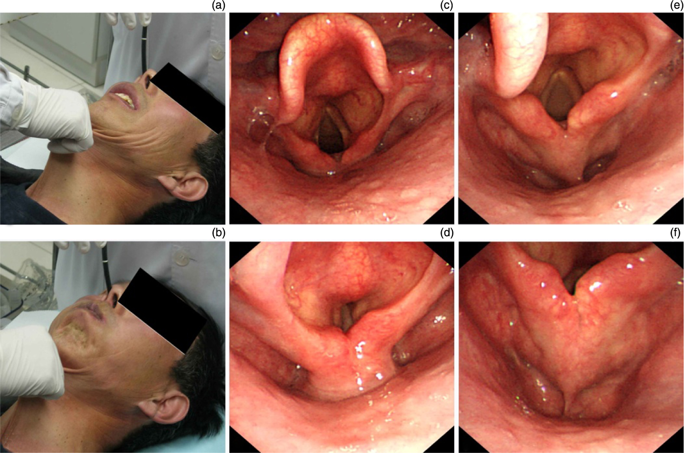
We used a triple manoeuvre to display the hypopharynx (Figure 1), incorporating the following elements.
Photographs and laryngoscopic views showing hypopharyngeal display strategies and outcomes. (a) Anterior cervical skin traction. (b) Cervical skin traction plus balloon-blowing manoeuvre. (c) & (d) Hypopharyngeal appearance while pronouncing ‘e'. (e) & (f) Hypopharyngeal appearance during anterior cervical skin traction plus balloon-blowing manoeuvre.

Firstly, the skin overlying the thyroid cartilage was pulled upward, while the patient lay in the supine position.
Secondly, during cervical skin traction, the patient was instructed to perform Valslava's balloon-blowing manoeuvre, i.e. to close the mouth and force air into the cheeks as if blowing a balloon, without leaking air through the mouth or nose.
Thirdly, oxygen was injected via the laryngoscope. An oxygen cylinder was connected to the laryngoscopic channel (Figure 2). When the laryngoscope tip arrived at the pyriform sinus, oxygen was injected via the laryngoscopic biopsy channel at high volume (4–5 l/minute), and at the same time the patient was instructed to swallow. The force of the oxygen opened the closed oesophageal entrance, enabling the laryngoscopist to examine the extent of neoplastic involvement of the oesophageal entrance.
Diagram showing equipment arrangement enabling injection of oxygen via the laryngoscopic biopsy channel.

The visualised site and extent of the primary tumour were recorded, firstly using conventional laryngoscopy with the patient pronouncing the letter ‘e', and secondly while performing the triple manoeuvre.
Results and analysis
Tumour site visualisation before and after triple manoeuvre
During pronunciation of ‘e', the hypopharynx and adjacent anatomical structures were poorly displayed in 80 patients (70.8 per cent) due to great difficulty in separating the posterior pharyngeal wall and the postcricoid area. In 33 patients (29.2 per cent), the primary tumour site was seen clearly, while in 11 patients (9.7 per cent) the primary tumour was not seen at all.
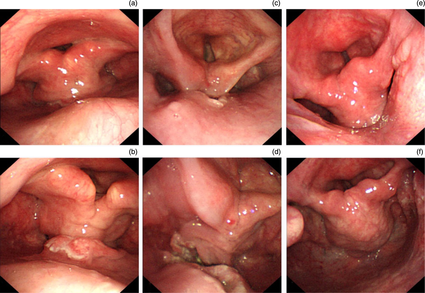
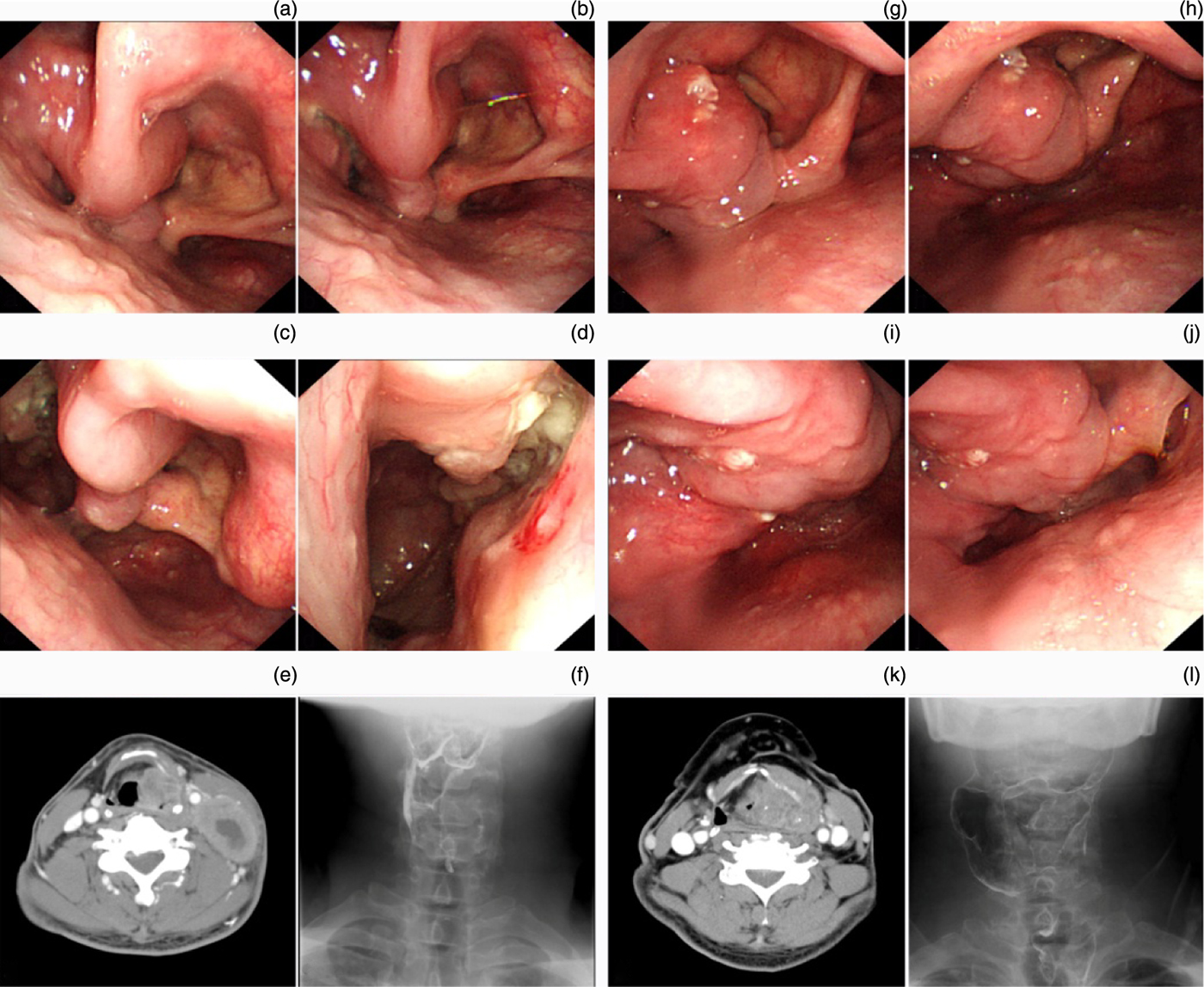
After anterior cervical skin traction plus Valsalva balloon-blowing manoeuvre, the posterior pharyngeal wall and postcricoid area were separated completely, and the hypopharynx was more clearly displayed in 106 patients (93.8 per cent); this difference was statistically significant (p < 0.0001) (Figures 3 and 4). In 7 patients (6.2 per cent), the primary site could not be evaluated owing to extensive invasion and a stiff hypopharynx.
Comparison of hypopharyngeal tumour visualisation under conventional laryngoscopic conditions and using cervical skin traction plus the balloon-blowing manoeuvre: case 1, (a) no significant lesion seen, and (b) protrusive neoplasm seen on the posterior pharyngeal wall, respectively; case 2, (c) ulcerative neoplasm seen on the posterior pharyngeal wall but postcricoid area obscured, and (d) two ‘kissing' ulcerative neoplasms on the posterior pharyngeal wall and in postcricoid area, respectively; and case 3, (e) no significant lesion seen, and (f) rough-surfaced neoplasm on the lateral wall of the right pyriform sinus, respectively.

Comparison of hypopharyngeal tumour site visualisation using different examination methods. In case 1, conventional laryngoscopy shows a significant swelling in the left aryepiglottic fold (a), cervical skin traction plus balloon-blowing manoeuvre reveals an ulcerated primary neoplasm in the left pyriform sinus and involving the left aryepiglottic fold along the submucosals (b–d), while hypopharyngeal contrast axial computed tomography (CT) and contrast anterior-posterior X-ray examinations show the neoplasm within the left pyriform sinus and involving the left aryepiglottic fold, as indicated by laryngoscopy (e, f). In case 2, conventional laryngoscopy shows significant left aryepiglottic fold swelling (g), while skin traction plus balloon-blowing manoeuvre reveals the primary neoplasm within the left aryepiglottic fold and infiltrating the inner wall of the left pyriform sinus (h–j), but hypopharyngeal contrast axial CT and anterior-posterior X-ray images suggest a left pyriform sinus cancer involving the left aryepiglottic fold (the wrong diagnosis) (k, l).

The total of 113 patients included 81 patients (71.7 per cent) with pyriform sinus cancer (50 in the right sinus and 31 in the left), 25 (22.1 per cent) with posterior pharyngeal wall cancer and 7 (6.7 per cent) with postcricoid area cancer.
Tumour extent visualisation before and after triple manoeuvre
The extent of hypopharyngeal cancer spread was recorded according to its anatomical divisions, namely the posterior pharyngeal wall, the postcricoid area, both pyriform sinuses and the larynx.
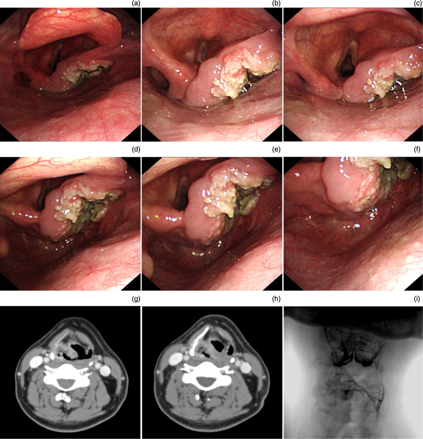
The observed extent of hypopharyngeal cancer was recorded before and after performance of the triple manoeuvre (see Table I). Of the 113 patients, the triple manoeuvre produced no change in visible tumour extent for 24 patients, but produced a noticeable change for 89 patients. Statistical analysis indicated no significant differences in the observed extent of pyriform sinus and laryngeal invasion, before and after performance of the triple manoeuvre (p > 0.05), but significant differences in the observed extent of posterior pharyngeal wall and postcricoid area invasion (p < 0.001) (Figure 5).
Comparison of hypopharyngeal tumour extent visualisation using different examination methods. (a–c) On conventional laryngoscopy, an ulcerative neoplasm is seen in the right pyriform sinus, involving the right arytenoid and aryepiglottic fold, but the lower margin of the tumour cannot be visualised due to cohesion of the posterior pharyngeal wall and the postcricoid area; the left pyriform sinus shows no invasion. (d–f) Following cervical skin traction plus the balloon-blowing manoeuvre, the posterior pharyngeal wall and the postcricoid area separate fully enabling involvement of the right half of the postcricoid area to be seen, with a lower margin at the apex of the right pyriform sinus; there is no invasion of the posterior pharyngeal wall or the oesophageal entrance. (g, h) Axial, contrast-enhanced computed tomography scans show an enhancing mass in the right pyriform sinus involving the mucosa and extending to the right aryepiglottic fold; the laryngopharynx is slightly compressed to the left. (i) Anterior-posterior, phase-contrast, hypopharyngeal X-ray suggesting damage to the right pyriform sinus mucosa, with apparent disappearance of the right pyriform sinus.

Visualisable tumour extent before and after triple manoeuvre

Data represent patient numbers unless otherwise specified. *Comparing visualisable extent before and after triple manoeuvre. L = left; R = right
Oesophageal inlet involvement: laryngoscopic vs radiological examination
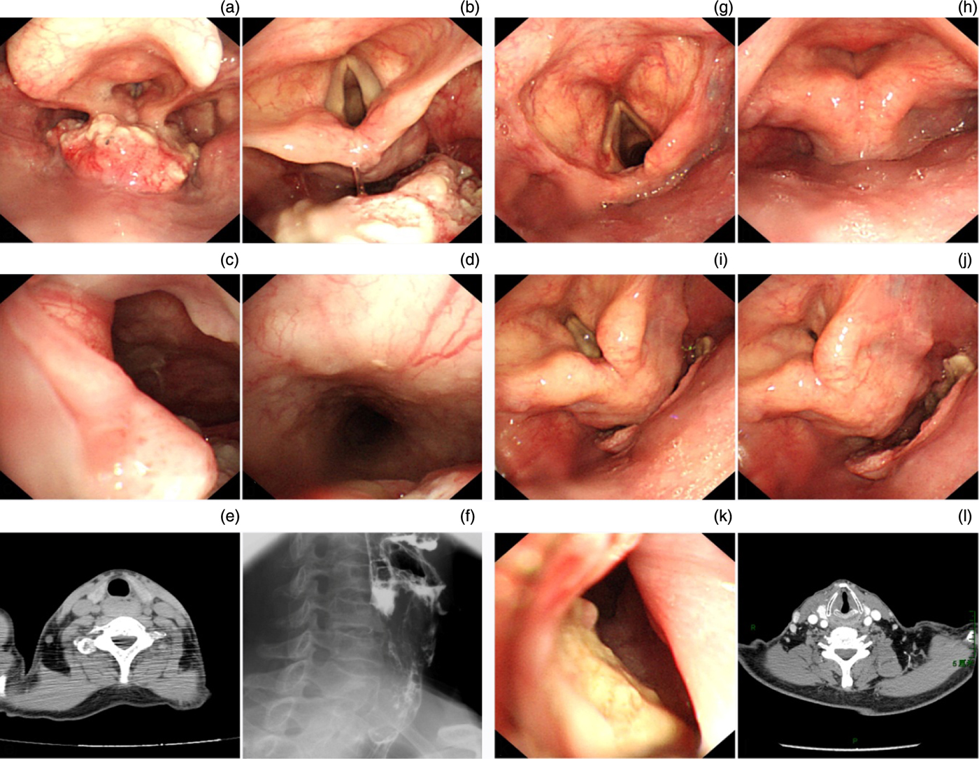
Conventional laryngoscopy was unable to evaluate oesophageal inlet involvement in any of the 113 study patients. After injecting oxygen through the laryngoscopic biopsy channel, the oesophageal inlet was not displayed in four patients with advanced stage cancer, because of a stiff hypopharynx. However, the oesophageal inlet was clearly displayed in the remaining 109 patients, of whom 33 had oesophageal inlet invasion and 76 did not. The 33 patients with invasion of the oesophageal inlet consisted of 19 patients with posterior pharyngeal wall cancer, 10 with pyriform sinus cancer and 4 with postcricoid area cancer.
Of the 33 patients with inlet invasion, 30 patients underwent computed tomography (CT) or magnetic resonance imaging (MRI), while 3 underwent hypopharyngeal radiographic examination. Imaging examinations confirmed invasion of the oesophageal inlet in 25 patients, but were unable to do so in 8 patients. Thus, laryngoscopy using triple manoeuvre was superior to radiological examination in determining invasion of the oesophageal inlet (p = 0.003) (Figure 6).
Comparison of hypopharyngeal tumour examination using different examination methods. Case 1: (a) conventional laryngoscopy shows a cauliflower-like neoplasm on the posterior pharyngeal wall, but the lower border of the tumour is obscured; (b) cervical skin traction plus the balloon-blowing manoeuvre fully separates the posterior pharyngeal wall and postcricoid area, showing the lesion located on the posterior pharyngeal wall and not invading the postcricoid area; (c, d) oxygen injection via the laryngoscope biopsy channel enables clear visualisation of the fully expanded oesophageal entrance and cervical oesophagus, and shows disease involving the cervical oesophagus; and (e, f) axial contrast computed tomography (CT) and lateral contrast X-ray images show neoplastic invasion of the oesophageal entrance and cervical oesophagus (as noted on laryngoscopy). Case 2: (g, h) conventional laryngoscopy shows no significant lesions; (i, j) upon skin traction plus balloon-blowing manoeuvre, an ulcerative neoplasm is seen on the posterior pharyngeal wall, not invading the postcricoid area; (k) oxygen injection shows that the lesion has invaded the lower oesophageal entrance; and (l) axial contrast CT fails to show any significant hypopharyngeal lesion or oesophageal entrance invasion.

Discussion
The early diagnosis of hypopharyngeal cancer is very challenging. Less than 5 per cent of patients with hypopharyngeal cancer are in stage I at diagnosis.Reference Sewnaik, Hoorweg, Knegt, Wieringa, van der Beek and Kerrebijn6 In most patients, the disease has already infiltrated a wide range of subsites and is accompanied by cervical lymphatic metastasis. The five-year survival rate is just 25–40 per cent.Reference Wycliffe, Grover, Kim and Simental7
Currently, the predominant recommended treatment for hypopharyngeal cancer is selective laryngeal function preservation surgery, radiotherapy or a combination of the two.Reference Mehta and Harrison8, Reference Hall, Groome, Irish and O'Sullivan9 Accurate determination of the extent and pretreatment clinical staging are of great value for laryngeal function preservation surgery and for determining the boundary of the radiotherapy-targeted area. Today, the diagnosis and staging of hypopharyngeal cancer are primarily determined by radiological examination (i.e. X-ray, CT and MRI) and laryngoscopy. High-resolution CT and MRI can provide information on tumour site, size, extent, infiltrative depth and lymphatic metastasis. Multiple planar reconstruction, produced using image postprocessing functions, can clearly display the relationship between the tumour and the peripheral structures; it is especially advantageous in judging the invasion of deep tissues, including the laryngeal cartilage, parapharyngeal space and pre-epiglottic space.Reference Keberle, Kenn and Hahn10 Laryngoscopy can clearly display the actual tumour site within the pharyngolaryngeal cavity, and can allow direct observation of the mucosal extent and easy identification of superficial mucosal lesions.Reference Watanabe, Hosokawa, Taniguchi, Tsujie and Sasaki11 It also allows the laryngoscopist to directly collect biopsy specimens for pathological diagnosis. However, laryngoscopy cannot visualise deep tumour infiltration or lymphatic metastasis. Therefore, the evaluation of hypopharyngeal cancer prior to surgery requires a combination of laryngoscopy and imaging modalities.
The hypopharynx is anatomically related to the larynx. The cavity is narrow and small. During quiet respiration, the posterior pharyngeal wall and the postcricoid area often cohere together, and cannot be fully separated simply by asking the patient to pronounce the letter ‘e' to mobilise the bilateral arytenoid cartilages. In this case, the anatomical subdivisions of the hypopharynx cannot be fully exposed; consequently, the extent of hypopharyngeal tumour is difficult to determine.
In recent years, the use of laryngeal function preservation hypopharyngectomy has increased due to the development of functional surgery techniques.Reference Joo, Cho, Park, Nam and Kim12 The key to successful surgical treatment is accurate determination of the primary tumour site and the involved extent. Therefore, pre-operative laryngoscopic examination requires maximal exposure of each hypopharyngeal subdivision in order to increase the accuracy of pre-operative tumour staging.
Some studies have reported that CT scanning using a modified Valsalva manoeuvre and trumpet manoeuvre can clearly display the hypopharynx and larynx and consequently increase the precision of determination of the primary tumour site(s) and extent.Reference Henrot, Blum, Toussaint, Troufleau, Stines and Roland13, Reference Hillel and Schwartz14 However, these manoeuvres are not used in conventional CT scanning because laryngeal motion is not observed under direct vision, and the accuracy and dynamics of patients' actions are difficult to co-ordinate well with CT scanning. In contrast, laryngoscopy allows such procedures to be observed under direct vision, and the triple manoeuvre described above enables the various hypopharyngeal subsites to be fully exposed.
In this study, the hypopharynx was displayed during laryngoscopy by pulling the anterior cervical skin and using the Valsalva balloon-blowing manoeuvre. The former method was used first in order to expose the hypopharynx, as it did not require the patient's active co-operation. If the hypopharynx could not be fully exposed by pulling the cervical skin, then the patient was asked to execute the balloon-blowing manoeuvre. Using these two methods, the anatomical subdivisions of the hypopharynx could be fully exposed.
We found it preferable that patients lay in the supine position while the triple manoeuvre was performed. This position enabled easy exposure of the hypopharyngeal subsites, as the patient's weight facilitated hypopharyngeal exposure and elevation of deep laryngeal structures when the anterior cervical skin was pulled. It would be difficult to perform this procedure in the seated position as the body would move forward when the anterior cervical skin was pulled, preventing the traction force from reaching the larynx and effecting sufficient hypopharyngeal exposure. However, it was possible to use the Valsalva balloon-blowing method to expose the hypopharynx, with the patient in the seated position.
• The hypopharynx and adjacent structures are difficult to visualise on conventional laryngoscopy
• Upon anterior cervical skin traction plus the Valsalva balloon-blowing manoeuvre, the posterior pharyngeal wall and postcricoid area separate, clearly displaying the hypopharynx
• Injecting oxygen via the laryngoscopy biopsy channel clearly displays the oesophageal entrance
• These two techniques could improve the accuracy of hypopharyngeal cancer staging
In cases of poor visualisation of the hypopharynx, conventional laryngoscopy and imaging examinations are unable to accurately determine the site of the primary neoplasm, and may miss small lesions between the posterior pharyngeal wall and the postcricoid area. In this study, radiological examination led to misdiagnosis of hypopharyngeal or laryngeal cancer in three cases and missed diagnosis in four cases, and could not identify the tumour origin in six cases.
When performing the triple manoeuvre in order to enhance visualisation of tumour extent, we found that the posterior pharyngeal wall and the postcricoid area were the most difficult sites to evaluate. Pulling the anterior cervical skin and using the balloon-blowing manoeuvre enabled full separation of the posterior pharyngeal wall and the postcricoid area, significantly increasing their observation outcomes and facilitating the determination of hypopharyngeal cancer extent.
Hypopharyngeal cancer can easily invade the cervical oesophagus.Reference Popescu, Bertesteanu, Mirea, Grigore, lonescu and Popescu15 In such cases, total laryngectomy or hypopharyngectomy is performed, in combination with oesophageal replacement using tissue from the jejunum or colon. Therefore, accurate determination of oesophageal invasion is particularly important prior to surgery. However, the oesophageal entrance is difficult to examine. Imaging examinations can correctly diagnose large lesions but are limited in detecting superficial lesions extending from the hypopharyngeal mucosa to the cervical oesophagus; detection of the latter lesions must depend on endoscopy. Conventional gastroscopy quickly passes through the oesophageal entrance to avoid the nausea induced by the pharyngeal reflex. Moreover, a thick gastroscope can induce significant discomfort, and visualisation of the oesophageal entrance is poor. For these reasons, we used high-flow oxygen injected through the thin laryngoscopic biopsy channel, in order to imitate gastroscopy and to enable optimal visualisation of the oesophageal inlet. Conventional laryngoscopy is unable to adequately observe the oesophageal inlet. However, using the oxygen injection method described above we were able to fully expose the oesophageal inlet and observe the cervical oesophagus. We found oesophageal inlet invasion in 76 per cent (19/25) of patients with posterior pharyngeal wall cancer. Therefore, clinicians should be constantly alert to the possibility of oesophageal invasion of posterior pharyngeal wall cancers.
Conclusion
The pre-operative staging of hypopharyngeal cancer primarily depends upon laryngoscopy and radiological examination. Both methods have their advantages and are complementary in determining the primary tumour site and extent. The key to improving the accuracy of pre-operative evaluation of specific hypopharyngeal structures is effective exposure of the anatomical divisions of the hypopharyngeal cavity. Using flexible endoscopy techniques, it is possible to fully expose the hypopharynx and larynx under direct vision, through the active movement of the laryngopharynx, in order to visualise the various anatomical subdivisions. In this study, cervical skin traction and Valsalva's balloon-blowing manoeuvre were used to expose the hypopharyngeal walls, while oxygen injection through the laryngoscopic biopsy channel was used to expose the oesophageal entrance and cervical oesophagus. This triple manoeuvre produced a favourable effect and significantly increased the accuracy of pre-treatment staging of hypopharyngeal cancer.