Introduction
CHD affects 1% of all live births in the United States.Reference Hasan, Abu Lehyah and Al Tarawneh1 Medical and surgical advancements have significantly increased the survival of all children with CHD.Reference Rao and Agarwal2–Reference Sood, Newburger and Anixt3 Approximately 50–75% of these children are affected by neurodevelopmental disabilities, which typically manifest as motor delays in infancy, behavioural challenges in toddlerhood, learning problems in childhood, and poor executive function and other adaptive impairments through adulthood .Reference Fourdain, Simard and Dagenais4–Reference Finkel, Sun and Jackson5 Children with critical CHD, such as those with single ventricle physiology and transposition of the great arteries, are at the greatest risk for neurodevelopmental disabilities.Reference Sood, Newburger and Anixt3,Reference Marino, Lipkin and Newburger6
Paediatric neurodevelopmental impairments negatively impact a child’s highest level of educational attainment, which limits employment opportunities in adulthood and likely contributes to unemployment rates twice that of the healthy population. These aftereffects negatively affect an individual’s quality of life, leading to a lack of independence that is costly for society.
There are socio-economic, medical, perioperative, and other surgical risk factors that may impact the developmental outcomes of children with CHD. These include race, ethnicity, APGAR scores, age at first surgery, time on cardiopulmonary bypass, aortic cross-clamp time, and hospital length of stay after surgery.
Socio-economic status
Socio-economic status is associated with children’s brain and behavioural development and is a known risk factor for children with CHD. Reference Robinson, Sahai, Pennacchio, Sharew, Chen and Karamlou7–Reference Xiang, Su and Liu9 For example, a recent study in children with hypoplastic left heart syndrome found that, when comparing scores on the Ages and Stages Questionnaire, the Vineland Adaptive Behavior Scales, and the Behavior Assessment System for Children, children with lower socio-economic status had more adverse neurodevelopmental outcomes by 6 years of age. Reference Bucholz, Sleeper and Goldberg8 In addition, in children with critical CHD, as measured by the Pediatric Quality of Life Inventory assessment, families with a lower socio-economic status reported lower quality of life than their counterparts. Reference Xiang, Su and Liu9
Children with critical CHD have worse outcomes across their lifespan if the individuals have racial and ethnic disparities. Reference Bayne, Garry and Albert10–Reference Dumornay, Lebois, Ressler and Harnett11 In a study observing the relationship between race/ethnicity to cognitive and language scores on the Bayley Scales of Infant and Toddler Development II (2nd edition) in extremely preterm toddlers, it was found that Black and Hispanic-White toddlers scored lower when compared to White toddlers. Reference Freeman Duncan, Watterberg and Nolen12
There are also relationships between payor status and neurodevelopment. In a recent study, it was shown that children with public insurance had neurodevelopmental disorders more than those with private insurance, and children with private insurance received an earlier diagnosis of neurodevelopmental disorders than those with public insurance. Reference Straub, Bateman and Hernandez-Diaz13
Medical/perioperative/surgical risk factors
Cardiopulmonary bypass is a transformative procedure that saves the lives of children with CHD by oxygenating blood outside the body and allowing the heart and lungs to rest perioperatively and postoperatively.
An aortic cross-clamp is an instrument that is used during open heart surgery to interrupt the blood flow of the aorta during surgical repair, which allows the surgeon to have a clear field during cardiac surgery. Reference Kumar14
There is a growing interest in determining relationships between perioperative factors (e.g., cardiopulmonary bypass, aortic cross-clamp time, and hospital length of stay) and neurodevelopment. Results continue to be mixed, with some studies finding no relationship between these factors and others finding significant relationships between these factors. Reference Shakya, Saxena and Gulati15–Reference Basgoze, Temur and Ozcan16 Early life stressors may have a lasting effect on neurodevelopment; therefore, it is important to consider opportunities that take advantage of the neural plasticity of the young brain.
Importance of early intervention
Neural plasticity helps to shape cognitive and behavioural developmental processes throughout the lifespan. In particular, the high plasticity of the infant brain provides opportunities for optimal benefit to early interventions that stimulate brain development. In children with neurodevelopmental disabilities, it has been shown that children who are < 3 years of age receive more benefits from neurodevelopmental interventions than their peers who received similar therapies at an older age. Reference Weyandt, Clarkin and Holding17–Reference Chorna, Cioni, Guzzetta, Gallagher, Bulteau, Cohen and of Michaud J.L.B.T.H.18 This stresses the importance of timely enrolment in developmental therapies.
Most children with critical CHD have neurodevelopmental disabilities. Reference Sood, Newburger and Anixt3 In the past, the primary goal in this population was survival through managing the heart defect. Because of the advancements in survival, there has been a shift in focus on addressing comorbidities such as neurodevelopmental disabilities. To best identify and treat neurodevelopmental disabilities in children with CHD, the American Heart Association recommends children with CHD be evaluated periodically, specifically at the following timepoints: 12–24 months of age, 3–5 years of age, and 11–12 years of age. Reference Marino, Lipkin and Newburger6 It has been suggested that hospitals implement a cardiac neurodevelopmental follow-up clinic where children with CHD can have these serial developmental assessments to identify areas of concern and refer to appropriate therapeutic interventions in a timely fashion. Reference Marino, Lipkin and Newburger6
Tools for early detection
When quantifying child development, many experts consider the Bayley Scales of Infant and Toddler Development III (3rd edition) to be the “gold standard” in research. Reference Rasheed, Kvestad, Shaheen, Memon and Strand19–Reference Ranjitkar, Kvestad and Strand20 Bayley-III scores are routinely used to quantify neurodevelopment in children starting in infancy and continuing through the lifespan.
The Bayley-III is a validated and repeatable measure that can be used for early identification of neurodevelopmental disabilities, can monitor progress over time, can assist in providing targeted interventions for children with developmental delays, and can assess multiple developmental domains (e.g. cognition, motor, language, social, and emotional). Reference Del Rosario, Slevin, Molloy, Quigley and Nixon21–Reference Balasundaram and Avulakunta22 Although this tool is well-accepted and reliable, there are some limitations to it, such as long administration time and dichotomous scoring. Despite its many strengths, administration of the Bayley-III requires specialised training and is time-intensive. Therefore, it is costly/impractical to incorporate its use in standard clinical settings.
To quantify development in infants and toddlers up to 36 months of age, Arnold Capute developed the Capute Scales for use in the real-world setting. The Capute Scales has two domains: (1) the Clinical Linguistic and Auditory Milestone Scale, which measures expressive and receptive language, and (2) the Cognitive Adaptive Test, which measures cognition through visual-motor skills. The Clinical Linguistic and Auditory Milestone Scale was originally developed to provide paediatricians with a rapid and efficient scale of linguistic and auditory milestones that can be conducted in a busy practice setting. Reference Accardo Pasquale and Capute23–Reference O.’Connor Leppert, Shank, Shapiro and Capute24 This tool can be used by physicians, nurses, and other health professionals, and it does not require specific training. Reference Semrud-Clikeman25
Using the Capute Scales, a clinician can determine the developmental age of a child at a single time point. To further characterise the developmental level (e.g., mild delay, moderate delay) of a child, the clinician uses this information to also determine the developmental quotient. Developmental quotients are calculated by dividing the developmental age by the chronologic age, and then multiplying by 100. An example of how this is calculated is as follows: a 3-year-old child who has a developmental age of 2 years has a developmental quotient of 67, which places the child in the range of mild language delay, as defined by a developmental quotient of 55–69.
Studies have shown that the Capute Scales developmental quotients are highly correlated with the Mental Development Index of the Bayley Scales II–III. Capute Scales developmental quotients correlate with the Bayley Scales II Mental Development Index scores at 12 months (r = 0.393, p = 0.008) and 30 months of age (r = 0.742, p = 0.0001). Reference Voigt, Brown and Fraley26
An assessment with the Capute Scales can be completed in 15–20 minutes by a trained clinician, whereas the Bayley Scales can take up to 90 minutes to complete. When evaluating children with complex medical conditions such as CHD, efficient and reliable evaluation is an important consideration.
Neurodevelopmental disabilities in children with CHD
Children with critical CHD often have mild deficits in cognitive, language, and behavioural skills, which are associated with academic problems. Our cardiac neurodevelopmental programme has successfully implemented systematic use of the Capute Scales in outpatient developmental surveillance in children at the highest risk for adverse neurodevelopmental outcomes. However, the use of the Capute Scales as a primary neurodevelopmental data collection tool for research purposes is limited. There remains limited early neurodevelopmental information on children with CHD. To remedy this, we decided to report our findings on early language development as measured by the Capute Scales.
Language development is a well-known predictor of future cognitive outcomes. Reference Allen, Marino, Abdulla, Berger, Backer and tal27 It is known that children with a history of developmental delays in language skills are more likely to develop behavioural problems than their typically developing peers. Reference Fourdain, Simard and Dagenais4 Because children with critical CHD often have behavioural problems in toddler years, we hypothesise that these children will have language delays when compared to age-appropriate normative values by three years of age. Our primary outcome was to visualise early developmental language trajectories in children with single ventricle physiology and transposition of the great arteries. We also included demographic, medical, and surgical information on our study population. To evaluate our hypothesis, we performed a retrospective cohort study of infants and toddlers with critical CHD who had serial developmental evaluations in our clinic.
Cardiac neurodevelopmental programme
At Arkansas Children’s Hospital, all children with CHD who meet American Heart Association criteria for developmental disorders risk category 1 (e.g., single ventricle physiology and transposition of the great arteries) are identified. Reference Sood, Newburger and Anixt3,Reference Marino, Lipkin and Newburger6 Because these children are at the highest risk for developmental disorders, they are enrolled in a Cardiac Neurodevelopmental Program. In particular, for children with single ventricle physiology and transposition of the great arteries, developmental surveillance is typically initiated in infancy. Clinicians (developmental neurologist and developmental paediatrician) evaluate these children approximately every 6 months to evaluate their neurodevelopmental progress, provide referrals to therapy, assess response to intervention, and treat children for comorbid or co-occurring conditions. As part of these scheduled visits, clinicians quantify language development in all children with the Clinical Linguistic and Auditory Milestone Scale subsection of the Capute Scales.
To evaluate developmental progress over time and assess response to intervention, the clinicians in the Cardiac Neurodevelopmental Program administer the Capute Scales serially on all children. As per the programme’s protocol, clinicians administer this assessment until the child reaches 3 years of age. After 3 years of age, children transition care, and they undergo a full battery of psychological assessments in preparation for school readiness.
Materials and methods
We identified all children with single ventricle physiology and transposition of the great arteries who were evaluated with the Capute Scales in our clinic prior to 3 years of age. Because all children with single ventricle physiology and transposition of the great arteries begin developmental surveillance using the Capute Scales starting in infancy, this cohort reduces the selection bias of our sample.
To isolate the effects of critical CHD on language development, we excluded infants with factors that may also contribute to developmental language delay. These factors were prematurity (< 37 weeks’ gestation), known genetic syndromes, and hearing impairment. We also excluded children who had a heart transplant in the first 3 years of life.
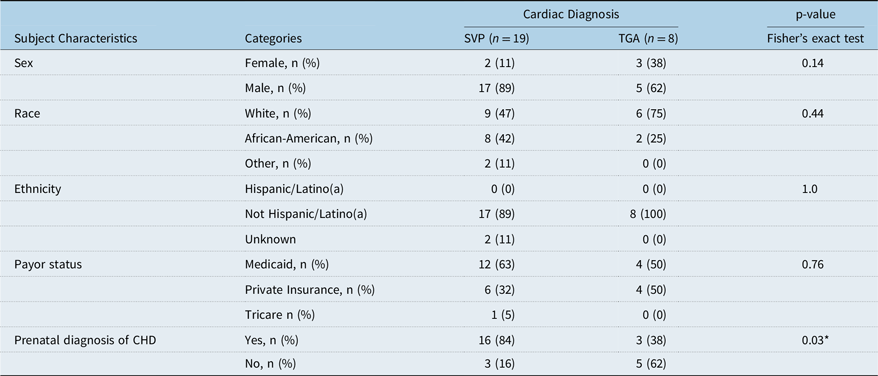
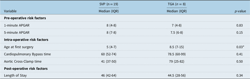
We gathered the following baseline demographic characteristics: sex, race, ethnicity, payor status, and prenatal diagnosis of CHD. We also gathered information on certain medical/pre-operative, intra-operative, and post-operative factors that may have an effect on the neurodevelopmental of children with single ventricle physiology and transposition of the great arteries. The pre-operative/medical factors were 1-minute and 5-minute APGAR scores. The intra-operative factors included age at first surgery, cardiopulmonary bypass, and aortic cross-clamp time. The primary post-operative factor was length of stay.
Our primary goal was to visualise language developmental trajectories in children with critical CHD. First, we created spaghetti plots for individual participants by tracking changes in the developmental age as a function of chronologic age at the time of assessment. Then, we treated participants with single ventricle physiology and with transposition of the great arteries as their own groups, and we created univariate models to determine what would be a general developmental trajectory in each group. Using these trajectories, we determined expected developmental level at 18, 24, and 30 months for both groups. Using our clinical experience, we hypothesised that children with critical CHD would have clinically significant delays by toddlerhood.
Statistical analysis
We compared demographic information between the two groups using Fisher’s exact test. We compared the pre-operative, intra-operative, and post-operative risk factors between the two groups. Because the factors did not follow a normal distribution, we used the Mann–Whitney Wilcoxon test to evaluate for differences.
We fitted a univariate linear regression model of group-wise data. To determine expected developmental levels at 18, 24, and 30 months, we calculated point estimates and 95% confidence intervals at each time point. All statistical analyses were conducted using R Studio, Reference Team28 version 2024.04.2 + 764. We used the following packages: car, ggpubr, ggfortify, gridExtra, knitr, readr, and tidyverse.
Results
With IRB approval, we identified a cohort of children with single ventricle physiology and transposition of the great arteries (a subgroup of children in risk category 1) who underwent developmental surveillance in the Cardiac Neurodevelopmental Program.
Our inclusion criteria yielded a total of 53 children. There were 39 children in the single ventricle physiology group and 14 children in the transposition of the great arteries group. In the single ventricle physiology group, we excluded 10 children for prematurity, 6 children for genetic syndromes, and 4 children who underwent a heart transplant. In the transposition of the great arteries group, we excluded three children for prematurity, two children for genetic syndromes, and one child who had a hearing impairment. This resulted in a total of 27 children who met our inclusion and exclusion criteria: 19 subjects with single ventricle physiology and 8 subjects with transposition of the great arteries.
Demographics
Baseline demographic characteristics are listed as frequency and percentage for categorical variables and mean and standard deviation for continuous variables (Table 1). There were more males in each group, most of the children were white and not hispanic, and most of the children had Medicaid. These characteristics were not significantly different.
Demographic characteristics

Abbreviations: SVP = single ventricle physiology; TGA = transposition of the great arteries.
However, we did find some differences between the groups in regards to prenatal diagnosis. In mothers of children with single ventricle physiology, 84% were aware of the diagnosis prenatally; whereas in mothers of children with transposition of the great arteries, 38% were aware of the diagnosis prenatally (p = 0.03). We attributed this difference to the historical dependence on the four-chamber view of the heart during the anatomy scan. Using this criterion, a diagnosis of transposition of the great arteries can easily be missed due to the four-chamber physiology. Hypoplastic left heart syndrome is a more obvious diagnosis using the four-chamber view than transposition of the great arteries in a routine obstetrics exam.
Surgical factors
Median 1-minute and 5-minute APGAR scores were between 7 and 8 in both groups. Median age at first surgery was 5 days old in children with single ventricle physiology and 8.5 days old in children with transposition of the great arteries (p = 0.03). This is consistent with standard practice for children with critical CHD. Reference Hampton Gray, Sorabella and Moellinger29 Median cardiopulmonary bypass time and cross-clamp times had wide variability in both groups. Median length of stay was similar in both groups. However, there were a few outliers with significantly longer length of stay in the single ventricle physiology group (Table 2).
Visual representation of language development
Individual trajectories elucidate changes in developmental age as a function of chronologic age (Figure 1). In infancy, language development in both groups is generally age-appropriate. None of the patients had developmental regression. Deviation from age-appropriate developmental trajectories becomes more significant by 18 months of age. This finding was similar in both groups.
A spaghetti plot depicting the language development of children with single ventricle physiology and transposition of the great arteries from 0 to 36 months of age. Each colour represents an individual and each dot of the same colour represents a clinic visit. The cirlces represent the single ventirc and the triangles represent children with transpostion of the great arteries. The deviation from age-appropriate developmental trajectories becomes more apparent by 18 months of age.

Quantification of language development
We fitted a group-wise linear regression of their developmental ages from infancy to toddlerhood for both groups. We conducted a residual analysis to confirm that this model was appropriate.
Similar to the individual trajectories, language development was age-appropriate in the first year of life. However, by 18 months, deviation from age-appropriate language development becomes apparent in both groups. (Figure 2).
Univariate linear regression model showing the language developmental trajectories per group. The single ventricle physiology group is shown in blue as circles and the transposition of the great arteries group is shown in red as triangles. The deviation from age-appropriate developmental trajectories becomes more apparent by 18 months of age in both groups.

We calculated and compared point estimates and 95% confidence intervals for developmental quotients at 18, 24, and 30 months for each group. In the single ventricle physiology group, the developmental quotients were 77 (70–83), 73 (67–69), and 71 (65–77), respectively. In the transposition of the great arteries group, the developmental quotients were 76 (66–85), 71 (62–80), and 68 (58–77), respectively. Because we fitted a linear model to our data, developmental quotients were similar and corresponded with borderline-mild delay.
Discussion
Using the Capute Scales, we quantified language development in infants and toddlers with two types of critical CHD. By creating language developmental trajectories, we observed that language development is age-appropriate in infancy, and deviations from the typical development curve only become apparent by 18 months. This deviation is measurable as a borderline-mild language delay that is evident by the child’s third birthday. Language delays are targets for early intervention, such as speech therapy, to help mitigate emerging behavioural problems. Reference Sood, Newburger and Anixt3,Reference Rogers, Malik and Fogel30 Our findings can help guide clinicians as they counsel families regarding optimisation of language outcomes through enrolment in multidisciplinary developmental surveillance programmes.
There are several factors that could contribute to increased risk of language impairments in children with critical CHD. These factors may include in utero hypoxia, medical, and surgical risk factors, and other factors related to CHD itself. Reference Leonetti, Back, Gallo and Ishibashi31 These factors may be associated with a final common pathway that results in brain dysmaturity. In support of this conjecture, children with CHD have brain volumes that are approximately 4–6 weeks behind their healthy same-age peers. Reference Leonetti, Back, Gallo and Ishibashi31 To assess the effects of these factors, we planned to evaluate them in a multivariable model. Unfortunately, due to the limitation of our small sample size, the multivariable model was unstable, so this was unsuccessful.
To avoid selection bias, we only selected children with single ventricle physiology and transposition of the great arteries because all of them begin developmental surveillance in infancy in our clinic. Although development in children with other types of CHD is also important, we only see the sickest of the other diagnoses within our clinic. Both attrition bias and survivorship bias may have influenced our longitudinal data collection and analysis.
Our work strengthens the impetus for recurrent developmental evaluations to identify and treat new delays in a timely manner. By showcasing the reliability and practical use of the Capute Scales, we hope others will use it in cardiac neurodevelopmental programmes at their respective institutions. We plan to continue using this tool to track early neurodevelopment in our clinic and in future longitudinal cohort studies. By providing targeted therapies and allocating resources strategically, we hope to improve neurodevelopmental outcomes and quality of life in children with critical CHD.
In the future, we would like to explore the contribution of the above risk factors on language development, using a multivariable model with a larger study population. Aetiologies of neurodevelopmental disabilities are not well understood and are multifactorial. Reference Verrall, Blue and Loughran-Fowlds32 Therefore, it is important to better identify factors that may pose a risk early on in development, we recommend multicenter collaborations to increase sample size and lead to new discoveries.
Pre-operative, intra-operative, and post-operative risk factors in children with CHD

Abbreviations: SVP = single ventricle physiology; TGA = transposition of the great arteries; IQR = interquartile range; Using a Mann–Whitney Wilcoxon test, the two groups were not significantly different in all variables except “surgery age” (P < 0.05).
Acknowledgements
The authors would like to acknowledge the guidance provided during preparation of this manuscript made possible by the Center for Childhood Obesity Prevention funded by the National Institute of General Medical Sciences of the National Institutes of Health under Award Number P20GM109096 (Arkansas Children’s Research Institute, PI: Weber). We would also like to acknowledge REDCap resources provided by the National Center for Advancing Translational Sciences of the National Institutes of Health under Award Number UL1 RR029884 (University of Arkansas for Medical Sciences, PI: James).
We would also like to thank the patients and their families for their participation in our study. We would also like to thank the following individuals for their assistance with our work: Meera Arunkumar, Gabrielle Gauthier, and Dr Julienne Thomas.
Financial support
This work was supported by the Arkansas Children’s Research Institute, the Arkansas Biosciences Institute, and the University of Arkansas for Medical Sciences Translational Research Institute: UL1 TR003107, KL2 TR003108
Competing interests
No relevant interests exist.
Ethical standard
The authors assert that all procedures contributing to this work comply with the ethical standards of the relevant national guidelines on human experimentation and with the Helsinki Declaration of 1975, as revised in 2008 and has been approved by the institutional committees.