Introduction
The design of compound eyes and the balance between different optical parameters have been discussed and reviewed in several studies (Snyder, Reference Snyder and Autrum1979; Land, Reference Land and Autrum1981; Warrant & McIntyre, Reference Warrant and McIntyre1993; Land & Nilsson, Reference Land and Nilsson2002). It is known that for an eye of a given size, sensitivity and spatial resolution are in conflict with each other, and a distinction between superposition eyes (useful under dim light conditions and characterized by a wide pigment-free clear zone between dioptric and light-perceiving structures) and apposition eyes (with no such clear zone and typical of diurnally active species) has been made ever since Exner (Reference Exner1891) described these two eye types. In the apposition eye, large facets, for example, can lead to an increase of the eye’s overall sensitivity but will decrease spatial resolution if the interommatidial angle is also large; smaller facets and the resultant smaller interommatidial angles, on the other hand, would favor higher resolution but then allow less light to enter each facet and, thus, lead to a reduction in sensitivity.
For butterflies, Rutowski et al. (Reference Rutowski, Gislen and Warrant2009) were able to show that an increase in the size of the eye was accompanied by an increase in sensitivity and resolution. The large radius of the eye allowed for a simultaneous decrease of the interommatidial angle (with the result of improved resolution) and large facet diameters (leading to increased sensitivity). Yet, how small insects with body sizes below 1 mm cope with the demands for optimal sensitivity and acuity in face of the extremely limited space for the eyes on the head are questions that to date have not been examined.
One consequence of reducing the size of the head in an insect is that the space for the eyes decreases as well and the question arises how far the size reduction of an eye and its components can be pushed before the reduction impacts on the function of the eye. As smaller facets are increasingly limited by diffraction (Barlow, Reference Barlow1952), one would expect a certain minimal facet size to exist that cannot be undercut. As the eye approaches this lower limit in size, it should be accompanied by a reduction in the total number of facets. However, one also needs to consider that the dioptric apparatus must have a certain size to focus (or at least channel) the impinging light on the underlying rhabdom and that the microvillar membrane fringes of the latter have to be sufficiently voluminous to house an adequate amount of visual pigment necessary to intercept incoming photons. How and to what extent in very small eyes neighboring facets are insulated from each other by screening pigment sleeves and how thin and curved a cornea can be made in order to serve equally well as a dioptric and a protective structure are further questions. Thus, in order to understand more fully the general features and functional limitations of compound eyes, we felt a look at the optical components, anatomical organization, and ultrastructure of very small eyes was needed. In this first paper on miniaturized compound eyes, we focus on the eye of the minute wasp Trichogramma evanescens Westwood (1833).
With a body size of only 0.3–0.4 mm, the parasitoid T. evanescens is one of the smallest insects known to possess compound eyes. Because of its use as a biological pest control agent against crop-impacting moths and butterflies, commercial rearing has made the species easily obtainable, and information on its biology and life cycle has been available for several decades (Schulze, Reference Schulze1926; Salt, Reference Salt1937; Quednau, Reference Quednau1958; Romeis et al., Reference Romeis, Brabendreier, Wäckers and Shanower2005). Since the females of this wasp react to the pheromones of a variety of lepidopteran species, special attention was given to chemosensory features of Trichogramma (e.g., Lewis et al., Reference Lewis, Sparks and Redlinger1971; Noldus & van Lenteren, Reference Noldus and van Lenteren1985). In contrast, not much is known about this species’ visual capacity and the role that vision plays in its life. Altogether missing is information on the anatomy and ultrastructure of its tiny eye.
Quednau (Reference Quednau1956) assumed that visual cues could be used by T. evanescens for host detection in the near field, but so far, this has not been confirmed. On the other hand, the functionality of the eyes of T. evanescens was demonstrated by Quednau (Reference Quednau1958), who showed that the species reacted positively phototactically and increased its activity during illumination, and by Brower and Cline (Reference Brower and Cline1984), who reported a stronger attraction to black (i.e., UV-A) than to white light. Romeis et al. (Reference Romeis, Shanower and Zebitz1998), furthermore, described a selective response to colored sticky traps that differed for males and females of various representatives of the genus Trichogramma. This suggests that there could be sexual differences in the optical devices of males and females.
Materials and methods
Transmission and scanning electron microscopy
T. evanescens wasps were purchased from a commercial distributor (Biologische Beratung, Berlin, Germany) and raised under natural day/night rhythm. Specimens, shielded from direct sunlight, were obtained from parasitized lepidopteran eggs that had been glued onto pieces of cardboard. Following determination of the sexes, the heads of specimens to be examined were decapitated and split in half during daytime hours under room light conditions before being fixed for 1 day in a modified Karnovsky’s (Reference Karnovsky1965) fixative solution, containing 2% paraformaldehyde and 2.5% glutaraldehyde, buffered with 0.1 m cacodylate (pH 7.4).
After a 2-h period of postfixation in 2% OsO4 solution, also buffered with 0.1 m cacodylate (pH 7.4), the samples were rinsed 3 times in the same buffer. The samples were dehydrated in a graded series of ethanol before being passed through different acetone/Epon mixtures (3:1, 1:1, 1:3, pure Epon). Pure Epon-812 resin was used for final embedding and the samples were then polymerized at a temperature of 60°C for 3 days. For light microscopy (LM), semithin sections were cut on an ultramicrotome (RMC, Boeckeler Instruments Inc., Tucson, AZ) with a glass knife and stained with 0.5% aqueous solution of toluidine blue on a hot plate for 30 s. Ultrathin sections were cut with a diamond knife and picked up on formvar-coated copper slot grids. The ultrathin sections were stained with lead citrate and 2% aqueous uranyl acetate for 20 min each (modified after Reynolds, Reference Reynolds1963). The sections were mainly examined under Zeiss EM 900 and Jeol JEM-1011 transmission electron microscopes (TEM), operated at 80 KV. The TEM was calibrated with a calibration grid (Plano S104) before any measurements commenced. For scanning electron microscopy (SEM), osmium-fixed heads were dehydrated in a graded series of ethanol and then subjected to critical-point-drying (EMITECH 850, Emitech Ltd., Ashford, UK). The dried samples were coated with a layer of approximately 15-nm gold in a sputter coater (Quorum Q150T S, Quorum Technologies Ltd., East Grinstead, UK) and observed under a JEOL JSM-5900 SEM, operated at 20 KV.
Morphometric analyses
SEM was used to determine dorsoventral and anterior–posterior lengths of the eye (i.e., height and width, respectively), total number of ommatidia per eye, facet diameters, and diameters of the ocelli. Longitudinal sections for LM were used for measurements of ommatidial lengths, radii of curvature of eye, and corneal facets as well as interommatidial angles. The radius of curvature of the eye was measured on the broadest anterior–posterior extension (eye width) of the eye, in sections done in dorsoventral direction, by laying a circular measurement tool over the eye surface in the image analysis software. The interommatidial angles were measured at the point of intersection of the normals to the eye surface through the ommatidial axes of central ommatidia. TEM micrographs of cross-sections were used to measure rhabdom diameters, shapes, and diameters of pigment granules of primary pigment cells (PPC) and secondary pigment cells (SPC) as well as retinula cells. Measurements of rhabdom diameters were taken at the distal tip of the rhabdom. Longitudinal sections were used to determine cone length, corneal thickness, and shapes and positions of the pigment granules within the retinula cells.
Five eyes of each sex were used for measurements under the SEM. For examinations involving LM and TEM, five eyes of either sex were used. All measurements were gathered by image analysis software (Image J, Rasband, W.S, U.S. National Institutes of Health, Berthesda, MD) and based on an identical number of objects per specimen to avoid a weighting in the statistics later.
Statistical analyses
In each set of data, normality of distribution was tested with the Shapiro–Wilk normality test. Comparisons between both sexes were performed with Mann–Whitney’s U Test for datasets that were not normally distributed, while the independent t-test was chosen for normally distributed data. The analytic software SPSS was used for all of the statistical tests.
Results
General organization of the eye
All measurements and observations reported in this paper are based on light-adapted specimens fixed under room light conditions during the day. A separate paper is planned that will deal specifically with light/dark adaptational changes in the eye of T. evanescens.
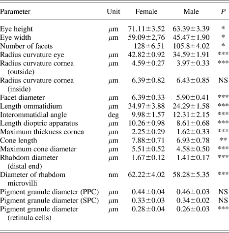
The reddish-colored compound eyes of T. evanescens are of an oval shape (Fig. 1a) and show a significant sexual dimorphism. Females have larger eyes than males: typical of the eye of a female are an eye height (dorsal–ventral length) of 71.11 μm, an eye width (anterior–posterior length) of 59.09 μm, and contains 128 facets of a relatively uniform diameter of 6.39 μm (Table 1). The eyes of the males are similarly shaped, but of smaller size (eye height of 63.39 μm and eye width of 45.47 μm), and consist of only 106 ommatidia with facet diameters of 5.90 μm. Semithin sections revealed a smaller radius of curvature of the eye in males (34.6 μm) than in females (42.8 μm). The facets are basically of hexagonal shape, but the strong curvature of the facets leads to an impression of somewhat circular outlines (Fig. 1b). In the eyes of both sexes, regularly arranged interfacetal hairs (up to 6–7 μm long and 0.45–0.70 μm thick) are present (Fig. 1c). Besides compound eyes, three ocelli are present, two smaller dorsally placed ones with diameters of approximately 11 μm and one larger median ocellus with a diameter of approximately 15 μm, the latter being nearly 3 times the diameter of a facet.
Scanning electron micrographs of the female of Trichogramma evanescens. (a) Overall appearance of T. evanescens, the compound eyes cover a large amount of the space of the head. (b) Detail of one compound eye showing the facets with a diameter of around 6.4 μm. Interfacetal hairs are present, distributed all across the eye. (c) Detail of an interfacetal hair in a corner between three facets.

Measurements of external and internal features of the compound eyes of male and female Trichogramma evanescens, fixed during the day under room light conditions

Data are expressed as means ± s.e.; NS = not significant, *P < 0.05, **P < 0.01, ***P < 0.001 in the independent t-test for normally distributed data and Mann–Whitney U Test for datasets that were not normally distributed.
Each ommatidium is made up of the distally located dioptric apparatus (i.e., multilayered cornea and tetrapartite crystalline cone) and the proximally placed photoreceptive retinula with its photoreceptive and screening pigment cells. Average ommatidial lengths differ between males and females and amount to 24.29 and 34.97 μm, respectively. Moreover, with a value of 9.84 deg, the interommatidial angle in the eyes of females is significantly smaller than the value of 12.17 deg for males. Regional differences in ommatidial shapes and sizes were not apparent in the eyes of either sex, and the rhabdoms also seemed to be arranged uniformly throughout the eyes (Fig. 2) of both sexes despite an occasional irregularly shaped rhabdom (Fig. 5c).
Transmission electron micrographs showing the ommatidial pattern within the compound eye of Trichogramma evanescens in an overview magnification: (a) Longitudinal section through the eye of the male (front to the right). (b) Cross-section through the distal part of the eye of the female, showing the regular arrangement of the rhabdoms, surrounded by the perirhabdomeric cisternae of the smooth endoplasmatic reticulum. The positions of the nuclei of adjacent retinula cells show the effective use of space as well as the large amount of space that is required by the nuclei.

Fine structural organization
Dioptric apparatus and primary pigment cells
In both sexes, the cornea is a transparent biconvex lens that consists of horizontally stacked microfibrils, typical for cuticle-covered euarthropods. In the female, the cornea is approximately 2.25 μm thick with an external radius of curvature of 4.63 μm, while in the male, it is 1.62 μm thick with a radius of curvature of 3.97 μm. The inner radius of curvature of the cornea does not vary between the sexes and measures approximately 6.40 μm. Due to the biconvex profile of the facets, the corneal thickness is least in the interommatidial areas.
Beneath the corneal lens of each ommatidium, we find two pigmented corneagenous cells, the so-called PPC (Fig. 4a–4c). Their moderately osmiophilic cytoplasm contains pigment granules of high electron density that measure approximately 0.46 μm in diameter in the eyes of males and 0.44 μm in the eyes of females (e.g., Fig. 4a and 4b). This difference is not statistically significant. The two PPC, having their large nuclei positioned at a level between cornea and proximal tip of the cone, contact the proximalmost corneal lamella by hemidesmosomes and envelop the cone cells down to their proximal tips (Fig. 4c). Emanating from the proximal sheath around the cone, the PPC narrow pigment-free extensions reach proximally for a few microns into spaces between the retinula cells close to the rhabdom (Fig. 4b). The PPC of two neighboring ommatidia are closely apposed to each other at the level of their nuclei but leave space elsewhere for the secondary pigment cells (SPC).
The cone is of the eucone type (cone matrix located inside the cone cell body); it has an average length of 7.88 μm and a maximum width of 5.51 μm in the female (corresponding values for males are 6.93 μm and 4.58 μm) and is formed by four cone cells, the so-called Semper cells. The cone matrix is rich in ribosomes and tiny particles of diameters below 2 nm that most likely consist of glycogen. Bundles of microtubuli, stabilizing the cone, are regularly encountered in the peripheral less-condensed cytoplasmic sheath of the cone cells when sectioned longitudinally. Furthermore, a ring of tiny, osmiophilic spherical structures lines the outer border of the cone as seen in cross-sections (Fig. 4b). The nuclei of the cone cells, characterized by high concentration of heterochromatin, can be found in the most distal region of the cone cells just below the cornea (Figs. 3 and 4c).
Semischematic drawings of one ommatidium of Trichogramma evanescens, showing the organization of the rhabdomeres of the retinula cells cut in different planes. (a) Longitudinal section of an ommatidium. Due to the complex interlocking arrangement of neighboring ommatidia, the cell borders of the retinula cells (dotted lines) have to be seen as purely schematic illustration. (b) Cross-section through the cone, built by four cone cells (CC), with its nuclei (NCC) and surrounding primary pigment cells (PPC) and secondary pigment cells (SPC). (c) Cross-section through the most distal part of the rhabdom showing the four duplets, each consisting of two neighboring rhabdomeres. The duplets are separated by the four cone cell projections (CP). The retinula cells contain nuclei (N), mitochondria of cristae type (M), and pigment granules. (d) In the upper third of the rhabdom, the eighth cell is replaced by a ninth cell. The beginning axonal process of the eighth cell is displaced to the periphery. (e) Proximal zone of the rhabdom. Rhabdomeres of the retinula cells 8,4,3, and 7 are the first to diminish in size and then turn into axons. In some axons, pigment granules or mitochondria are present. (f) Cross-section through region directly below the basement membrane. Axonal processes (AX) prior to entering the visual neuropil, are combined to form a primary axon bundle (one per ommatidium), surrounded by a sheath of glial cells (SGC).

Transmission electron micrographs of the compound eye of Trichogramma evanescens: (a) The dioptric apparatus made up of biconvex cornea (C) and crystalline cone (CC) is surrounded by primary pigment cells (PPC) and secondary pigment cells (SPC). The cone, showing its nuclei (NCC) in the longitudinal section, possesses four cone cell projections (white arrow) that join the rhabdom down to the basement membrane. (b) Oblique section through the cone revealing a peripheral ring of osmiophilic, dense spherical structures (arrow) and two adjacent retinula cells (RC) with their nuclei. (c) The nuclei of the cone cells (NCC) are situated below the cornea. At the contact zone of the four cone cells adhering junctions (maculae adhaerentes) are visible (white arrow). (d) Longitudinal section of the rhabdom showing the dense packing of the retinula cells with their nuclei (N) and the retinula cell pigment granules (RCP) of ovoid shapes and long axes parallel to the ommatidial axis. (e) The rhabdom (RH) ends near the basement membrane (BM) and is surrounded by the swollen tips of the cone cell projections (white arrows). Below the basement membrane pigment granules are found next to, and sometimes also within, the axons (AX). (f) Cross-section of two axon bundles of neighboring ommatidia, surrounded by a sheath of glial cells (SGC), each containing nine axons. At this level, some of the axons contain pigment granules and mitochondria. The axons are enclosed by basal pigment cells (BPC).

Proximally, the cone tapers down to the diameter of the distal tip of the rhabdom. In the transition zone between the cone and subjacent rhabdom, the four cone cells separate and each of them sends a long process running proximally between the retinula cells (infraretinular space) and alongside the rhabdom all the way down to the basement membrane, where the processes end in four swollen tips just above the basement membrane (Fig. 4e).
Secondary pigment cells
Six accessory pigment cells, here termed SPC, are present per ommatidium. Unlike the situation in other more typical apposition eyes, the SPC in T. evanescens do not accompany the retinula cells down to the basement membrane and can thereby not be classified as interommatidial pigment cells.
Measuring only 0.33–0.34 μm in diameter, the pigment granules of the SPC are of a significantly smaller size than those of the PPC, but with regard to optical density or chemical composition (judging by their coloration), there seems no difference between the granules of the two cell types. Granule diameters were the same in males and females. The nuclei of the SPC can be found near the cornea, close to the plane of the widest extension of the cone.
Pigment granules with diameters similar to those of the PPC are numerous below the basement membrane. The same holds true for pigment shielding the eye from the rest of the head (Fig. 2a). In both areas, tracheoles can be found that are missing in the eye itself. All pigments exhibit a reddish color in toluidine blue–stained semithin sections.
Retinula cells and rhabdom
The rhabdom, with a length of approximately 25 μm in females and 14 μm in males, extends from the proximal tip of the cone to just above the basement membrane. Longitudinal sections of the eye of T. evanescens reveal that the nuclei of the retinula cells are not situated in one plane but make use of almost the entire length of the retinula cells, from the distal end of the rhabdom down to the lower third. No retinula cell nuclei, tracheoles, or basal pigment cells were detected in the region above the basement membrane. Two retinula cells of neighboring ommatidia rarely both have a nucleus at the same level, at any given plane (Fig.2b). In fact, the limited space is used more effectively by alternating the positions of the nuclei in transverse as well as longitudinal planes. This results in somewhat “wavy” interdigitating outlines of neighboring retinula cells.
Most distally, where the rhabdom has its widest expansion, the nuclei of up to three retinula cells per ommatidium can be found in one horizontal plane, but further proximally, owing to the diminishing radius of the ommatidium, the number of nuclei seen at any given plane decreases. Typical cell organelles of the retinula cells, like mitochondria of the cristae type, are predominantly found close to the cell borders (e.g., Fig. 5c).
Transmission electron micrographs of the rhabdom of Trichogramma evanescens. (a) Transverse section of the most distal part of the rhabdom, showing a regular arrangement of four duplets, consisting of two neighboring rhabdomeres with the same microvilli orientation. The duplets are separated by the four cone cell projections (CP). In the cisternae of the endoplasmatic reticulum, enveloping the rhabdom, large bulges can be noticed in the intercellular spaces between the retinula cells in the distal part of the rhabdom (arrows). (b) Oblique sections reveal that the pigment-free extensions of the primary pigment cells (PPC) reach into these bulges. (c) Cross-section through the eye of a male. In the upper third, the ninth retinula cell (white arrow) replaces the eighth cell. Due to the irregular position of the proximal processes of the cone cells (CP), one of which can be seen to the left of the rhabdom, this rhabdom is of a different (not the normal) appearance. To the right side, a neighboring rhabdom is visible, with irregularly orientated microvilli of retinula cell R4. At this level, the eighth cell (R8) had already been replaced by the ninth cell (R9) in this rhabdom. Mitochondria of the cristae type (M) are usually present near the borders of all retinula cells. (d) The ninth cell shows a different microvillar orientation from that of the eighth cell, which is dislocated from the rhabdom in this plane.

The centrally fused columnar rhabdom of T. evanescens is formed throughout its entire length by eight retinula cells, all of them contributing rhabdomeres. In agreement with the finding of nine axons per axon bundle, a ninth retinula cell has been identified, which participates in the formation of the rhabdom over two third of its length, replacing the eighth retinula cell.
At the distal end of the rhabdom, the microvilli of the rhabdomeres of two opposite sets of two neighboring retinula cells each (involving four retinula cells) are aligned in the same direction. The microvilli of the rhabdomeres of the other four retinula cells are also aligned in parallel but at right angle to the microvilli of the aforementioned two sets of rhabdomeres; thus, transverse sections through rhabdoms at this level show microvilli running in just two orthogonal directions (Fig. 5a). These retinula cell duplets are separated by the four proximal projections of the cone cells that accompany the retinula cells down to the basement membrane. A divergent position of the cone cell projections can be found sporadically in some rhabdoms of the males (Fig. 5c). In the distal part, just below the cone, the rhabdom has its greatest diameter, measuring 1.67 μm in the female and 1.41 μm in the male. Apart from the difference in rhabdom diameter, there was also a significant sexual difference with regard to the diameter of the microvilli (62 nm in the female vs. 58 nm in the male).
The rhabdom is surrounded by cisternae of the smooth endoplasmic reticulum (ER). This so-called perirhabdomeric or “palisade” ER becomes enlarged as the eye becomes dark adapted (not shown in this paper). In eyes fixed under room light, the development of perirhabdomeric cisternae is restricted to a pigment-free sleeve of approximately 0.3–0.7 μm width around the rhabdom. Pigment granules of the retinula cells, measuring about 0.28 μm in diameter in the female and 0.26 μm in the male, are most abundant close to the cisternae. Considering the elongated ovoid shape of the pigment granules, the mathematically calculated significance may have been influenced by unavoidable inconsistencies in the measurements. As longitudinal sections show (Fig. 4d and 4e), the pigment granules are oriented predominantly lengthwise along the perirhabdomeric ER cisternae parallel to the ommatidial axis.
Considering the presence of an irregular ninth retinula cell, it is possible to number and identify individual retinula cells (cf., Fig. 3d and 3e). Starting with the ninth cell, the retinula cells are counted from one to eight in the direction of the adjacent duplet partner irrespective of whether it is on the right or on the left (thus in a direction away from the cone cell projection). The ninth cell starts in the upper third of the rhabdom, replacing the eighth retinula cell. The eighth cell is initially displaced to the periphery and its axon can later be found near the cone cell projection (Figs. 3d, 3e, and 5c). The different alignment of the microvilli of the ninth cell’s rhabdomere, compared with the eighth cell, leads to a situation, in which three neighboring rhabdomeres all have their microvilli oriented in the same direction (Fig. 5d), thus forming a rhabdomeric triplet.
In some rhabdoms of the eyes of females, and even more commonly in the males, an irregular rotation in the orientation of the microvilli of the fourth cell (oppositely placed to the ninth cell) may be seen, but such irregular rhabdoms are not restricted to particular regions of the eye. In situations like these, single cells with identical microvillar orientations separate two opposite triplet rhabdomeres that have their microvilli aligned at right angle to those of the two cells occupying the space between the two triplets (Fig. 5c). Arrangements intermediate between this and the arrangement of four orthogonally arranged cell duplets (see above) can also be found (Fig. 6b).
Cross-sections through the basal part of the rhabdom of Trichogramma evanescens in more proximal section planes. (a) Cross-section through different planes near the lower end of the rhabdom. In some rhabdoms (RH), the microvilli of the rhabdomeres show a different orientation to each other, so that the adjacent rhabdomeres are not arranged at an angle of 90 degree to each other anymore (arrows). The dislocated eighth cell (R8), as well as the axons (AX), is positioned in between the two cone cell projections (CP) of adjacent rhabdoms. (b) The position of the cone cell projections (CP) facilitates the identification of the changed orientation of the microvilli of one rhabdomere each of the two duplets in some of the rhabdoms. In these rhabdoms also, the fourth retinula cell (R4) exhibits a changed orientation of its microvilli. (c) At the basal end of the rhabdom, only two cells are facing each other (R2 and R6), surrounded by the axons (AX) of the other retinula cells and the cone cell projections (white arrows).

Further proximally, the retinula cells 4, 3, and 7 are the first to diminish in size and turn into axons. In this lower part of the rhabdom, a dislocation of some retinula cells can be observed, leading to minor shifts in the microvillar alignments (Fig. 6a). The next two retinula cells changing into axons are those placed opposite to each other but with identical microvillar orientations, namely retinula cells 1 and 5; the ninth retinula cell follows. At the very end of the rhabdom, only retinula cells 2 and 6, facing each other, remain. They are the longest retinula cells and contribute to the rhabdom from its most distal to its most proximal end (Fig. 6c).
At the basement membrane, axon bundles, each containing nine fibers, leave the ommatidium through separate holes in the thin and structurally homogenous basement membrane. In each bundle, usually two larger fibers are observed to be enveloped by three smaller fibers on either side with one small fiber in the center of the axon bundle (Figs. 3 and 4f). The nine axonal processes are surrounded by glial cells. Pigment granules and mitochondria were noticed in some axons.
Dark/light adaptational changes
Although we intend to describe in detail structural and ultrastructural responses of the eye of T. evanescens, caused by an exposure to light and darkness, in a separate paper, it may suffice to mention here that photomechanical events do occur. We observed displacements of the osmiophilic screening pigment granules along the ommatidial axis. This pigment migration reduces the amount of light reaching the rhabdom upon an exposure to bright light. Additionally, we noticed a progressive formation of perirhabdomeric ER cisternae around the edge of the rhabdom, accompanying an adaptation to lower ambient light levels. Total lengths of the ommatidia or interommatidial angles were apparently not affected by exposures to different environmental brightnesses.
Discussion
General remarks
Compared with the compound eyes (just termed eyes in the following) of other common nonparasitic Hymenoptera, the eyes of T. evanescens are rather small. With a dorsoventral height of 60–70 μm and facet numbers between 100 and 135, they are nearly 1/100 the size of the eye of the largest known hymenopteran, the Japanese giant hornet Vespa mandarinia ssp. japonica (whose bean-shaped eyes extend dorsoventrally over 5.8 mm and consist of at least 8000 facets), and approximately 1/40 the size of the eye of the common wasp Paravespula vulgaris, in which the eye contains approximately 3000 facets and the dorsoventral eye height is 2.5 mm (Fischer et al., unpublished). As eye size depends on the available space for the eyes on the head, eye size has to diminish with decreasing body size. A relation between eye size and body size is not only apparent during the postembryonic growth of an insect (Meyer-Rochow & Keskinen, Reference Meyer-Rochow and Keskinen2003; Keskinen & Meyer-Rochow, Reference Keskinen and Meyer-Rochow2004) but has also been demonstrated for the adults of various insect species of different body size (e.g., Jander & Jander, Reference Jander and Jander2000; Rutowski et al., Reference Rutowski, Gislen and Warrant2009).
Barlow (Reference Barlow1952) carried out measurements on the eyes of 27 different species of Hymenoptera and showed that there was a relationship between the diameters of their ommatidia and the square root of the height of the eye. T. evanescens falls below the size of the smallest species that Barlow included in his study and both size of the eye and facet diameters in T. evanescens are smaller than any of Barlow’s data; yet they are in line with Barlow’s general conclusion.
Adaptations of the ultrastructural organization to the small size of the eye
The eye of T. evanescens displays a compact and well-organized usage of space. With a total length of only 39.97 μm (24.29 in the males), a single ommatidium of T. evanescens is considerably shorter than that found in eyes of larger hymenopteran species (Apis drone: 400 μm; Perrelet, Reference Perrelet1970), Formica polyctena 140 μm (Menzel, Reference Menzel and Wehner1972). Space available in one ommatidium is not only limited due to ommatidial length but also restricted by the small facet diameter; yet each ommatidium must provide sufficient space for the retinula cells (including the rhabdom) and other cells, their nuclei, pigment granules, mitochondria, and other cell organelles involved in or influenced by photomechanical adaptations (see summary, Eguchi, Reference Eguchi, Eguchi and Tominaga1999).
The uniquely modified arrangement of the nuclei of adjacent retinula cells, dividing up available space in a most efficient way, must be understood as a direct effect of miniaturization in the Trichogramma eye. This arrangement allows more ommatidia per eye to be developed and results in more densely packed and larger rhabdoms than if retinula cells and their nuclei were conventionally arranged. The smallest amount of space in the retinula cells is obviously present around the most proximal part of the rhabdom, where no nuclei at all occur and the whole of the small available space is occupied by pigment granules and cell organelles aligned in parallel to the ommatidial axis. The kinds of cell organelles present, their dimensions, and their relative abundance, however, are not unlike those seen in the much larger eyes of the honeybee Apis mellifera (Perrelet, Reference Perrelet1970; Skrzipek & Skrzipek, Reference Skrzipek and Skrzipek1971). As the size of the pigment granules is not critically smaller than that seen in larger hymenopterans, it appears obvious that, just as predicted by Meyer-Rochow and Reid (Reference Meyer-Rochow and Reid1996), there is a minimal limit for the diameter of screening pigment granules. Whether chemical, physical, or developmental factors are involved in setting the size limit of the pigment granules is still unclear.
Given that a minimum size exists for cell organelles, then retinula cells, too, cannot infinitely be reduced in size, for they need to accommodate not only pigment granules but also nuclei, mitochondria, ER, and other essential organelles. Furthermore, considering the virtually ubiquitous ability of organelles to translocate and cells to change shape during light/dark adaptation in insect photoreceptors, space and conditions need to exist to allow such adjustments to changing ambient light levels to occur.
Contrary to what has been described in the eyes of other Hymenoptera, the SPC in T. evanescens do not reach the basement membrane. Only extensions of the PPC, although free of pigments, were found in between the intercellular spaces of adjacent retinula cells. With regard to the limited space between cones of neighboring ommatidia, nearly all that space is filled by the PPC; the space for the SPC is restricted to a small area close to the cornea. It is not possible to decide whether this tight packing of the PPC is the reason why extensions of the SPC into the retina do not occur or if it is the interlocking nature of the retinula cells of neighboring ommatidia. At least, it would seem that the loss of interommatidial (secondary) pigment cells (assumed to be a ground pattern component of mandibulate ommatidia generally: Müller et al., Reference Müller, Rosenberg, Richter and Meyer-Rochow2003) is a further result of miniaturization.
Organization of the retinula cells and the rhabdom
The rhabdomere arrangement into four duplets in the distal part of the rhabdom conforms to that of the larger worker honeybee (Skrzipek & Skrzipek, Reference Skrzipek and Skrzipek1971, Reference Skrzipek and Skrzipek1974). The eye of T. evanescens also shares the presence of a ninth retinula cell with other hymenopterans (ants: Formica; Menzel & Lange, Reference Menzel and Lange1971), Cataglyphis (Brunnert & Wehner, Reference Brunnert and Wehner1971), and bees: workers and drones of the honeybee A. mellifera (Perrelet, Reference Perrelet1970; Grundler, Reference Grundler1974; Skrzipek & Skrzipek, Reference Skrzipek and Skrzipek1974) and halictid bee Megalopta (Greiner et al., Reference Greiner, Ribi and Warrant2004). What is different, though, is that in the eye of T. evanescens, the ninth cell starts earlier, that is, more distally, probably due to the short total length of the rhabdom, than in workers of A. mellifera and Megalopta. Nevertheless, the retinula cells in T. evanescens still show the same order in which the cells are contributing to the rhabdom as in A. mellifera (Skrzipek & Skrzipek, Reference Skrzipek and Skrzipek1974). This finding underscores the conservative nature of compound eye organization in pterygote taxa generally (cf., Paulus, Reference Paulus and Gupta1979) and the Hymenoptera in particular, but it could also suggest that in evolutionary terms, eyes of T. evanescens have not diverged much from the original type inherited from the last common ancestor of the Hymenoptera. Our results support Paulus (Reference Paulus and Gupta1979), who suggested that a corona made of nine retinula cells belong to the hymenopteran ommatidial ground pattern.
Irregularities, similar to those that were occasionally seen to involve the organization of the rhabdom in T. evanescens, were also reported from other hymenopterans (e.g., Skrzipek & Skrzipek, Reference Skrzipek and Skrzipek1974). In T. evanescens, some of the irregular rhabdoms were of similar shapes to those described from the dorsal rim area of the eye of Megachile rotundata (Wachmann et al., Reference Wachmann, Richter and Schricker1973), but in T. evanescens, the presence of the irregular rhabdoms was not limited to any particular eye region. It is thus unlikely that T. evanescens possesses regionally differently specialized rhabdoms, and it is more likely that the presence of the relatively large number of irregularly shaped rhabdoms indicates that selective pressure on retaining high visual acuity has not been strong in this tiny eye.
It is conceivable that the decreasing space in the lower part of the rhabdom has an influence on the orientation of the retinula cells. This could explain why the irregular orientation of the fourth cell is found more often in the smaller eyes of the males. The fact that other rhabdomeres also frequently show a change in their microvillar orientation toward the lower end of the rhabdom shortly before they terminate supports this hypothesis. With diameters of 1.4–1.6 μm, the rhabdoms of T. evanescens are no more slender than many known from much larger insects. Actually, rhabdom diameters in T. evanescens are wider than those of rhabdomeres R7 and R8 (1 μm) in the open rhabdom of the fly (Kirschfeld & Snyder, Reference Kirschfeld and Snyder1976). Thus, even in the tiny eye of T. evanescens, the minimum possible rhabdom diameter may not yet have been reached.
Optical features
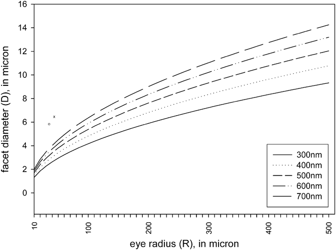
Facets in the compound eye of T. evanescens are not only much smaller than those of all other hymenopterans examined to date but also to the best of our knowledge, they are smaller than the facets of any other insect compound eye. With regard to the diffraction limit, described by Barlow (Reference Barlow1952), one would assume that it is optically counterproductive to have a compound eye with facets as small as those seen in T. evanescens. However, if we consider the small eye radius, it would seem that diffraction may not be a problem for the T. evanescens eye. The diffraction limit is given by DΔΦ ≈ 0.58 λ (Snyder, Reference Snyder1977) with facet diameter D, interommatidial angle ΔΦ, and the wavelength λ. According to Snyder (Reference Snyder1977), DΔΦ (the P-formula) can equally well be expressed by the square of the facet diameter divided by the eye radius (D 2/R) since the interommatidial angle ΔΦ equals D/R. Transposing this formula, it is possible to plot the theoretical minimal facet diameter D for different eye radii for different wavelengths, showing the impact of a small eye radius on the minimum possible facet size for a diffraction limited eye (Fig. 7).
Diffraction limit plotted for different facet diameters, wavelengths, and eye radii. The values for Trichogramma evanescens are illustrated for males (o) and females (x).

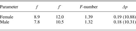
A comparison with the measured interommatidial angles and facet diameters reveals that the eyes of Trichogramma are not diffraction limited (DΔΦ > 0.58 λ). Minimal optical limits are therefore set by the minimal functional size of the lens in order to fulfill the requirements of a short focal length, the reduced amount of light entering the dioptric apparatus due to the decrease in facet diameter and rhabdom length as well as diameter. In order to reduce focal length, lens power must increase. And this is achieved by a smaller radius of curvature of the lens. Focusing the light on the tip of the rhabdom would mean that the focal length must be equal to the distance between the cornea and the tip of the rhabdom, resulting in a lens power of around 100,000–120,000 diopters (diopters = 1/f, in meters). This can be tested against the calculation of the focal length of the lens using the thick lens approximation (Jenkins & White, Reference Jenkins and White1976). As the real refractive indices are not known for the components of the eye of T. evanescens, approximations (taken from measurements on the compound eye of A. melifera; Varela & Wiitanen, Reference Varela and Wiitanen1970) were used for the lens (n l = 1.452) and for the cone (n′ = 1.348). Taking into account the limitations of the formula, namely that the refractive indices may not be homogenously distributed in the lens (Varela & Wiitanen, Reference Varela and Wiitanen1970; Stavenga et al., Reference Stavenga, Kruizinga and Leertouwer1990), the calculated image focal lengths (Table 2) for both sexes lead to a focal plane behind the tip of the rhabdom: 1.74 μm behind the tip in the females and 1.93 μm in the males. As the distance from the principal point to the front surface of the cornea must be added to the image focal length (see Stavenga, Reference Stavenga2003, for optical diagram), the distance to the tip of the rhabdom is slightly longer.
Optical parameters calculated on basis of measurements on the eye of Trichogramma evanescens and approximations for refractive indices of the dioptric apparatus. Parameters f and f´ describe the focal length and the image focal length (both in micrometer), respectively. The F-number gives the ratio of the focal length to the facet diameter and serves as a value for comparing optical properties of different compound eyes. The acceptance angle of the ommatidium (Δp) is presented in radians and in degree (in brackets)

Nevertheless, as approximations were used and the values for the refractive index of the cornea (but not cone) showed a gradient in A. melifera (Varela & Wiitanen, Reference Varela and Wiitanen1970), it is likely that the dioptric apparatus is able to focus the light on the tip of the rhabdom. Already, a slightly higher corneal refractive index (e.g., 1.49, the highest value mentioned for A. mellifera, and not an uncommon value for the cornea of other insects: Meyer-Rochow, Reference Meyer-Rochow and Horridge1975, Reference Meyer-Rochow1978) would bring the lens even more into focus (image focal point 0.49 μm behind the tip of the rhabdom in females and 0.86 in males).
The rhabdom
The amount of light that can be absorbed by the rhabdom is defined by its diameter and the total length of the receptor (Warrant & McIntyre, Reference Warrant and McIntyre1993; Warrant & Nilsson, Reference Warrant and Nilsson1998). A decrease in diameter as well as in the total length will cause a decrease in the amount of absorbed light. The amount of absorbed light can be calculated with the sensitivity formula, modified for white light perception by Warrant and Nilsson (Reference Warrant and Nilsson1998) using a value of 0.0067 μm−1 for the absorption coefficient of the rhabdoms for invertebrates. This gives a value of S w = 0.040 μm2/sr for the female and 0.026 μm2/sr for the male of T. evanescens. According to Stavenga (Reference Stavenga2003), the formula is weak for eyes with F-numbers < 2 and rhabdoms that function as waveguides (rhabdom diameter < 2 μm). Since both limitations would apply to the eye of T. evanescens (Tables 1 and 2), the results have to be treated with caution. However, they nevertheless show that the amount of absorbed light in comparison with that of larger species, for example, Apis (0.1 μm2/sr Greiner et al., Reference Greiner, Ribi and Warrant2004) is somewhat smaller.
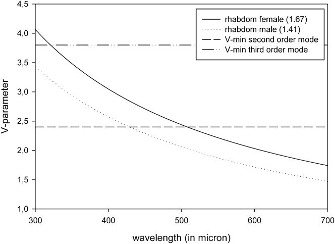
Light propagation in rhabdoms that act as waveguides cannot be explained by geometrical optics (Snyder, Reference Snyder and Autrum1979; van Hateren, Reference van Hateren, Stavenga and Hardie1989; Warrant & McIntyre, Reference Warrant and McIntyre1993). Unlike the situation in larger rhabdoms, light is not guided simply by total reflection in the rhabdom; moreover, stable waveguide modes travel on the inside and partly on the outside of the rhabdom as well. How many modes are possible for a certain rhabdom diameter and wavelength can be calculated by the V-parameter (Snyder, Reference Snyder and Autrum1979); more modes increase the total light intensity as well the receptive angle over which light can be perceived (Stavenga, Reference Stavenga2003). Fig. 8 shows the plotted results for V of the rhabdoms of male and female T. evanescens for different wavelengths using a value of 1.36 for the refractive index for the rhabdom and 1.34 for the surrounding. The graph indicates that the second order mode is only possible for wavelengths below λ = 500 nm in the female and below approximately λ = 400 nm in the male. For the female, the third order mode is also possible in the far UV. As part of the light power is traveling also on the outside of the waveguide (amount of that fraction increases with mode order), the fraction of light inside the rhabdom (n p) has to be calculated in order to determine the part that is effective for absorption. Stavenga (Reference Stavenga2003) calculated n p for different wavelengths and rhabdom diameters and showed that the light fraction within the rhabdom decreases with increasing wavelength for all modes.
V-Parameter plotted against the wavelength for the rhabdom diameter of the female (1.67 μm) and the male (1.41 μm) of Trichogramma evanescens. Dashed lines indicate the minimal limits for the second (V = 2.4) and third (V = 3.8) order mode for the waveguide.

This means for T. evanescens that sensitivity increases in the blue, violet, and ultraviolet and that these wavelengths furthermore represent the largest fractions of light power inside the rhabdom. These results fit very well with the findings of Brower and Cline (Reference Brower and Cline1984) who showed a stronger preference of T. evanescens for black light (=UV-A) rather than white light. We therefore predict that T. evanescens has UV-sensitive photoreceptors, which is not exactly surprising as other hymenopterans have also been shown to be UV sensitive (Peitsch et al., Reference Peitsch, Fietz, Hertel, de Souza, Fix Ventura and Menzel1992).
What do the findings tell us about vision in Trichogramma?
Despite its small size, T. evanescens possesses a typical apposition eye, the functionality of which as a photoreceptor was demonstrated as long ago as 1958 by Quednau (Reference Quednau1958). The small rhabdom, in connection with the small facet diameter, limits T. evanescens to bright light conditions, and its vision seems to be already light limited under room light conditions, given the typical ultrastructural signs of dark-adapted apposition eyes like, for example, the swelling of perirhabdomeric ER cisternae in the photoreceptive cells and positions of screening pigment granules (unpublished observations). Due to the even shorter rhabdoms in the eye of the male, the latter is expected to be especially light insensitive.
Differences in the structural organization of the eyes can often be linked to different life styles or visual requirements (Land, Reference Land, Stavenga and Hardie1989; Meyer-Rochow & Nilsson, Reference Meyer-Rochow, Nilsson, Eguchi and Tominaga1999; Greiner et al., Reference Greiner, Ribi and Warrant2004; Kelber et al., Reference Kelber, Warrant, Pfaff, Wallen, Theobald, Wcislo and Raguso2006). On the other hand, the size of the eye can also be simply the consequence of the limited space for it in a given insect head.
Behavioral studies of T. evanescens males and females have shown that males show a lower activity than females (Romeis et al., Reference Romeis, Shanower and Zebitz1998) around a smaller radius of the hatching site, where most of the copulations take place. Females instead are more active and disperse over larger areas searching for hosts to deposit their eggs in (Martel & Boivin, Reference Martel and Boivin2004). Therefore, the presence of smaller eyes in the males could be linked to the lower activity and a lesser need of vision of the males.
Except for a few solitary species, Hymenoptera have trichromatic vision with peaks at wavelengths of 340, 430, and 535 nm (Menzel, Reference Menzel1971; Menzel & Blakers, Reference Menzel and Blakers1976; Meyer-Rochow, Reference Meyer-Rochow1981). Some species, furthermore, appear to possess a fourth peak in the red (>600 nm: Peitsch et al., Reference Peitsch, Fietz, Hertel, de Souza, Fix Ventura and Menzel1992). With regard to the calculations of the V-parameters of the rhabdoms of male and female T. evanescens, it seems feasible that the first two orders of modes can be used by the females in connection with the peak near 430 nm, while the third mode would involve the UV. The males instead would only benefit from the second order mode in the UV. This suggests sexually different spectral sensitivities in the mid range of the wave spectrum, although we must not hastily conclude that the V-parameters alone explain the different attractiveness of differently colored sticky traps for females (white > clear > green) and males (yellow > green > white) as reported by Romeis et al. (Reference Romeis, Shanower and Zebitz1998) for Trichogramma spp.
The V-parameter shows that peak sensitivity in the UV is a property shared by both sexes but that the females are even more sensitive to light generally than the males. The use of UV in insects can help them find food as in nectar-seeking species (Manning, Reference Manning1956), be involved in partner selection (Meyer-Rochow, Reference Meyer-Rochow1991), or be of use in navigational tasks (Möller, Reference Möller2002). For the small eye of T. evanescens, it is difficult to relate a specific function to its presumed ability to perceive UV, keeping in mind the small amount of light that can be absorbed in the short rhabdom. Since UV can indicate open space better than any other wavelength (Mazokhin-Porshnyakov, Reference Mazokhin-Porshnyakov1969), it could possibly lead T. evanescens into open areas, when entrapped by foliage. Many insects appear to use UV in this way. Quite common among Hymenoptera is also the use of UV radiation in connection with the e-vector (Duelli & Wehner, Reference Duelli and Wehner1973; Wehner, Reference Wehner1989, Horvath & Varju, Reference Horvath and Varju2004), but preliminary behavioral tests with T. evanescens by us did not reveal any polarization sensitivity (unpublished observations).
General functional conclusions relevant for small compound eyes
As insects evolved smaller and smaller species, increasingly less space had to remain in the head capsule for essential structures like the musculature to operate mouthparts and antennae and the brain to integrate sensory inputs. Small insects frequently do not feed and have degenerate mouthparts. A reduction in eye size had to be one consequence of smaller heads. However, giving up photoreception completely (in contrast to feeding) has not been an option. A total disappearance of eyes is only known from some (and then not always minute) cavernicolous species (Meyer-Rochow & Nilsson, Reference Meyer-Rochow, Nilsson, Eguchi and Tominaga1999), which demonstrates that maintaining a compound eye makes sense even in the smallest members of insects, provided sufficient light is available to enter the facets and excite the visual pigments in the rhabdom.
To have small eyes means first and foremost a reduction of the diameter of the eye, one consequence of which a reduction of the total length of an ommatidium is. A shorter ommatidium by necessity will have a rhabdom of reduced length and this in turn is likely to result in a decrease in absolute sensitivity because a small rhabdom houses fewer visual pigment molecules in its microvillar membranes. The effects outlined above are independent of the size of the diameter of the facet lens, but a larger facet diameter does, of course, allow more light to enter the facet. Yet, facet diameters can only become enlarged when interommatidial angles concomitantly increase, and this leads to a decrease in the resolving power of the eye.
Furthermore, an isometric relationship between cone and total ommatidium length is deleterious; yet, a reduction of the total length of the ommatidium will affect somehow the total length of the cone (reduction of the length between the lens and the tip of the rhabdom (=f). As long as the lens still focuses the light onto the tip of the rhabdom, a decrease in the F-number (f/D) is the consequence. Generally, small F-numbers refer to highly photosensitive eyes (or optical systems), and larger F-numbers are typical of eyes (or optical systems) with reduced photosensitivity. In larger compound eyes, small F-numbers result from an increase in facet diameters, but small F-numbers can equally well be achieved given similarly sized f and D values (≈1). In order to achieve a short focal length, the curvature of the lens has to increase. A maximum limit for the curvature is set by the facet diameter, as the radius of curvature of the lens (r) should not go below D = 2 × r, as otherwise only a part of and not the whole facet would be covered.
Further size restrictions of a facet are set by the diffraction limit and the need of the underlying ommatidium to accommodate all the essential retinal cell types and their organelles. As Fig. 7 shows, the evolution of smaller eye radii is possible with smaller facet diameters without reaching the diffraction limit. But decreasing facet diameters will result in increasingly smaller interommatidial angles and, thus, less and less space for retinula cells and other ommatidial cell types, including the wealth of essential organelles dispersed therein. As the extent to which rhabdom diameter can be reduced is limited in respect to its light gathering function, sufficient place for the retinula cell nuclei will remain only in the upper somewhat wider third of the ommatidium. Thinner rhabdoms, with shorter microvilli to house the visual pigment, will be able to absorb fewer photons than larger rhabdoms, and there has to be a limit below which the gain for the insect obtained from photoreception no longer outweighs the cost to maintain photoreceptive cell specializations.
Thus, the minimum eye size seems to depend on the rhabdom length and diameter in combination with eye radius and facet diameter. To avoid crossing the limit (given through the interaction of these eye characteristics), one should expect a reduction in the number of facets, in which the latter can either be of the minimum possible dimensions or may have larger diameters. In T. evanescens, facet sizes appear to have reached their functionally possible minimum size limit, but does this hold true for the rhabdoms as well? Even if somewhat narrower rhabdoms do exist in the eyes of some larger insect species, such rhabdoms are considerably longer than the slightly wider ones seen in the eye of T. evanescens. Considering it is photon absorption, based on the amount of visual pigment molecules incorporated into each rhabdomere, which limits rhabdom size, the rhabdom dimensions encountered in the eye of T. evanescens most probably also represent the functional limit of this structure. What is worthy of note, however, is that the diameters of the microvilli in the rhabdoms of T. evanescens are no different from those making up the rhabdoms of larger species. As with the diameters of the screening pigment granules, the exact reason for this uniformity across vastly differently sized species of insects remains unknown.
Perspective
As there are other and even smaller Hymenoptera besides Trichogramma that still possess compound eyes, like, for example, the chalcid species Chiloe micropteron (Gibson & Huber, Reference Gibson and Huber2000), further investigations on insects with small compound eyes are needed to test whether the theoretical minimal size limits and conclusions on compound eye miniaturizations advanced in this paper are generally valid. As in this paper approximations were used for the refractive index gradients, measurements of the real indices by interference microscopy would be helpful in order to carry out more precise calculations. However, how realistic it is to expect such measurements to be obtained on the minute dioptric structures of the eye of T. evanescens is anyone’s guess at this moment. It would seem easier to carry out behavioral tests to determine absolute and spectral sensitivities as well as flicker fusion frequencies. It would also be interesting to determine whether temporal summation events occur in the eye of T. evanescens to compensate for its short rhabdom lengths.
Acknowledgments
We would like to thank Prof. Dr. Gabriele Uhl (Department for General Zoology and Zoological Systematics, Ernst-Moritz-Arndt-University Greifswald, Germany) and Mrs. Gabriele Ladwig (Electron Microscopic Unit within the Institute for (Neuro-)Pathology, University Hospital Essen, Germany) for having provided us generously with session time at their TEM machines. Dr. Giovanni Talarico (Department for Evolutionary Neuroethology, Max Planck Institute for Chemical Ecology, Jena, Germany) gave numerous useful technical comments for improving our fixation protocol and photodocumentation of the material. V.B.M.-R. and S.F. gratefully acknowledge the financial support received from the German Science Foundation (DFG) toward this project.