Many hospitals, clinics, schools, and summer camps in the United States conduct annual employee testing for tuberculosis infection, regardless of risk factors. A tuberculosis infection diagnosis based on a positive result, particularly when interpreted as an occupational conversion, can trigger unwarranted, expensive, and time-consuming investigations, radiography, and potentially dangerous chemotherapy.
During serial testing, seemingly spontaneous interferon gamma release assay “reversions” (a positive QuantiFERON Gold In-Tube tuberculosis test [QFT] followed by a negative test) are observed.Reference Dorman, Belknap and Graviss 1 , Reference Thanassi, Noda and Hernandez 2 Reversions cause confusion and consternation for individuals and their healthcare providers. The imposition of a binary cut-point on a continuum of interferon-gamma release measurements (range, 1.0–10.0 international units [IU]/mL) underlies QFT reversions. The Food and Drug Administration endorsed the QFT cut-point of tuberculosis-antigen-minus-nil (TBag-nil) at 0.35 IU/mL to impart a specificity of 99%, on the basis of receiver operating characteristic (ROC) analyses.Reference Mori, Sakatani and Yamagishi 3 The range encompassing most reversions likely arises from inter- and intralaboratory processing variability,Reference Whitworth, Hamilton and Goodwin 4 – Reference Doberne, Gaur and Banaei 6 combined with the low positive predictive value that is mathematically inherent when serially screening any low-risk population for a low-prevalence disease. Thus it is critical that labs include TBag-nil values when reporting results.
In a previous publication,Reference Thanassi, Noda and Hernandez 2 our group delineated, using multivariate analyses of 862 QFT-positive US healthcare workers (HCW) from 3 cities, a QFT TBag-nil peak reversion zone between 0.35 and 1.11 IU/mL. In this evaluation we expanded the sample from 862 to 1,094 serially tested, QFT-positive US HCW from 7 cities as we sought verification of the previously reported peak reversion range (cohort 1, analysis 1). Additionally, we analyzed a 400-subject subset with known tuberculin skin test (TST) results to discern whether TST status is a predictor of reversion (cohort 2, analysis 2).
METHODS
A total of 1,094 HCW from 7 states with positive QFT participated in cohort 1. All were more than 17 years old. Cohort 2 is a 400-HCW subset of cohort 1 for whom TST results were known.
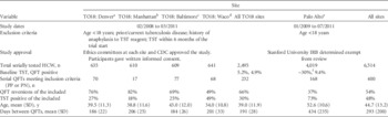
Four sites (Table 1) contributed in the Centers for Disease Control and Prevention–supported prospective Task Order 18 trial,Reference Dorman, Belknap and Graviss 1 where HCW had QFT and TST at baseline, 6, 12 and 18 months. Study inclusion began with the first positive QFT and included the next test in the series. A total of 232 HCW met inclusion criteria for cohorts 1 and 2. TST was considered positive at greater than 10 mm.
Cohort 2 Characteristics

NOTE. CDC, Centers for Disease Control and Prevention; HCW, healthcare workers; IRB, institutional review board; PN, 1 positive QuantiFERON tuberculosis test (QFT) followed by 1 negative QFT; PP, 2 consecutive positive QFTs; TO18, Task Order 18 trial; TST, tuberculin skin test.
a Denver Health and Hospital Authority and the Denver Health Department, Colorado.
b Columbia University Medical Center, New York.
c Johns Hopkins University Medical Centers, Maryland.
d Baylor College of Medicine, Texas.
e Palo Alto Veterans Affairs Health Care System, California.
f Precise data not available.
The fifth site was the Palo Alto Veterans Affairs (VA) Health Care System in California, where HCW were serially tested with QFT. TST results were extracted from medical records. A total of 186 HCW from Palo Alto VA Health Care System met inclusion criteria and are included in cohort 1; of these, 168 met inclusion criteria for cohort 2. Table 1 presents information for the combined 400 HCW from the 5 sites in cohort 2.
Sites 6 and 7 include HCW from the University of Illinois, Chicago and the Cleveland Clinic, where 613 and 63 HCW, respectively, met criteria for cohort 1. Since all sites did not contribute evenly to cohort 1, results are skewed toward University of Illinois, Chicago’s sizeable data contribution. TST results were not available for these participants so they are not in cohort 2.
The interferon gamma release assay QuantiFERON Gold In-Tube (Qiagen) was performed according to manufacturer’s instructions. 7 For Task Order 18 trial sites, QFT tubes were transported to on-site laboratories within 8 hours. Stimulated plasma was either tested immediately or incubated, then stored at 2°C–8°C for up to 2 weeks before manual enzyme-linked immunoassay testing. At Palo Alto VA Health Care System and University of Illinois, Chicago, stimulated plasma was incubated before analysis using the DS2 and DSX Automated ELISA System (Diamedix), respectively. The Cleveland Clinic used an ELx800 Absorbance Microplate Reader (BioTek). All reported results met nil, mitogen, and equation criteria for test validity.
Without a gold standard against which to evaluate latent tuberculosis infection, 2 consecutive positive tests were employed as a proxy for the probable presence of infection, the result in question. In seeking the best predictor of an individual reverting on their second QFT, the first positive TBag-nil value in the sequential series was evaluated as the predictor variable. Other variables included as potential predictors were age, time between tests, and, for analysis 2, TST result.
Statistical analyses were conducted using ROC analysis software developed at Palo Alto VA Health Care System and Stanford University.Reference Thanassi, Noda and Hernandez 2 , Reference Yesavage 8 Equal weight was placed on sensitivity and specificity for the defined criterion, reversion. The criterion for ROC progression was a critical χ2 value with a probability less than .00001.
We used ROC to analyze the 2 cohorts separately. In cohort 1 (1,094 HCW) the ROC identifies the initial TBag-nil separation point that best predicts reversion; in cohort 2 (400 HCW) the ROC also determined whether TST status influenced reversion rates.
RESULTS
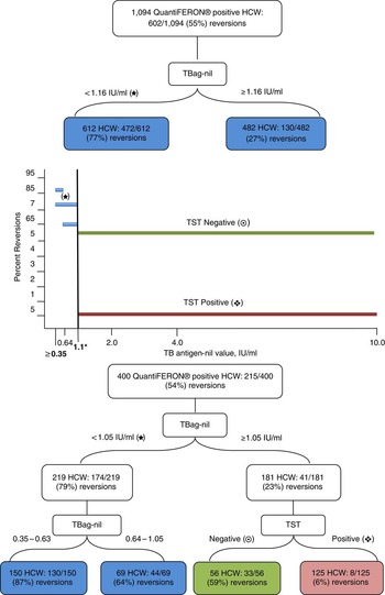
In cohort 1, comprising 1,094 HCW with 55% reverters, the ROC identified an initial TBag-nil of less than 1.16 IU/mL (kappa, 0.50; χ2, 274.1; P <.00001) as the most significant predictor of reversion, very similar to our previous findingReference Thanassi, Noda and Hernandez 2 of a separation point of 1.11 IU/mL. For 612 HCW with TBag-nil less than 1.16 IU/mL, 77% reverted; whereas for 482 HCW with TBag-nil at least 1.16 IU/mL, only 27% reverted (Figure 1). Neither the HCW age nor the time between tests statistically significantly predicted reversion (data not shown).
Percent reversions in 1,094 QuantiFERON tuberculosis tests (QFT)–positive US healthcare workers (HCW) and 400 HCW in whom the tuberculin skin test (TST) status is known. Recursive partitioning trees for analysis 1, cohort 1 (above bar graph), and analysis 2, cohort 2 (below bar graph), show the corresponding values for each bar, representing results with P <.00001. Asterisk indicates 1.1 as average between 1.05 and 1.16, results of the 2 receiver operating characteristic analyses.

For cohort 2 (400 HCW with known TST results) the ROC identified the most significant predictor of reversion as TBag-nil less than 1.05 IU/mL (hereafter rounded to 1.1; kappa, 0.57; χ2, 128.6; P <.00001), which closely matches the results of analysis 1. Here 80% of the 219 HCW with TBag-nil less than 1.1 IU/mL reverted. HCW age and time between tests did not reach statistical significance as predictors of reversion.
Additionally, for the 181 HCW (23% reversions) with TBag-nil greater than 1.1 IU/mL, the ROC found that TST result is a predictor of reversion (kappa, 0.57; χ2, 60.9; P<.00001). Of 56 HCW with a negative TST and TBag-nil at least 1.1 IU/mL, 59% reverted; whereas among 125 HCW with a positive TST and with TBag-nil at least 1.1 IU/mL, only 6% reverted (Figure 1).
DISCUSSION
Our previously reported finding that the range of highest variability occurs between TBag-nil of 0.35 and 1.11 IU/mL has been repeated in Occupational Health guidelines.Reference Thanassi, Noda and Hernandez 2 , Reference Behrman, Budnick and Hodgson 9 , Reference Schablon, Nienhaus, Ringshausen, Preisser and Peters 10 In this expanded cohort of 1,094 QFT-positive HCW, the separation point identified was nearly identical, though it should be noted that 862 of these HCW were in our original study.
Again, the time between tests did not meet criteria for significance, so although waiting 30 days before retesting patients has been suggested by some, this approach lacks documented impact. Practitioners could maximize testing compliance without sacrificing result integrity by retesting at any mutually convenient date.
In analysis 2 we found a strong association between QFT reversion and TST results when the initial TBag-nil was at least 1.1 IU/mL. Note that below 1.1 IU/mL, TST response was not found to be a significant predictor of reversion, implying variability around the cut-point is more likely attributable to mathematic and laboratory influences than patient characteristics.
Our results show that being TST-negative with a TBag-nil of at least 1.1 IU/mL predicts a negative follow-up QFT in 59% of cases. This has important clinical implications because it suggests all QFT-positive/TST-negative HCW should have a repeat QFT before further evaluation. Conversely, since TST-positive HCW with TBag-nil of at least 1.1 IU/mL had only 6% reversions, repeat testing is unlikely to change their outcome.
In this study we identified a TST-negative subgroup of QFT-positive HCW with significantly higher reversion rates when QFT values are at least 1.1 IU/mL. Retesting more QFT-positive HCW is likely to improve diagnostic accuracy.
ACKNOWLEDGMENTS
Financial support. Department of Veterans Affairs, Veterans Health Administration, Palo Alto; Department of Veterans Affairs Sierra-Pacific Mental Illness Research, Education, and Clinical Center; Centers for Disease Control and Prevention Tuberculosis Epidemiologic Studies Consortium.
Potential conflicts of interest. All authors report no conflicts of interest relevant to this article.