




No CrossRef data available.
Article contents
rDNA phylogeny of Intrapredatorus barri (Microsporida: Amblyosporidae) parasitic to Culex fuscanus Wiedemann (Diptera: Culicidae)
Published online by Cambridge University Press: 12 July 2001
Abstract
Intrapredatorus barri, a microsporidian parasite of the predator mosquito Culex fuscanus Wiedemann has been shown to be heterosporous. In many features this species has been reported to be similar to Amblyospora trinus, another microsporidian parasite of a different predator mosquito which was thus proposed for reassignation to the genus Intrapredatorus. In this report small subunit rDNA has been sequenced from I. barri and phylogenetic analysis of 42 microsporidia sequences has been performed.Nucleotide sequence data reported in this paper are available in the Gen Bank™ database under the accession number AY013359. The molecular data show that I. barri can not be a member of the genus Amblyospora. Furthermore, the phylogenetic analysis shows, with high confidence, that the Amblyospora spp. sequences in the present study are not monophyletic.
Information
- Type
- Research Article
- Information
- Copyright
- © 2001 Cambridge University Press
INTRODUCTION
The microsporidia are obligate intracellular parasites with a broad host spectrum. The Insecta is a common host group and more than 90 species of microsporidia have been reported from at least 79 mosquitoes species in probably more than 8 genera (Hazard & Chapman, 1977; Castillo, 1980; Larsson, 1988; Sprague, Becnel & Hazard, 1992; Andreadis, 1994a, 1999).
Intrapredatorus barri, a new species of microsporidium infecting Culex fuscanus, was recently described from Liu-Chiu islet in Southern Taiwan (Chen, 1998). This is, in addition to Amblyospora trinus from C. falifaxi, the second species of microsporidia reported from a predator mosquito belonging to the genus Culex (Becnel & Sweeney, 1990; Chen, Kuo & Wu, 1998). Although these 2 species are different in spore size and certain ultrastructural features, as well as infecting different species of predator mosquitoes (Becnel & Sweeney, 1990; Chen et al. 1998), they are similar in most ultrastructures of the spore and the developmental sequence, including concurrent heterosporogony in their life-cycles. Based on all features described thus far, Chen et al. (1998) suggested that A. trinus should be placed in the genus Intrapredatorus.
In addition to ultrastructural studies, molecular methods have become useful to understand the taxonomy and phylogeny of microsporidia. Several studies utilizing molecular phylogeny have been performed on various groups of microsporidia (Vossbrinck et al. 1993; Baker et al. 1994; Zhu et al. 1994; Moser et al. 1998; Nilsen, Endresen & Hordvik, 1998; Nilsen, 1999, 2000; Cheney et al. 2000) including microsporidia infecting insects (Malone et al. 1994; Malone & McIvor, 1996; Gatehouse & Malone, 1998). In addition, Baker et al. (1998) have recently studied the small subunit (SSU) rDNA phylogeny of some species belonging to the Amblyosporidae that are primarily parasites of mosquitoes. In the present study, we compare SSU rDNA sequences between Intrapredatorus and other species typically belonging to the genus Amblyosporidae, including the type species Amblyospora californica.
MATERIALS AND METHODS
Mosquitoes and parasites
All larvae of C. fuscanus infected by the microsporidium Intrapredatorus barri used in this study were collected from Liu-Chiu islet in southern Taiwan. Classic symptoms of a swollen and porcelain appearance were seen in infected larvae. Release of meiospores from dissected mosquito larvae was used to confirm the microsporidian infection of the mosquitoes (Chen, 1998). Infected larvae were stored in 80% ethanol. Infected specimens were homogenized and the suspension of spore and host tissue filtered through a nylon mesh to remove the majority of host tissue. The spore suspension was washed in double distilled water (ddH2O) 3 times by centrifugation in an Eppendorf Microfuge at 4000 g for 2 min and finally resuspended in 100 μl of ddH2O and stored at 4 °C.
Nucleic acid preparation
The procedures for nucleic acid preparation followed the description by Nilsen (1999). The above-described spore suspension was centrifuged at 6000 g for 5 min and dissolved in a digestion buffer (100 mM NaCl, 10 mM Tris–HCl, 25 mM EDTA, 0.5% SDS, pH 8.0) containing 0.5 mg/ml proteinase K and incubated overnight at 37 °C. Nucleic acid was then extracted twice with an equal volume of phenol/chloroform followed by an equal volume of chloroform, and then ethanol precipitated. The precipitated DNA was dissolved in 50 μl of ddH2O. The DNA concentration was estimated by running a 5 μl aliquot on a 1% agarose gel.
Polymerase chain reaction (PCR)
The PCR was carried out as described elsewhere (Nilsen, 1999). Briefly, a set of primers (V1f = 5′-CACCAGGTTGATTCTGCC-3′ and 1492r = 5′-GGTTACCTTGTTACGACTT-3′) was selected to amplify the SSU rDNA gene. PCR was carried out in 100 μl reaction volume using 10 pmol of each primer, 20 nmol of each dNTP, 10 μl of 10×Taq polymerase buffer (Pharmacia, Uppsala, Sweden), 2.5 units of Taq polymerase and approximately 25 ng of the above-prepared genomic DNA. The reactions were run on a Techne thermocycler for 35 cycles at 94 °C for 1 min, 50 °C for 1 min and 72 °C for 2 min. After completion of 35 cycles, 10 min of extension at 72 °C was applied. PCR products were visualized by running 20 μl aliquots on a 1% agarose gel and the band of desired size (approximately 1400 bp) was then excised from the gel.
Sequencing
Purified PCR product was manually sequenced in both directions using the Thermo sequenase kit from US Biochemicals (Cleveland, OH). Approximately 20 ng of PCR product was used and the sequence reactions were done in accordance with the manufacturer's recommendations. The sequence reactions were run on a Teche thermocycler.
Phylogenetic analysis
The SSU sequences were aligned by eye to an alignment of microsporidia obtained from the rRNA WWW server at the Department of Biochemistry, Molecular Biology, University of Antwerp (UIA), Belgium (http://www-rrna.uia.ac.be/rrna/index.html) (Van De Peer et al. 1999). This alignment is based on the secondary structure model by Van De Peer et al. (1996), and was used in phylogenetic analysis using PAUP* 4.0b2 (Swofford, 1999). The alignment was analysed using parsimony, distance methods and maximum likelihood (ML). Different distance models were used to generate the distance matrix, i.e. HKY85 and TamNei (Swofford, 1999). The ML analysis was run assuming a gamma distribution where the kappa value was estimating from the data set. The analysis was run using the HKY85 model in PAUP* with rate heterogeneity. Tree topology was tested by bootstrapping (parsimony and distance method) and the quarter puzzle method for ML analysis. A total of 1000 replicates were run for each test.
The I. barri SSU rDNA sequence has been deposited in the GenBank and the accession number is AY013359. In addition, the following species have been included in the phylogeneic analysis (species and (accession number)): Antonospora scoticae (AF024655), Amblyospora californica (U68473), A. connecticus (AF025685), A stimuli (AF027685), A. salinaria (U68474), Ameson michaelis (L15741), Bacillidium sp (AF104087), Culicosporella lunata (AF027683), Edhazardia aedis (AF027684), Encephalitozoon cuniculi (L39107), E. hellem (L39108), Endoreticulatus schubergi (L39109), Enterocytozoon bieneusi (L07123), Giardia ardeae (Z17210/S53313), G. lamblia (M54878/M19500), Glugea anomala (AF104084), G. americanus (AF056014), G. atherinae (U15987), G. stephani (AF056015), Hexamita inflata (L07836), Ichthyosporidium sp. (139110), Loma sp. (AF104081), Microsporidium prosopium (AF151529), Nosema bombycis (D85504), Nucleospora salmonis (U78176), Pleistophora anguillarum (U47052), P. mirrandellae (AF104085), P. typicalis (AF104080), Pleistophora sp. 2 (AF104083), Pleistophora sp. 3 (AF104082), Pleistophora sp. A (U10342), Pleistophora sp. B (D85500), Parathelohania anophaelis (AF027682), Septata intestinalis (L39113), Spraguea lophii (AF104086), S. lophii (AF033197), Thelohania solenopsae (AF134205), Thelohania sp. (AF031537), Trachipleistophora hominis (AJ002605), Vairimorpha necatrix (Y00266), Vavraia oncopera (X74112), Visvesvaria algerae (AF024656), V. acridophagus (AF024658), Vittaforme corneae (L39112).
RESULTS
The sequence data showed that the primer pair produced a PCR product of 1337 bp with a GC content of 49.6%. The primer 1492r anneals about 25 bp downstream from the 3′ end of the SSU rRNA gene which makes the coding region about 1362 bp. Searching the GeneBank with the I. barri sequence, Edhazardia aedis gave the highest score followed by 4 Amblyospora spp. sequences.
Phylogenetic analyses were performed on an alignment consisting of 42 microsporidia sequences. The I. barri sequence occurred in a group consisting of 4 Amblyospora spp. sequences, E. aedis, Culicosporella lunata and Parathelohania anophelis (Fig. 1). The first branch on the microsporidia tree is Antonospora scotciae, followed by the Amblyospora group. The bootstrap support for the Amblyospora group was 76% for maximum likelihood (ML) (using quarter puzzle) and 99% using parsimony. The branching order in the Amblyospora group varied somewhat between the different tree-building methods used (Fig. 2) but some species showed a stable branching pattern. Amblyospora salinaria and A. californica grouped together supported by high bootstrap values. Furthermore, the position of P. anophelis and C. lunata was the same in all the trees (Figs 1 and 2). In all the analyses, I. barri grouped together with A. stimuli, E. aedes and A. connecticus. Using ML bootstrapping with Puzzle I. barri and A. connecticus scored 80% support. Alternative groupings are shown in Fig. 2 A and B.

Fig. 1. Maximum likelihood tree with some selected bootstrap values. Branch lengths are proportional to changes. Details of the Amblyospora group are shown in Fig. 2. ML, bootstrap values obtained by the quartet puzzle method; Pa, bootstrap values obtained using parsimony. The tree is rooted by using Giardia ardeae, G. lamblia and Hexamita inflata as outgroup. Pleistophora sp. 2 and Pleistophora sp. 3 corresponds to the same species as reported by Nilsen et al. (1998).

Fig. 2. Different tree topologies obtained for the Amblyospora group using different analysis methods. Only the species in the Amblyospora group are shown. (A) Distance tree using minimum evolution as optimal criterion and distances measured with the logdet/paralinear method. (B) Tree obtained using maximum parsimony as optimal criterion. Random addition of sequence with 100 subreplicates generated a tree with tree length of 5967 steps. (C) Puzzle quartet tree with maximum likelihood as optimal criterion. The numbers on the branches refer to percentage support obtained by the puzzle analysis. See text for explanations.
The number of nucleotide differences between the different members in the Amblyospora group was relatively large (Table 1) and all species contained several signature sequences (i.e. the occurrence of an unique sequence motif). The number of nucleotide differences between the 2 closest related species (A. californica and A. salinaria) was 34. The number of nucleotide differences between I. barri and the other species was between 129 and 262 (Table 1).

DISCUSSION
Although the microsporidia belonging to Intrapredatorus shared some features with species of Amblyospora, the uniqueness for the former species was that they developed in a manner of simultaneous heterosporogony and were parasites of the predator mosquitoes. Almost all known heterosporous microsporidia other than species in the Intrapredatorus normally require 2 successive host generations to complete their life-cycle (Hazard, Fukuda & Becnel, 1984; Becnel et al. 1987, 1989; Andreadis 1994a, b; Garcia & Becnel, 1994; Becnel & Andreadis, 1998). We now know that both microsporidia infecting predacious mosquitoes exhibit simultaneous heterosporogony in the larvae of their own hosts.
The present phylogenetic analysis confirms that Intrapredatorus is a valid genus and the molecular data show that this genus is different from the type species of Amblyospora, A. californica. The number of nucleotide differences between I. barri and the other species in the Amblyospora group is relatively large (from 129 to 262), supporting the conclusion of Chen et al. (1998) that Intrapredatorus is a new genus.
The phylogenetic analysis shows with high confidence that the Amblyospora spp. present in the Genbank do not form a monophyletic group. A. californica and A. salinaria group together and this group is supported by high bootstrap values. The other Amblyospora species (i.e. A. connecticus and A. stimuli) occur outside this group and they are more closely related to E. aedis and I. barri than to A. californica (i.e. the type species of the genus). This is in agreement with Baker et al. (1998) who discussed the phylogeny of Amblyospora and other related genera. In addition to the species used by Baker et al. (1998), we have the sequence from 2 additional species that are present in the Amblyospora group (I. barri and A. connecticus).
It is important to obtain as complete a sample of species as possible when performing phylogenetic analysis. This is clearly illustrated when comparing the trees obtained in the present study and the tree Baker et al. (1998) presented. Baker et al. (1998) obtained 99% bootstrap support for a group containing E. aedis and A. stimuli. In the present study we get a 68% support for this group when using ML. Furthermore, we only obtained 76% support for the Amblyospora group in our ML analysis (99% using parsimony) whereas Baker et al. (1998) found 100% support using a significantly smaller data set (only 10 sequences) than the present study (42 microsporidia sequences).
The number of differences in the sequences between the different members of the Amblyospora group is large compared to other groups of microsporidia that have been studied (e.g. the fish-infecting species, Nilsen et al. 1998, Nilsen, 2000). Characteristic of the species in the Amblyospora group is that most of the species contain 1 or more significant insertion/deletion in the SSU rDNA sequence. The reason for the higher level of difference in this group can be due either to a higher mutation rate for these species or that the time since divergence is larger than for other groups of related microsporidia species that have been studied.
The species present in the Amblyospora group all have a complex developmental cycle comprising 2 or more sporulation sequences. Baker et al. (1998) discussed the molecular phylogeny in relation to morphology/ultrastructure and life-cycle characteristics of the included species. However, they had difficulties finding characters that explained/supported the molecular phylogeny. In our analysis with 2 more species in the Amblyospora group we obtained similar trees to them and we can identify 4 groups that occur in the analysis. The branching pattern varies only for the species in group 4. The grouping pattern of the species in the Amblyospora group makes it difficult to use the life-cycle to explain the phylogeny. P. anophaelis, the first species to branch off from this group has a 2 host life-cycle whereas C. lunata has a 1-host life-cycle. Assuming that a 2-host life-cycle is a pleomorphic character for the Amblyospora group, 3 independent losses of this character are necessary to explain the molecular phylogeny. This is not a parsimonious explanation and the number of hosts in the life-cycle does not seem to be a useful character to explain the phylogeny of these species.
P. anophaelis and Amblyospora spp. are very similar in life-cycle and morphology of their meiospores (Avery & Undeen, 1990; Becnel, 1992). The main difference is that P. anophaelis possess a posterior collar-like prolongation on the exterior surface (Larsson, 1988; Garcia & Becnel, 1994) and probably Parathelohania possesses an additional sporulation sequence in adult female (Garcia, Hazard & Fukuda, 1993). In addition, they preferentially infect different genera of mosquitoes. The hosts of Parathelohania are somehow limited to Anophelinae; while the species belonging to Amblyospora mostly infect Culicinae (Culex and Aedes) (Sprague, Becnel & Hazard, 1992; Baker et al. 1998). Judged from the morphology, one would expect that species possessing so many similar features would be closely related genetically as well. Nevertheless, comparing the SSU rDNA sequences between P. anophaelis and A. californica notable differences are present. The length of the gene is different, being 1287 bp and 1338 bp in P. anophaelis and A. califonica respectively. The sequence identity is only 70.4% when aligning these 2 sequences. The genetic data suggest that P. anophaelis and A. californica should be in different families. This can be explained in several ways. One is that the morphological and life-cycle similarities observed are due to convergent evolution where genetically distant species show similarity due to adaptation to such an occurrence in a similar environment. If the mutation rate for the species in the Amblyospora group is similar to that observed in other related microsporidia, it would be a long time since P. anophaelis and A. californica diverged from a common ancestor. The species in the Amblyospora group appears to be host specific (Andreadis, 1989; Garcia & Becnel, 1994), and perhaps a long period of anagenesis may explain the large genetic difference observed between the different species in this group.
This work was supported by a grant from the National Science Council, ROC. The authors would like to thank Chih-Fong Dong for his assistance in mosquito collection.
References
ANDREADIS, T. G. (1989). Host specificity of Amblyospora connecticus (Microsporida: Amblyosporidae), a polymorphic microsporidian parasite of Aedes cantator (Diptera: Culicidae). Journal of Medical Entomology 26, 140–145.CrossRefGoogle Scholar
ANDREADIS, T. G. (1994a). Ultrastructural characterization of meiospores of six new species of Amblyospora (Microspora: Amblyosporidae) from northern Aedes (Diptera: Culicidae) mosquitoes. Journal of Eukaryotic Microbiology 41, 147–154.Google Scholar
ANDREADIS, T. G. (1994b). Host range tests with Edhazardia aedis (Microsporida: Culicosporidae) against northern nearctic mosquitoes. Journal of Invertebrate Pathology 64, 46–51.Google Scholar
ANDREADIS, T. G. (1999). Epizootiology of Amblyospora stimuli (Microsporida: Amblyosporidae) infections in field populations of a univoltine mosquito, Aedes stimulans (Diptera: Culicidae), inhabiting a temporary vernal pool. Journal of Invertebrate Pathology 74, 198–205.CrossRefGoogle Scholar
AVERY, S. W. & UNDEEN, A. H. (1990). Horizontal transmission of Parathelohania anopheles to the copepod, Microcyclops varicans and the mosquito, Anopheles quadrimaculatus. Journal of Invertebrate Pathology 56, 98–105.CrossRefGoogle Scholar
BAKER, M. D., VOSSBRINCK, C. R., MADDOX, J. V. & UNDEEN, A. H. (1994). Phylogenetic relationships among Vairimorpha and Nosema species (Microspora) based on ribosomal RNA sequence data. Journal of Invertebrate Pathology 64, 100–106.CrossRefGoogle Scholar
BAKER, M. D., VOSSBRINCK, C. R., BECNEL, J. J. & ANDREADIS, T. G. (1998). Phylogeny of Amblyospora (Microsporida: Amblyosporidae) and related genera based on small subunit ribosomal DNA data: a possible example of host parasite cospeciation. Journal of Invertebrate Pathology 71, 199–206.CrossRefGoogle Scholar
BECNEL, J. J. (1992). Horizontal transmission and subsequent development of Amblyospora californica (Microsporida: Amblyosporidae) in the intermediate and definitive hosts. Diseases of Aquatic Organisms 13, 17–28.CrossRefGoogle Scholar
BECNEL, J. J. & ANDREADIS, T. G. (1998). Amblyospora salinaria n. sp. (Microsporida: Amblyosporidae), parasite of Culex salinarius (Diptera: Culicidae): its life cycle stages in an intermediate host. Journal of Invertebrate Pathology 71, 258–262.Google Scholar
BECNEL, J. J. & SWEENEY, A. W. (1990). Amblyospora trinus n. sp. (Microsporida: Amblyosporidae) in the Australian mosquito Culex halifaxi (Diptera: Culicidae). Journal of Protozoology 37, 584–592.Google Scholar
BECNEL, J. J., HAZARD, E. I., FUKUDA, T. & SPRAGUE, V. (1987). Life cycle of Culicospora magna (Kudo, 1920) (Microsporida: Culicosporidae) in Culex restuans Theobald with special reference to sexuality. Journal of Protozoology 34, 313–322.CrossRefGoogle Scholar
BECNEL, J. J., SPRAGUE, V., FUKUDA, T. & HAZARD, E. I. (1989). Development of Edhazardia aedis (Kudo, 1930) N. G., N. Comb. (Microsporida: Amblyosporidae) in the mosquito Aedes aegypti (L.) (Diptera: Culicidae). Journal of Protozoology 36, 119–130.Google Scholar
CASTILLO, J. M. (1980). Microsporidian pathogens of Culicidae (Mosquitos). In Bibliography on Pathogens of Medically Important Arthropods: 1980 (ed. Roberts, D. W. & Castillo, J. M.). Bulletin of World Health Organization (Suppl.) 58, 33–46.
CHEN, W. J. (1998). A microsporidium of the predacious mosquito Culex fuscanus Wiedemann (Diptera: Culicidae) from southern Taiwan. Journal of Invertebrate Pathology 71, 179–181.CrossRefGoogle Scholar
CHEN, W. J., KUO, T. L. & WU, S. T. (1998). Development of a new microsporidian parasite, Intrapredatorus barri n.g., n. sp. (Microsporida: Amblyosporidae) from the predacious mosquito Culex fuscanus Wiedemann (Diptera: Culicidae). Parasitology International 47, 183–193.Google Scholar
CHENEY, S. A., LAFRANCHI-TRISTEM, N. J. & CANNING, E. U. (2000). Phylogenetic relationships of Pleistophora-like microsporidia based on small subunit ribosomal DNA sequences and implications for the source of Trochipleistophora hominis infections. Journal of Eukaryotic Microbiology 47, 280–287.CrossRefGoogle Scholar
GARCIA, J. J. & BECNEL, J. J. (1994). Eight new species of microsporidia (Microspora) from Argentine mosquitoes (Diptera: Culicidae). Journal of Invertebrate Pathology 64, 243–252.CrossRefGoogle Scholar
GARCIA, J. J., HAZARD, E. I. & FUKUDA, T. (1993). Light and electron microscopy studies on the development of Parathelohania anophelis (Microspora: Amblyosporidae) in female Anopheles quadrimaculatus (Diptera: Culicidae). Journal of Invertebrate Pathology 61, 85–89.CrossRefGoogle Scholar
GATEHOUSE, H. S. & MALONE, L. A. (1998). The ribosomal RNA gene region of Nosema apis (Microspora): DNA sequence for small and large subunit rRNA genes and evidence of a large tandem repeat unit size. Journal of Invertebrate Pathology 71, 97–105.CrossRefGoogle Scholar
hazard, e. i. & chapman, h. c. (1977). Microsporidian pathogens of Culicidae (Mosquitos). In Pathogens of Medically Important Arthropods, (ed. Robert, D. W. & Strand, M. A.). Bulletin of World Health Organization (Suppl.) 55, 63–107.
HAZARD, E. I., FUKUDA, T. & BECNEL, J. J. (1984). Life cycle of Culicosporella lunata (Hazard & Savage 1970) Weiser, 1977 (Microspora) as revealed in the light microscope with a redescription of the genus and species. Journal of Protozoology 31, 385–391.CrossRefGoogle Scholar
LARSSON, R. (1988). Identification of microsporidian genera (Protozoa, Microspora)–a guide with comments on the taxonomy. Archiv für Protistenkunde 136, 1–37.CrossRefGoogle Scholar
MALONE, L. A. & MCIVOR, C. A. (1996). Use of nucleotide sequence data to identify a microsporidian pathogen of Pieris rapae (Lepidoptera, Pieridae). Journal of Invertebrate Pathology 68, 231–238.CrossRefGoogle Scholar
MALONE, L. A., BROADWELL, A. H., LINDRIDGE, E. T., MCIVOR, C. A. & NINHAM, J. A. (1994). Ribosomal RNA genes of two microsporidia, Nosema apis and Pavraia oncoperae are very variable. Journal of Invertebrate Pathology 64, 151–152.CrossRefGoogle Scholar
MOSER, B. A., BECNEL, J. J., MARUNIAK, J. & PATTERSON, R. (1998). Analysis of the ribosomal DNA sequences of the microsporidia Thelohania and Vairimorpha of fire ants. Journal of Invertebrate Pathology 72, 154–159.CrossRefGoogle Scholar
NILSEN, F. (1999). Small subunit rDNA phylogeny of Bacillidium sp. (Microsporidia, Mrazekiidae) infecting oligochaets. Parasitology 118, 553–558.Google Scholar
NILSEN, F. (2000). Small subunit ribosomal DNA phylogeny of microsporidia with particular reference to genera that infect fish. Journal of Parasitology 86, 128–133.CrossRefGoogle Scholar
NILSEN, F., ENDRESEN, C. & HORDVIK, I. (1998). Molecular phylogeny of microsporidians with particular reference to species that infect muscles of fish. Journal of Eukaryotic Microbiology 45, 529–537.CrossRefGoogle Scholar
SPRAGUE, V., BECNEL, J. J. & HAZARD, E. I. (1992). Taxonomy of Phylum Microspora. Critical Reviews in Microbiology 18, 285–395.CrossRefGoogle Scholar
SWOFFORD, D. L. (2000). PAUP*. Phylogenetic Analysis Using Parsimony (*and Other Methods), Version 4. Sinauer Associates, Sunderland, Massachusetts.
VAN DE PEER, Y., NICOLAI, S., DE RIJK, P. & DE WACHTER, R. (1996). Database on the structure of small ribosomal subunit rRNA. Nucleic Acids Research 24, 86–91.CrossRefGoogle Scholar
VAN DE PEER, Y., ROBBRECHT, E., DEHOOG, S., CAERS, A., DERIJK, P. & DEWACHER, R. (1999). Database on the structure of small subunit ribosomal RNA. Nucleic Acids Research 27, 179–183.CrossRefGoogle Scholar
VOSSBRINCK, C. R., BAKER, M. D., DIDIER, E. S., DEBRUNNER-VOSSBRINCK, B. A. & SHADDUCK, J. A. (1993). Ribosomal DNA sequences of Encephalitozoon hellem and Encephalitozoon cuniculi: Species identification and phylogenetic construction. Journal of Eukaryotic Microbiology 40, 354–362.CrossRefGoogle Scholar
ZHU, X., WITTNER, M., TANOWITZ, H. B., CALI, A. & WEISS, L. M. (1994). Ribosomal RNA sequences of Enterocytozoon bienusi, Septata intestinalis and Ameson michaelis: Phylogenetic construction and structural correspondence. Journal of Eukaryotic Microbiology 41, 204–209.CrossRefGoogle Scholar
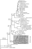
Fig. 1. Maximum likelihood tree with some selected bootstrap values. Branch lengths are proportional to changes. Details of the Amblyospora group are shown in Fig. 2. ML, bootstrap values obtained by the quartet puzzle method; Pa, bootstrap values obtained using parsimony. The tree is rooted by using Giardia ardeae, G. lamblia and Hexamita inflata as outgroup. Pleistophora sp. 2 and Pleistophora sp. 3 corresponds to the same species as reported by Nilsen et al. (1998).
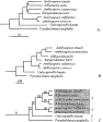
Fig. 2. Different tree topologies obtained for the Amblyospora group using different analysis methods. Only the species in the Amblyospora group are shown. (A) Distance tree using minimum evolution as optimal criterion and distances measured with the logdet/paralinear method. (B) Tree obtained using maximum parsimony as optimal criterion. Random addition of sequence with 100 subreplicates generated a tree with tree length of 5967 steps. (C) Puzzle quartet tree with maximum likelihood as optimal criterion. The numbers on the branches refer to percentage support obtained by the puzzle analysis. See text for explanations.
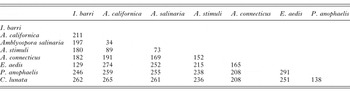
Table 1. Number of nucleotide differences between the different species in the Amblyospora group
 You have
Access
- 8
- Cited by