


%20--%3e%3c!DOCTYPE%20svg%20PUBLIC%20'-//W3C//DTD%20SVG%201.0//EN'%20'http://www.w3.org/TR/2001/REC-SVG-20010904/DTD/svg10.dtd'%3e%3csvg%20version='1.0'%20id='Layer_1'%20xmlns='http://www.w3.org/2000/svg'%20xmlns:xlink='http://www.w3.org/1999/xlink'%20x='0px'%20y='0px'%20width='64px'%20height='64px'%20viewBox='5.5%20-3.5%2064%2064'%20enable-background='new%205.5%20-3.5%2064%2064'%20xml:space='preserve'%3e%3cg%3e%3ccircle%20fill='%23FFFFFF'%20fill-opacity='0'%20cx='37.785'%20cy='28.501'%20r='28.836'/%3e%3cpath%20fill='%23595959'%20d='M37.441-3.5c8.951,0,16.572,3.125,22.857,9.372c3.008,3.009,5.295,6.448,6.857,10.314%20c1.561,3.867,2.344,7.971,2.344,12.314c0,4.381-0.773,8.486-2.314,12.313c-1.543,3.828-3.82,7.21-6.828,10.143%20c-3.123,3.085-6.666,5.448-10.629,7.086c-3.961,1.638-8.057,2.457-12.285,2.457s-8.276-0.808-12.143-2.429%20c-3.866-1.618-7.333-3.961-10.4-7.027c-3.067-3.066-5.4-6.524-7-10.372S5.5,32.767,5.5,28.5c0-4.229,0.809-8.295,2.428-12.2%20c1.619-3.905,3.972-7.4,7.057-10.486C21.08-0.394,28.565-3.5,37.441-3.5z%20M37.557,2.272c-7.314,0-13.467,2.553-18.458,7.657%20c-2.515,2.553-4.448,5.419-5.8,8.6c-1.354,3.181-2.029,6.505-2.029,9.972c0,3.429,0.675,6.734,2.029,9.913%20c1.353,3.183,3.285,6.021,5.8,8.516c2.514,2.496,5.351,4.399,8.515,5.715c3.161,1.314,6.476,1.971,9.943,1.971%20c3.428,0,6.75-0.665,9.973-1.999c3.219-1.335,6.121-3.257,8.713-5.771c4.99-4.876,7.484-10.99,7.484-18.344%20c0-3.543-0.648-6.895-1.943-10.057c-1.293-3.162-3.18-5.98-5.654-8.458C50.984,4.844,44.795,2.272,37.557,2.272z%20M37.156,23.187%20l-4.287,2.229c-0.458-0.951-1.019-1.619-1.685-2c-0.667-0.38-1.286-0.571-1.858-0.571c-2.856,0-4.286,1.885-4.286,5.657%20c0,1.714,0.362,3.084,1.085,4.113c0.724,1.029,1.791,1.544,3.201,1.544c1.867,0,3.181-0.915,3.944-2.743l3.942,2%20c-0.838,1.563-2,2.791-3.486,3.686c-1.484,0.896-3.123,1.343-4.914,1.343c-2.857,0-5.163-0.875-6.915-2.629%20c-1.752-1.752-2.628-4.19-2.628-7.313c0-3.048,0.886-5.466,2.657-7.257c1.771-1.79,4.009-2.686,6.715-2.686%20C32.604,18.558,35.441,20.101,37.156,23.187z%20M55.613,23.187l-4.229,2.229c-0.457-0.951-1.02-1.619-1.686-2%20c-0.668-0.38-1.307-0.571-1.914-0.571c-2.857,0-4.287,1.885-4.287,5.657c0,1.714,0.363,3.084,1.086,4.113%20c0.723,1.029,1.789,1.544,3.201,1.544c1.865,0,3.18-0.915,3.941-2.743l4,2c-0.875,1.563-2.057,2.791-3.541,3.686%20c-1.486,0.896-3.105,1.343-4.857,1.343c-2.896,0-5.209-0.875-6.941-2.629c-1.736-1.752-2.602-4.19-2.602-7.313%20c0-3.048,0.885-5.466,2.658-7.257c1.77-1.79,4.008-2.686,6.713-2.686C51.117,18.558,53.938,20.101,55.613,23.187z'/%3e%3c/g%3e%3c/svg%3e)
%20--%3e%3c!DOCTYPE%20svg%20PUBLIC%20'-//W3C//DTD%20SVG%201.0//EN'%20'http://www.w3.org/TR/2001/REC-SVG-20010904/DTD/svg10.dtd'%3e%3csvg%20version='1.0'%20id='Layer_1'%20xmlns='http://www.w3.org/2000/svg'%20xmlns:xlink='http://www.w3.org/1999/xlink'%20x='0px'%20y='0px'%20width='64px'%20height='64px'%20viewBox='5.5%20-3.5%2064%2064'%20enable-background='new%205.5%20-3.5%2064%2064'%20xml:space='preserve'%3e%3cg%3e%3ccircle%20fill='%23FFFFFF'%20fill-opacity='0'%20cx='37.637'%20cy='28.806'%20r='28.276'/%3e%3cg%3e%3cpath%20fill='%23595959'%20d='M37.443-3.5c8.988,0,16.57,3.085,22.742,9.257C66.393,11.967,69.5,19.548,69.5,28.5c0,8.991-3.049,16.476-9.145,22.456%20C53.879,57.319,46.242,60.5,37.443,60.5c-8.649,0-16.153-3.144-22.514-9.43C8.644,44.784,5.5,37.262,5.5,28.5%20c0-8.761,3.144-16.342,9.429-22.742C21.101-0.415,28.604-3.5,37.443-3.5z%20M37.557,2.272c-7.276,0-13.428,2.553-18.457,7.657%20c-5.22,5.334-7.829,11.525-7.829,18.572c0,7.086,2.59,13.22,7.77,18.398c5.181,5.182,11.352,7.771,18.514,7.771%20c7.123,0,13.334-2.607,18.629-7.828c5.029-4.838,7.543-10.952,7.543-18.343c0-7.276-2.553-13.465-7.656-18.571%20C50.967,4.824,44.795,2.272,37.557,2.272z%20M46.129,20.557v13.085h-3.656v15.542h-9.944V33.643h-3.656V20.557%20c0-0.572,0.2-1.057,0.599-1.457c0.401-0.399,0.887-0.6,1.457-0.6h13.144c0.533,0,1.01,0.2,1.428,0.6%20C45.918,19.5,46.129,19.986,46.129,20.557z%20M33.042,12.329c0-3.008,1.485-4.514,4.458-4.514s4.457,1.504,4.457,4.514%20c0,2.971-1.486,4.457-4.457,4.457S33.042,15.3,33.042,12.329z'/%3e%3c/g%3e%3c/g%3e%3c/svg%3e)
Introduction
Toxoplasmosis, caused by Toxoplasma gondii, usually persists as lifelong latent infection. Reactivation or donor-derived infection can cause severe disease in immunocompromised hosts, especially people with advanced Human Immunodeficiency Virus (HIV) infection and solid-organ transplant recipients. Reference Elsheikha, Marra and Zhu1–Reference Derouin and Pelloux3 Central Nervous System (CNS) disease is common in HIV, whereas disseminated disease can occur in transplant recipients and other immunocompromised hosts.
Diagnosis of CNS toxoplasmosis is often presumptive. In HIV, neuroimaging classically shows multiple ring-enhancing lesions, but the differential includes primary CNS lymphoma and other opportunistic infections. Reference Vidal2 Serology documents exposure but cannot distinguish latent infection from reactivation; after transplantation, early infection may occur before seroconversion. Reference Derouin and Pelloux3 Cerebrospinal fluid (CSF) T. gondii polymerase chain reaction (PCR) is highly specific but has modest sensitivity, highest before or within the first week of therapy, and lumbar puncture or brain biopsy may be delayed or contraindicated in critically ill patients. Reference Vidal2,Reference Cingolani, De Luca and Ammassari4
Plasma microbial cell-free DNA next-generation sequencing (mcfDNA NGS) is an unbiased assay that detects and quantifies microbial DNA fragments in plasma and can identify a broad range of pathogens without targeted primers. Reference Blauwkamp, Thair and Rosen5 In multicenter studies, plasma mcfDNA testing has been associated with changes in antimicrobial management, but interpretation requires diagnostic stewardship because co-detection of organisms and uncertain negative predictive value are common. Reference Hogan, Yang and Garner6,Reference Park, Chang and Ledeboer7 Evidence specific to toxoplasmosis is limited to case reports and small series. Reference Roy, Siddique, Bathina and Ahmad8,Reference Goren, Lehman, Drozdov and Thielen9 We describe a single-center five-patient case series of plasma mcfDNA NGS identifying T. gondii and compare turnaround time (TAT) and clinical impact with conventional testing.
Methods
We performed a retrospective case series of patients with plasma mcfDNA NGS (Karius, Redwood City, CA) positive for T. gondii at Baylor St. Luke’s Medical Center (Houston, Texas) from January 1, 2017, through December 31, 2025.
A result was considered positive when T. gondii was reported by the assay; quantitative concentrations (molecules per microliter [MPM]) were recorded when provided. The assay’s analytical validation supports a reporting threshold of >10 MPM. Reference Blauwkamp, Thair and Rosen5
We abstracted demographics, immunocompromising conditions, presenting syndrome, imaging findings, diagnostic testing, treatment decisions, and outcomes from the electronic medical record. Clinical TAT was defined as the time from test order entry to result availability.
Toxoplasma IgG testing was performed in-house; IgM testing was sent to a reference laboratory (Quest Diagnostics, San Juan Capistrano, CA); and T. gondii PCR testing (when performed) was sent to the University of Washington (Seattle, WA).
This study was reviewed and approved by the Baylor College of Medicine Institutional Review Board (protocol H-45143) with a waiver of informed consent for de-identified retrospective analysis.
Results
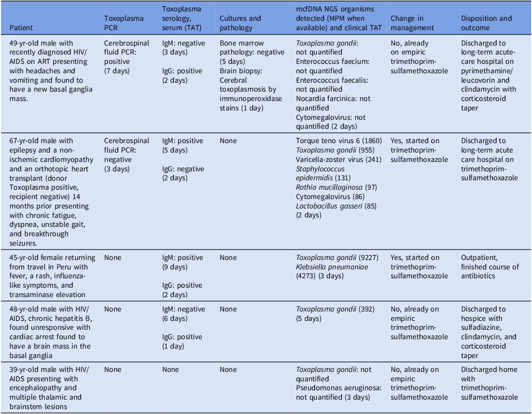
Five patients were identified (median age, 48 yr; range, 39–67); four were male. Three had HIV/AIDS (CD4 range, 3–106 cells/mm3), one was a heart transplant recipient (donor seropositive/recipient seronegative), and one was immunocompetent. Three presented with new neurologic symptoms and ring-enhancing brain lesions, one had neurologic symptoms without compatible lesions, and one had prolonged febrile systemic illness with hepatitis after travel. Patient-level findings are summarized in Table 1.
Clinical characteristics and diagnostic testing among patients with plasma mcfDNA NGS positive for Toxoplasma gondii (2017–2025)

Note: Co-detection of additional organisms by mcfDNA NGS occurred in four of five patients.
MPM values were recorded when reported; “not quantified” indicates the organism was detected but no quantitative MPM value was provided on the clinical report. Abbreviations: ART, antiretroviral therapy; CSF, cerebrospinal fluid; D+/R−, donor seropositive/recipient seronegative; LTACH, long-term acute-care hospital; mcfDNA NGS, microbial cell-free DNA next-generation sequencing; MPM, molecules per microliter; PCR, polymerase chain reaction; TAT, turnaround time (order to result); TMP-SMX, trimethoprim-sulfamethoxazole.
Plasma mcfDNA NGS detected T. gondii in all five cases. Median TAT for mcfDNA NGS was 3 days (range 2–5), compared with 2 days (range 1–2) for Toxoplasma IgG and 5.5 days (range 3–9) for IgM. Two patients underwent CSF T. gondii PCR testing (TAT, 3 and 7 days), no patient underwent blood T. gondii PCR, and one patient had diagnostic confirmation by brain biopsy.
In two cases, mcfDNA NGS directly prompted anti-Toxoplasma therapy (one transplant recipient with neurologic symptoms and one outpatient with systemic illness). In three cases with compatible CNS imaging, mcfDNA NGS supported ongoing empiric therapy. Co-detection of additional organisms occurred in four of five patients.
Discussion
In this five-patient case series, plasma mcfDNA NGS returned with a median TAT of 3 days and, in our institutional workflow, preceded send-out IgM and CSF PCR testing when obtained. In two patients, the result initiated targeted anti-Toxoplasma therapy, including a donor-seropositive/recipient-seronegative heart transplant recipient with neurologic symptoms but non-diagnostic imaging and an outpatient with prolonged febrile hepatitis. In patients with classic ring-enhancing lesions, plasma mcfDNA supported continued empiric therapy while confirmatory studies were pending.
Our findings complement prior reports suggesting that plasma mcfDNA sequencing can detect both CNS and non-CNS toxoplasmosis. Roy et al described toxoplasma pneumonitis in AIDS diagnosed by plasma NGS before later recognition of CNS lesions. Reference Roy, Siddique, Bathina and Ahmad8 Goren et al identified eight cases of T. gondii infection by plasma cfDNA sequencing in immunocompromised hosts, including clinically unsuspected and non-CNS presentations. Reference Goren, Lehman, Drozdov and Thielen9
Plasma mcfDNA may be most useful when conventional diagnostics are equivocal or infeasible. In advanced HIV, toxoplasmosis is often diagnosed presumptively based on compatible imaging and clinical response, with brain biopsy generally reserved for non-responders after 10–14 days. Reference Vidal2 CSF PCR is highly specific but variably sensitive, with highest yield before or within the first week of therapy. Reference Vidal2,Reference Cingolani, De Luca and Ammassari4 In our series, mcfDNA NGS often returned before send-out IgM or CSF PCR and provided actionable confirmation without lumbar puncture or brain biopsy.
Transplant recipients represent a distinct diagnostic context. Donor-derived toxoplasmosis is strongly associated with donor-seropositive/recipient-seronegative mismatch, particularly after heart transplantation, and disease can occur before seroconversion, limiting the utility of serology in early infection. Reference Derouin and Pelloux3 The transplant case in our cohort illustrates this challenge: IgG remained negative, and IgM returned later, whereas plasma mcfDNA identified T. gondii within 2 days and prompted initiation of therapy. No patient underwent blood T. gondii PCR. In allogeneic hematopoietic cell transplantation, serial blood qPCR has been proposed for preemptive early detection, although blood PCR can remain negative in established disease. Reference Aerts, Mehra and Groll10
The open-ended nature of plasma mcfDNA testing necessitates diagnostic stewardship. Co-detection of additional taxa was common in our patients and is widely reported in clinical practice, where interpretation depends on pretest probability and syndrome compatibility. Reference Hogan, Yang and Garner6,Reference Park, Chang and Ledeboer7 In our series, additional detections did not meaningfully change management because the T. gondii result was interpreted alongside host factors, imaging, and conventional testing rather than as a stand-alone diagnosis. A positive plasma mcfDNA result should therefore not be considered diagnostic in isolation, and false-positive or clinically irrelevant detections remain possible, particularly when orthogonal confirmation is unavailable or multiple organisms are reported. This contextual interpretation may be more difficult outside infectious diseases-supported or multidisciplinary care.
Plasma mcfDNA also has inherent limitations: it cannot localize infection, quantitative values (MPM) have not been validated as a marker of toxoplasmosis disease burden, and false-negative results can occur, particularly in isolated CNS disease or after therapy. A negative plasma mcfDNA result should therefore not dissuade empiric treatment when clinical suspicion is high. Our study is limited by retrospective design, small sample size, possible false-positive attribution in unconfirmed cases, selection bias toward patients in whom clinicians already pursued plasma mcfDNA testing, and TAT comparisons that reflect local laboratory workflows. Prospective multicenter studies are needed to define optimal patient selection and timing and to evaluate whether plasma mcfDNA meaningfully reduces invasive procedures, antimicrobial exposure, or time to appropriate therapy.
Data availability statement
De-identified data supporting the findings of this study are available from the corresponding author upon reasonable request.
Acknowledgments
None.
Financial support
None reported.
Competing interests
All authors report no conflicts of interest relevant to this article.

 Open access

