




No CrossRef data available.
Article contents
Toxoplasmosis in Pallas' cats (Otocolobus manul) raised in captivity
Published online by Cambridge University Press: 21 October 2004
Abstract
Manuls or Pallas' cats (Felis manul, syn. Otocolobus manul) are endangered wild cats from Central Asia kept and bred in many zoos. Despite good breeding success young cats frequently die from acute toxoplasmosis. From 1998 to 2002, a breeding pair in the Schönbrunn Zoo in Vienna, Austria, gave birth to 24 kittens; 58% of kittens died between the 2nd and the 14th week of life, mostly due to acute toxoplasmosis. The epidemiology of toxoplasmosis in Pallas' cats was examined and a control strategy to protect the kittens from fatal toxoplasmosis was developed. One 12-week-old kitten from a litter of 6 born in 2001 died of generalized toxoplasmosis. This kitten had shed T. gondii oocysts that were bioassayed in mice. Toxoplasma gondii was isolated in tissue culture inoculated with tissues of these mice. The surviving animals were immediately treated with clindamycin for 16 weeks; they acquired a natural infection and seroconverted by the end of this time without clinical signs.
Keywords
Information
- Type
- Research Article
- Information
- Copyright
- 2004 Cambridge University Press
INTRODUCTION
The Pallas' cat (Felis manul, syn. Otocolobus manul) is a threatened small-sized felid species that inhabits steppes, rocky terrain and cold deserts in Central Asia. A special effort is being made world-wide to breed this species in captivity. Between 1995 and 2000 Pallas' cats were imported from Russia and Mongolia into North American zoos, but a high (>50%) mortality of newborn kittens within 4 months of birth was recorded (Brown et al. 2002; Ketz-Riley et al. 2003). In the majority of deceased kittens toxoplasmosis was identified as the cause of death (Swanson, 1999). Pallas' cats held in captivity appear to be highly susceptible to toxoplasmosis and can shed T. gondii oocysts (Riemann et al. 1974; Dubey et al. 1988a; Dubey & Beattie, 1988; Löschenberger et al. 2000; Zenker et al. 2001; Kenny et al. 2002). The objectives of this investigation were to study pathogenesis, epidemiology and course of natural T. gondii infection in captive Pallas' cats.
MATERIALS AND METHODS
In 1997, a breeding pair of Pallas' cats purchased from the Novosibirsk Zoo, Novosibirsk, Russia, has been held in captivity at the Schönbrunn Zoo, Vienna, Austria. Both cats were born in captivity at the Russian zoo (the female in April 1996, the male in April 1995). At the Vienna zoo the female gave birth to 3 successive litters with a high neonatal mortality rate: 1998: 80% (4/5), 1999: 75% (3/4), 2000: 40% (2/5). Toxoplasmosis was confirmed in the litter born in 2000 (Löschenberger et al. 2000; Zenker et al. 2001).
In March 2001 the original breeding pair gave birth to 6 apparently healthy kittens at the Schönbrunn Zoo, 1 male (kitten no. 5) and 5 females (kitten nos. 1, 2, 3, 4 and 6). The queen and her kittens were housed together in an 8×5×2·5 m cage. At the age of 186 days, kittens were separated from their mother and housed in a new cage. The queen was also transferred to a new cage, which had never before housed felids. The adult male was separated from the queen before kittens were born.
The cats were fed fresh mice and rats raised at the breeding unit of the Zoo. At the age of 68 and 87 days the kittens were vaccinated against feline rhinotracheitis, calicivirus and panleucopenia infection (Fevaxyn i-CHP® Fort Dodge, Ireland).
Case report
At the age of 84 days, kitten no. 5 was found to be weak, ataxic and anorexic. The kitten was treated for toxoplasmosis with single doses of 62·5 mg sulfadoxin and 12·5 mg trimethoprim (Borgal® 7·5%, Intervet, Austria) administered subcutaneously. However, the kitten died 2 days later. At this time the remaining 5 litter mates and their parents exhibited respiratory signs, characterized by rhinitis and conjunctivitis. The parents showed more severe signs than the kittens, and were admitted to the Intensive Care Unit of the Zoo.
Kitten no. 5 was examined at necropsy, and samples of heart blood taken to detect antibodies against T. gondii, feline herpes virus-1 (FHV-1) and feline immunodeficiency virus (FIV). Samples from lung, spleen and liver were collected for FHV-1 isolation and DNA detection by the PCR.
Tissue samples from central nervous system (CNS), lung, heart, liver, spleen, kidney, pancreas, mesenteric lymph nodes and striated muscle were fixed in 10% formalin for histopathological and immunohistochemical studies. Paraffin-embedded tissues were sectioned and stained with haematoxylin-eosin (HE) or examined for T. gondii-antigen using the Vectastain®-avidin biotin complex test (ABC Kit; Vector Laboratories, Burlingame, USA) (Uggla, Sjöland & Dubey, 1987). Native and Diff-Quik® (Dade AG, Düdingen, Switzerland) stained impression smears from the brain were also examined.
Prophylaxis
As prophylaxis clindamycin (Antirobe®, Pharmacia, Austria; 15 mg/mouse) was administered orally to the remaining kittens and their parents via medicated mice from 87 days of age/post-parturition for 15 weeks.
Serological examination against T. gondii
Blood samples from the kittens were collected from the cephalic vein between the 45th and 295th day of age (Table 1) and from the breeding pair on days 87 and 186 post-parturition. Sera were stored at −20 °C until examined for anti-T. gondii-antibodies using the direct agglutination test (DAT; Merial, Lyon, France) (Desmonts & Remington, 1980; Dubey, 1996) and an immunofluorescence antibody test (IFAT) (Omata et al. 1990). The DAT was performed according to the manufacturer's instructions, using 2-fold dilutions starting from 1[ratio ]40 to 1[ratio ]40960.
The IFAT was performed with culture-derived tachyzoites of the RH strain at 2-fold dilutions starting at a dilution of 1[ratio ]16. A goat-anti cat-IgG conjugate (Jackson Lab. Inc., West Grove, Pennsylvania, USA) was used at a dilution of 1[ratio ]50. A complete peripheral fluorescence of the tachyzoites was interpreted as positive, while polar or lack of fluorescence was judged as negative.

Eighty mice and 60 rats from the Zoo breeding unit were serologically tested for T. gondii-antibodies using the DAT test at a dilution of 1[ratio ]40.
Fecal examination for T. gondii and bioassay in mice
Fecal samples from the queen were collected daily from the first day post-partum over a period of 8 months. The first fecal samples from the kittens were obtained at 44 days of age. Before this, no fecal deposits were found due to the coprophageous behaviour of the queen. Each fecal sample was examined for T. gondii oocysts using a sucrose solution (specific gravity 1250) (Dubey, Swann & Frenkel, 1972; Dubey & Beattie, 1988). Oocysts were sporulated in a 2·5% potassium dichromate solution at room temperature (20 °C) for 3 days. For the bioassay, oocysts were washed with PBS to remove potassium dichromate and fed to 4 NMRI mice at doses of 250000 and 500000 oocysts (2 mice each).
Six days later, 1 mouse was dead and the other 3 displayed weakness, tremors and ataxia and were euthanized and necropsied. Samples from brain, lungs and spleen from the surviving mice were collected under sterile conditions, homogenized individually in a sterile mortar with 5 ml of Iscove's modified Dulbecco's medium with L-glutamine containing 1000 IU/ml penicillin, 100 μg/ml streptomycin, 250 μg/l amphotericin B and 3% horse serum. The homogenates were passed through hypodermic needles of decreasing diameter (15 G, 19 G and 23 G) to disrupt the tissue and release the tachyzoites. The homogenates were inoculated onto Vero cell monolayers that had grown in the above-mentioned medium with 10% horse serum and incubated at 37 °C in a 5% CO2 atmosphere for 1 h. The medium was then changed and the flasks incubated for a maximum of 2 months. The medium was changed weekly. The growth of T. gondii-tachyzoites was monitored every second day.
The peritoneal cavity of each of the 3 mice was washed with 3 ml of a 0·5% sterile NaCl solution and the fluids were collected. Peritoneal fluids were either inoculated in Vero cell cultures as above or inoculated intraperitoneally into 2 NMRI mice which were bled 20 days post-inoculation and examined for T. gondii-antibodies by DAT at a serum dilution of 1[ratio ]40. Diff-Quik®-stained impression smears from brain, lungs, spleen and liver samples of the mice and native peritoneal fluids were microscopically examined for the presence of T. gondii-tachyzoites.
Isolation of T. gondii from tissues from kitten no. 5
Samples from brain, lung and mesenteric lymph nodes were collected, processed individually and inoculated into mice and Vero cell cultures as described above. Brains with Toxoplasma-cysts of these infected mice were subcultured in Vero cells as above.
Virological and serological investigations
Lung, liver and spleen tissues of kitten no. 5 were homogenized and inoculated onto Crandell Feline Kidney (CRFK) cells using Eagle's Minimum Essential Medium and were additionally submitted to PCR for the detection of feline herpesvirus 1 (FHV-1) specific DNA. PCR was performed as described by Volopich et al. (2004) using the following primers: forward: 5′-CTCGACGACCAACCATTACC-3′, position 3469-3488, reverse: 5′-TCCGATGACCGCAAGAGTAG-3′, position 4233-4214; accession number S72415. The blood samples of kitten no. 5 on day 68 and obtained at necropsy were tested for antibodies against FHV-1 using a virus neutralization assay. Briefly 100 TCID50 (tissue culture infectious doses 50%) of an FHV-1 isolate were incubated with 2-fold serial dilutions of the heat-inactivated serum samples and inoculated onto CRFK cells. The cells were monitored daily for the occurrence of cytopathic effects. Additionally the serum samples were tested for antibodies against feline immunodeficiency virus using the Witness FIV test kit (AGEN Biomedical Ltd, Brisbane, Australia).
RESULTS
Histopathological and immunhistochemical studies of kitten no. 5
Microscopically, generalized toxoplasmosis was diagnosed in kitten no. 5 with most severe involvement of the striated muscle, liver and spleen. Non-suppurative encephalitis, myositis, hepatitis and pneumonia were associated with T. gondii-tissue cysts and tachyzoites. T. gondii-tissue cysts were also present in sections of heart and kidney. The diagnosis was confirmed immunohistochemically (Figs 1 and 2). T. gondii-tissue cysts and tachyzoites were also seen in brain smears (Fig. 3).

Fig. 1. Toxoplasmosis in the Pallas' cat kitten no. 5. Non-suppurative encephalitis with focal gliosis and mononuclear cell infiltration (HE staining).

Fig. 2. Toxoplasma-tachyzoites in the lung of the Pallas' cat kitten no. 5 (immunostaining).

Fig. 3. Tissue cyst of T. gondii in Diff Quik®-impression smears of brain of the Pallas' cat kitten no. 5.
Serological examinations
The queen had antibody titres to T. gondii of [ges ]1[ratio ]10240 (DAT) and 1[ratio ]32000 (IFAT) and of [ges ]1[ratio ]1280 (DAT) and 1[ratio ]1024 (IFAT) at 87 and 186 days post-parturition, respectively. The breeding male cat had been bled at the same time and had a titre of [ges ]1[ratio ]10240 (DAT) and 1[ratio ]1024 (IFAT); and of [ges ]1[ratio ]1280 (DAT) and 1[ratio ]1024 (IFAT), respectively. The queen had been previously bled in 1999 and had at that time a titre of 1[ratio ]2560 (IFAT) (Löschenberger et al. 2000).
All 6 kittens had antibodies to T. gondii (Table 1).
Fecal examinations
T. gondii-like oocysts were detected in fecal samples of at least 1 of the kittens from 72 to 86 days of age and from 198 to 219 days of age (Fig. 4). In fecal samples of the queen Toxoplasma-oocysts were observed from the 59th and to 86th day and from the 192th to the 199th day post-parturition.

Fig. 4. Oocysts of Toxoplasma gondii in the feces of the Pallas' cat kitten no. 5.
Parasites from mice or rats were sometimes observed in the feces of the queen and the kittens during fecal examinations. Oocysts of Cryptosporidium parvum were detected in the feces of at least one of the kittens at 72 and 162 days of age. Oocysts of Isospora rivolta and Isospora felis were occasionally detected, while eggs of Toxocara mystax and Toxascaris leonina were frequently demonstrated in the feces of the queen and the kittens during this study.
Bioassays of oocysts in mice and cultivation in Vero cells
Tachyzoites of T. gondii were detected in Diff-Quik®-stained impression lung smears from 1 mouse and in native- and Diff-Quik®-stained specimen from peritoneal fluid from 2 mice. T. gondii-tachyzoites were first observed at 21 days post-inoculation in all flasks inoculated with mouse tissues, and were cryopreserved.
Mice inoculated with peritoneal liquid from one mouse had a Toxoplasma-antibody titre of [ges ]1[ratio ]40 by DAT at 20 days post-inoculation.
Serological examination of mice and rats from the zoo breeding unit
No antibodies to T. gondii were detected in the sera of 60 rats and 80 mice (DAT titre<1[ratio ]40).
Virological studies of kitten no. 5
After an incubation period of 24 h the CRFK cells inoculated with lung material of kitten no. 5 showed a cytopathic effect with plaques typical for herpesviruses. Additionally, a positive PCR result was achieved with lung tissue by PCR for FHV-1 specific DNA. The antibody titres against FHV-1 were 1[ratio ]16 on day 68, but <1[ratio ]8 (negative) at time of necropsy. No antibodies against FIV could be detected.
DISCUSSION
The rising antibody titres in kittens found in the present study are likely due to active infection because passively-transferred anti-T. gondii antibodies decline considerably in the kittens by 10 weeks of age (Dubey, Lappin & Thulliez, 1995: Omata et al. 1994). The decline of the anti-Toxoplasma maternal antibodies between days 45 and 130 and subsequent increase indicates active synthesis of T. gondii antibodies by kittens.
Co-infection with FHV-1 in kitten no. 5 probably contributed to the severe course of T. gondii-infection in this animal. The other 5 kittens showed no clinical signs, probably influenced by the clindamycin treatment.
Kitten no. 3 and no. 6 seroconverted during clindamycin medication but the first T. gondii-oocysts were detected in the feces 12 days after the end of therapy. Clindamycin reduces the level of replication of T. gondii by inhibiting the protein synthesis of the parasite and impairs the ability of tachyzoites to infect host cells (Dubey & Yeary, 1977; Gross & Pohl, 1996). Cats excrete T. gondii-oocysts in feces 3 to 10 days after ingesting bradyzoites, [ges ]18 days after ingesting sporulated oocysts and tachyzoites (Dubey, 2002b).
The bradyzoite-induced cycle in cats is considered the most efficient as nearly all cats fed tissue cysts excrete oocysts, whereas <30% of cats fed tachyzoites or oocysts excrete oocysts (Dubey, 1998). It is hypothesized that after oocyst ingestion, T. gondii invades extraintestinal tissues and bradyzoites produced in these tissues return to the intestine to initiate the intestinal cycle (Freyre et al. 1989). The proposed infrequent rupture of cysts may account for the complete coccidian cycle occurring only in a low percentage of cats (Dubey, 1998).
If T. gondii prepatent periods observed in the domestic cat are also applicable to Pallas' cats, it is tempting to assume that kitten no. 5 became infected post-natally by the ingestion of T. gondii from the environment; it had no antibodies (DAT titre <1[ratio ]40) at 68 days of age but a positive titre 18 days later (DAT titre 1[ratio ]10240). The presence of T. gondii-oocysts could be demonstrated in the intestinal content of kitten no. 5 at necropsy (at 86 days of age) and oocyst shedding from one of the kittens was recorded between 72 and 86 days of age (time-point at which kitten no. 5 was found dead). After that day no T. gondii-oocysts were observed for 112 days. Anti-T. gondii seroconversion was documented in the remaining kittens at different points in time after this oocyst excretion period, suggesting a post-natally acquired infection. The first of the remaining kittens (no. 3) developed anti-T. gondii-antibodies at 130 days of age. Two weeks after infection either with T. gondii-oocyst or cysts, specific anti-T. gondii-IgG can be demonstrated (Omata et al. 1990; Dubey et al. 1995). We therefore assume that kitten no. 3 was infected at a later stage and the oocyst shedding phase observed corresponded to kitten no. 5. In the remaining kittens the source of infection could not be determined. In spite of the fact that congenitally-infected kittens can excrete T. gondii oocysts 1–3 months after birth (Dubey & Carpenter, 1993), we have evidence from the serological data (post-natal seroconversion after the 10th week of life) that in the examined litter congenital infection did not occur.
The queen which had a chronic T. gondii-infection (Löschenberger et al. 2000) shed oocysts in two stages during this study: between 59 and 86 days and between 192 and 199 days post-parturition. The stress associated with a co-FHV-1 infection (about which will be reported separately), and moving to a new cage might have led to shedding of oocysts by the queen.
During microscopical examinations of fecal samples from the kittens we often found murine parasites including eggs of Capillaria hepatica, a common helminth in the free-living murine population of the Schönbrunn Zoo (Junker-v.Voss, Kübber-Heiss & Prosl, 1998). The first fecal samples of the kittens collected at 44 days of age already contained parasites of the free-living mice. This finding suggests that the Pallas' cats had occasional access to wild mice which could have been a source of a T. gondii-infection. A serological and coproscopical survey of a greater number of mice caught in the breeding unit would be desirable. The presence of passerine birds in the Pallas' cats' cage, mainly house sparrows (Passer domesticus), was often noticed. Toxoplasmosis in wild birds is well-documented (Dubey, 2002a).
Dubey & Beattie (1988) reported that T. gondii-oocysts are generally excreted before the cat (Felis catus) becomes clinically ill, and fecal examination could be unrewarding. In contrast to this, we demonstrated the presence of T. gondii-oocysts in intestinal contents at the necropsy of Pallas' cat kitten no. 5, simultaneously with extraintestinal lesions and stages of this protozoan in accordance with the acute and fatal toxoplasmosis case reported by Dubey et al. (1988a) in a 6-year-old female Pallas' cat in the Milwaukee County Zoo, Wisconsin, USA. The authors reported entero-epithelial stages and oocysts of T. gondii in the small intestine of the queen present simultaneously with enteritis, hepatitis, splenic necrosis and pneumonia due to T. gondii.
While some authors consider the pre-natal infection with T. gondii as an important cause of neonatal mortality in Pallas' cats (Dubey et al. 1988; Swanson, 1999; Kenny et al. 2002) in this study it was demonstrated that the infection of the whole litter occurred post-natally.
The cause of the particular susceptibility of this species to T. gondii is unknown. Preliminary studies from Ketz-Riley et al. (2000, 2003) on T. gondii- and FHV seropositive Pallas' cats suggested an immunodeficiency that mimics FIV infection. In that study the animals showed a decreased lymphoproliferation response to mitogens and a high interleukin 10 to interleukin 12 ratio when compared to healthy control cats, as occurs in FIV infected cats, but a FIV infection could not be confirmed.
Brown et al. (2002) compared wild and captive Pallas' cat populations and reported that 87% (13/15) of the wild Pallas' cats captured in Mongolia had no antibodies to T. gondii, while 100% (9/9) of the captive population kept at 3 American Zoos was positive. These findings suggested that wild Pallas' cats frequently become infected in captivity.
Until adequate vaccines are developed for Pallas' cats, a prophylactic treatment is useful, mainly for kittens born in captivity. The therapeutic management carried out in the present study could be of use in the prophylaxis of toxoplasmosis in captive neonates preventing the clinical manifestations of this infection and also in the treatment of clinically ill adults.
Further studies on immunological mechanisms related to T. gondii, FHV-1 and FIV interactions in Pallas' cats are needed to improve the breeding success of this species in captivity.
References
REFERENCES
BROWN, M.,
LAPPIN, M. R.,
BROWN, J. L.,
MUNKHTSOG, B. &
SWANSON, W. (
2002).
Exploring the ecological basis for extreme susceptibility of Pallas' cats (Otocolobus manul) to fatal toxoplasmosis: Comparison of wild and captive populations.
Proceedings of the American Association of Zoo Veterinarians, pp.
12–15.Google Scholar
DESMONTS, G. &
REMINGTON, J. S. (
1980).
Direct agglutination test for diagnosis of Toxoplasma gondii infection: method for increasing sensitivity and specificity.
Journal of Clinical Microbiology 11,
562–568.Google Scholar
DUBEY, J. P. (
1996).
Infectivity and pathogenicity of Toxoplasma gondii oocysts for cats.
Journal of Parasitology 82,
957–961.CrossRefGoogle Scholar
DUBEY, J. P. (
1998).
Advances in the life cycle of Toxoplasma gondii.
International Journal for Parasitology 28,
1019–1024.CrossRefGoogle Scholar
DUBEY, J. P. (
2002 a).
A review of toxoplasmosis in wild birds.
Veterinary Parasitology 106,
121–153.Google Scholar
DUBEY, J. P. (
2002 b).
Tachyzoite-induced life cycle of Toxoplasma gondii in cats.
Journal of Parasitology 88,
713–717.Google Scholar
DUBEY, J. P. &
BEATTIE, C. P. (
1988).
Toxoplasmosis of Animals and Man, 1st Edn. CRC Press Inc,
Boca Raton,
Florida.
DUBEY, J. P. &
CARPENTER (
1993).
Neonatal toxoplasmosis in littermate cats.
Journal of the American Veterinary Association 203,
1546–1549.Google Scholar
DUBEY, J. P.,
GENDRON-FITZPAZTRICK, A. P.,
LENHARD, A. L. &
BOWMAN, D. (
1988).
Fatal toxoplasmosis and enteroepithelial stages of Toxoplasma gondii in a Pallas' cat (Felis manul).
Journal of Protozoology 35,
528–530.CrossRefGoogle Scholar
DUBEY, J. P.,
LAPPIN, M. R. &
THULLIEZ, M. D. (
1995).
Diagnosis of induced toxoplasmosis in neonatal cats.
Journal of the American Veterinary Medical Association 207,
179–185.Google Scholar
DUBEY, J. P.,
SWAN, G. V. &
FRENKEL, J. K. (
1972).
A simplified method for isolation of Toxoplasma gondii oocysts from the feces of cats.
Journal of Parasitology 58,
1005–1006.CrossRefGoogle Scholar
DUBEY, J. P. &
YEARY, R. A. (
1977).
Anticoccidial activity of 2-sulfamoyl-4-4-diaminodiphenylsulfone, sulfadiazine, pyrimethamine and clindamycin in cats infected with Toxoplasma gondii.
Canadian Veterinary Journal 18,
51–57.Google Scholar
FREYRE, A.,
DUBEY, J. P.,
SMITH, D. D. &
FRENKEL, J. K. (
1989).
Oocyst-induced Toxoplama gondii infections in cats.
Journal of Parasitology 75,
750–755.CrossRefGoogle Scholar
GROSS, U. &
POHL, F. (
1996).
Influence of antimicrobial agents on replication and stage conversion of Toxoplasma gondii.
Current Topics in Microbiology and Immunology 219,
235–245.CrossRefGoogle Scholar
JUNCKER-V.VOSS, M.,
KÜBBER-HEISS, A. &
PROSL, H. (
1998).
Zum Vorkommen von Capillaria hepatica bei Hausmäusen (Mus musculus) in Österreich.
Mitteilungen der Österreichischen Gesellschaft für Tropenmedizin und Parasitologie 20,
137–144.Google Scholar
KENNY, D. E.,
LAPPIN, M. R.,
KNIGHTLY, F.,
BAIER, J.,
BREWER, M. &
GETZY, D. M. (
2002).
Toxoplasmosis in Pallas' cats (Otocolobus felis manul) at the Denver Zoological Gardens.
Journal of Zoo and Wildlife Medicine 33,
131–138.Google Scholar
KETZ-RILEY, C. J.,
RITCHEY, J.,
BARRIE, M. T. &
HOOVER, J. (
2000).
Preliminary investigation of suspected immunodeficiency in Pallas' cats (Otocolobus manul) with concurrent toxoplasmosis and herpes virus infection.
Proceedings of the 3rd Scientific Meeting of the European Association of Zoo and Wildlife Veterinarians,
Paris, France, p.
29.
KETZ-RILEY, C. J.,
RITCHEY, J. W.,
HOOVER, J. P.,
JOHNSON, C. M. &
BARRIE, M. T. (
2003).
Immunodeficiency associated with multiple concurrent infections in captive Pallas' cats (Otocolobus manul).
Journal of Zoo and Wildlife Medicine 34,
239–245.CrossRefGoogle Scholar
LÖSCHENBERGER, K.,
KLEIN, D.,
PESCHKE, R.,
VORACEK, T.,
ZENKER, W.,
EDELHOFER, R. &
PROSL, H. (
2000).
Hohe Mortalitätsraten bei Felis manul Welpen durch Toxoplasma gondii.
Proceedings of the XXIV Tagung der Österreichischen Gesellschaft für Tropenmedizin und Parasitologie, Innsbruck, Austria, p.
15.
OMATA, Y.,
OIKAWA, H.,
KANDA, M.,
MIKAZUKI, K.,
NAKABAYASHI, T. &
SUZUKI, N. (
1990).
Experimental feline toxoplasmosis: Humoral immune responses of cats inoculated orally with Toxoplama gondii cysts and oocysts.
Japanese Journal of Veterinary Science 52,
865–867.CrossRefGoogle Scholar
OMATA, Y.,
OIKAWA, H.,
KANDA, M.,
MIKAZUKI, K.,
DILORENZO, C.,
CLAVERIA, F. G.,
TAKAHASHI, M.,
IGARASHI, I.,
SAITO, A. &
SUSUKI, N. (
1994).
Transfer of antibodies to kittens from mother cats chronically infected with Toxoplasma gondii.
Veterinary Parasitology 52,
211–218.CrossRefGoogle Scholar
RIEMANN, H. P.,
FOWLER, M. E.,
SCHULZ, T.,
LOCK, A.,
THILSTED, J.,
PULLEY, L. T.,
HENRICKSON, R. V.,
HENNESS, A. M.,
FRANTI, C. E. &
BEHYMER, D. E. (
1974).
Toxoplasmosis in Pallas' cats.
Journal of Wildlife Diseases 10,
471–477.CrossRefGoogle Scholar
SWANSON, W. F. (
1999).
Toxoplasmosis and neonatal mortality in Pallas' cats: a survey of North American zoological institutions.
Proceedings of the American Association of Zoo Veterinarians, pp.
347–350.
UGGLA, A.,
SJÖLAND, L. &
DUBEY, J. P. (
1987).
Immunhistochemical diagnosis of toxoplasmosis in fetuses and fetal membranes of sheep.
American Journal of Veterinary Research 48,
348–351.Google Scholar
VOLOPICH, S.,
BENETKA, V.,
SCHWENDENWEIN, I.,
MÖSTL, K.,
SOMMERFELD-STUR, I. &
NELL, B. (
2004).
Assessment of cytologic findings and feline herpesvirus DNA and Chlamydophila felis antigen detection rates in normal cats and cats with conjunctival and corneal lesions.
Veterinary Ophthalmology (in the Press).Google Scholar
ZENKER, W.,
LÖSCHENBERGER, K.,
MÖSTL, K.,
VORACEK, T.,
BASSO, W. &
PROSL, H. (
2001).
Toxoplasmose bei Manuls (Felis manul): Ein Fallbericht.
Proceedings of the XXI Arbeitstagung der Zootierärzte im deutschsprachigen Raum, Halle, Germany, pp.
118–122.
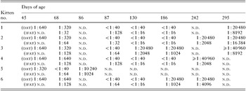
Table 1. Toxoplasma gondii-specific IgG antibody titre development in the serum of Pallas' cat kittens as determined by DAT and IFAT (N.D., not done)

Fig. 1. Toxoplasmosis in the Pallas' cat kitten no. 5. Non-suppurative encephalitis with focal gliosis and mononuclear cell infiltration (HE staining).
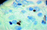
Fig. 2. Toxoplasma-tachyzoites in the lung of the Pallas' cat kitten no. 5 (immunostaining).
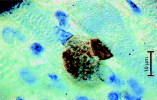
Fig. 3. Tissue cyst of T. gondii in Diff Quik®-impression smears of brain of the Pallas' cat kitten no. 5.
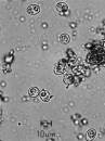
Fig. 4. Oocysts of Toxoplasma gondii in the feces of the Pallas' cat kitten no. 5.
 You have
Access
- 54
- Cited by