Introduction
Experimental and epidemiological studies have shown that fetal growth restriction is correlated with later obesity and metabolic disorders, implying that fetal nutritional deprivation may be a strong programming stimulus for adverse health outcomes in later life.Reference Barker, Winter, Osmond, Margetts and Simmonds1–Reference Yajnik and Deshmukh3 However, in many societies, maternal and postnatal nutrition are either sufficient or excessive and, as a result, excessive weight gain and/or obesity are common nutritional problems complicating pregnancy in developed countries and those transitioning to first world economies. The rising prevalence of obesity in pregnancy and the association with gestational diabetesReference Ehrenberg, Dierker, Milluzzi and Mercer4 have led to an increasing interest in the detrimental influence of maternal obesity and excess maternal nutrition on disease risk in childhood and beyond. Increasing evidence from both humansReference Catalano and Ehrenberg5 and animalsReference Guo and Jen6–Reference Samuelsson, Matthews and Argenton9 highlights the consequences of maternal high-fat diet on offspring adiposity and metabolic function.
Recently, we demonstrated in rats that offspring of mothers fed a high fat (HF) diet during pregnancy and lactation were obese as adults, irrespective of their postnatal diet, associated with higher circulating leptin and insulin concentrations.Reference Howie, Sloboda, Kama and Vickers10 These data highlight the fact that later metabolic health can be altered by events that occur early in development. Although the mechanisms contributing to the programming of altered metabolic function and obesity have yet to be fully described, the hypothalamic–pituitary–adrenal (HPA) axis has been implicated.Reference Bjorntorp11 HPA function is altered in overweight and obese adultsReference Marin, Darin and Amemiva12–Reference Rutters, Nieuwenhuizen, Lemmens, Born and Westerterp-Plantenga15 and in offspring exposed to altered nutrition as fetusesReference Lesage, Blondeau, Grino, Breant and Dupouy16, Reference Bloomfield, Oliver and Giannoulias17 or whose mothers endured stressful situations during pregnancy.Reference Entringer, Kumsta, Hellhammer, Wadhwa and Wust18 Glucocorticoids (GC) have been shown to play a role in the development of obesity,Reference King and Smith19 a phenotype that can be averted by adrenalectomy in the obese Zucker rat.Reference Freedman, Horwitz and Stern20
Leptin has been shown to have an inhibitory effect on adrenal steroidogenesisReference Cherradi, Capponi, Gaillard and Pralong21, Reference Hsu, Chang and Chiu22 and a stimulatory effect on epinephrine synthesisReference Shibuya, Utsunomiya and Toyohira23, Reference Trevenzoli, Valle and Machado24 and the ObRb isoform of the leptin receptor has been localized to both the adrenal corticalReference Glasgow, Haidan and Hilbers25–Reference Chelikani, Glimm and Kennelly27 and medullaryReference Glasgow, Haidan and Hilbers25, Reference Cao, Considine and Lynn28, Reference Takekoshi, Motooka and Isobe29 cells. These observations may provide evidence for the direct influence of leptin on GCs as well as catecholamine synthesis and secretion.Reference Malendowicz, Rucinski, Belloni, Ziolkowska and Nussdorfer30 Leptin stimulates the rate-limiting enzyme tyrosine hydroxylase (TH)Reference Shibuya, Utsunomiya and Toyohira23, Reference Trevenzoli, Valle and Machado24 and subsequent increase in epinephrine synthesis, mediated by the terminal enzyme phenylethanolamine N-methyl transferase (PNMT).Reference Kelner and Pollard31 This increased medullary epinephrine synthesis and output increases satiety, thermogenesis and blood glucose, inhibits insulin secretion and exerts a lipolytic effect on adipocytes.Reference Cao, Considine and Lynn28, Reference Vander Tuig, Kerner and Romsos32 In addition, it has long been known that GCs mediate the PNMT-induced increased conversion of norepinephrine to epinephrine, while adrenocorticotrophic hormone (ACTH) and direct input from the sympathetic nervous system also stimulate enzymes in the catecholamine-synthesizing cascade.Reference Kelner and Pollard31, Reference Wurtman and Axelrod33, Reference Thoenen, Mueller and Axelrod34
Recent evidence has demonstrated that elevated plasma levels of homocysteine (HCY) may be associated with increased body mass index and obesity.Reference Zhu, Huang, Li and Neubauer35, Reference Karatela and Sainani36 HCY, a sulfur-containing amino acid, is remethylated to methionine, primarily by the folate-dependent enzyme methionine synthase in the remethylation pathway, or metabolized to form cysteine in the pyridoxine (vitamin B6)-dependent transsulfuration pathway.Reference Austin, Lentz and Werstuck37 When either of these pathways is impaired, either through a reduction in the amount of available vitamin cofactorsReference Franken, Boers, Blom, Trijibels and Kloppenborg38, Reference Selhub, Jacques, Wilson, Rush and Rosenberg39 or by enzyme deficiency,Reference Mudd, Havlik, Levy, McKusick and Feinleib40, Reference Frosst, Blom and Milos41 HCY accumulates and is excreted in the blood. The HCY metabolic pathway plays an important role in methyl balance, in which the major biological methylating agent S-adenosylmethionine (SAM) is demethylated to HCY by methyltransferases.Reference Austin, Lentz and Werstuck42 This reaction is compulsory for the synthesis of epinephrine from norepinephrine and requires PNMT.Reference Martin, Begun, McLeish, Caine and Grunewald43 Thus, like leptin, HCY metabolism may play a permissive role in increasing catecholamine synthesis from the adrenal medulla. Therefore in this study, we hypothesized that our previously demonstrated maternal HF diet-induced obesity and hyperleptinemia in offspring are associated with reduced adrenal steroidogenic and increased catecholaminergic enzyme mRNA levels and may have downstream effects on circulating corticosterone and/or HCY concentrations. This study also investigated whether maternal nutritional history before pregnancy (periconceptional) affected offspring adrenal phenotype in a manner that was different from nutritional challenges restricted to pregnancy and lactation alone.
Methods
Animal model
All experiments were approved by the Animal Ethics Committee at the University of Auckland. At weaning (P22), Wistar rats were randomized to receive either standard control rat chow (n = 16, diet 2018, Harlan Teklad, Blackthorn, Bicester UK, http://www.harlan.com/research_models_and_services/laboratory_animal_diets; soybean oil fat source) or HF diet (n = 8, 45% kcal as fat; D12451, Research Diets, New Brunswick, NJ, USA, http://www.researchdiets.com/pdf/Data%20Sheets/D12451.pdf; lard and soybean oil fat sources) to be fed ad libitum. At P100, females were mated as previously described.Reference Howie, Sloboda, Kama and Vickers10 Upon confirmation of mating, three maternal dietary groups were established: (1) control (CON): dams were fed a standard control chow diet throughout their life and during pregnancy and lactation; (2) maternal high fat diet (MHF): dams were fed an HF diet through their life and during pregnancy and lactation; and (3) pregnancy and lactation high fat diet (PLHF): dams were fed a standard control chow diet throughout their life until conception and then fed a HF diet throughout pregnancy and lactation. All dams were weighed daily and food intakes recorded as previously described.Reference Howie, Sloboda, Kama and Vickers10 At weaning, male offspring were randomly assigned to receive either standard control chow (con) or HF diet (hf) ad libitum until the end of the study resulting in six groups of male offspring: CON-con (n = 18), CON-hf (n = 18), MHF-con (n = 16), MHF-hf (n = 14), PLHF-con (n = 14) and PLHF-hf (n = 12). At P160, offspring were fasted overnight, killed and blood was collected as previously described.Reference Howie, Sloboda, Kama and Vickers10 Adrenal glands were rapidly dissected, one snap frozen in liquid nitrogen and stored at −80°C until analyzed and the other fixed in 4% paraformaldehyde for later embedding in paraffin before sectioning. We focused our analyses on male offspring because significant interactions between maternal and postweaning diets, with respect to diet-induced obesity, were observed in males but not in females.Reference Howie, Sloboda, Kama and Vickers10
Plasma analyses
Corticosterone concentrations were analyzed using mass spectrometry. The internal standard (IS) was corticosterone-d8. Briefly, 100 μl of IS (20 ng/ml in water) was added to 200 μl plasma or standard mixture in a glass tube. Steroids were immediately extracted into ethyl acetate (1 ml). The supernatant was removed to a new tube, then dried in a centrifugal vacuum drier. The contents were resuspended in 100 μl of mobile phase (80% methanol and 20% water) and transferred into high performance liquid chromatography (HPLC) injector vials. In all, 30 μl aliquots were injected into an HPLC mass spectrometer system consisting of a Waters Alliance 2690 Separations Module (Waters Corporation, Milford, MA, USA), followed by an Ion Max APCI source on a Finnigan TSQ Quantum Ultra AM triple quadrapole mass spectrometer controlled by Finnigan Xcalibur software (Thermo Electron Corporation, San Jose, CA, USA). The mobile phase flow rate was 600 μl/min through a Luna 3 μ C18(2) 100A 250 × 4.6 mm column held at 40°C (Phenomenex, Auckland, New Zealand). The retention time (Rt) was 7.0 min for corticosterone-d8. Ionization was in positive mode and Q2 had 1.2 mTorr of argon. The mass transition followed was 355.3→125.2 at 24 V for corticosterone-d8. The intra- and inter-assay coefficients of variation were 4.2% and 9%, respectively. HCY concentrations were analyzed using a commercially available kit for the Axsym System as described by the manufacturer’s protocol (Abbott Diagnostics 5F51-20, Abbott Park IL, USA).
Measurement of mRNA expression levels by quantitative (q) RT PCR
The effects of maternal and postnatal HF diets on expression of key enzymes involved in adrenal steroidogenesis, catecholamine synthesis and leptin receptor expression were determined by real-time PCR. Specifically, we examined expression of ACTH receptor (ACTH-R) and the key enzymes in adrenal steroidogenesis StAR, P450scc, 3β-hydroxysteroid dehydrogenase (3βHSD), 21β-hydroxylase (P450C21) and 11β-hydroxylase (P450C11) to determine changes in adrenal steroidogenesis, expression of TH and PNMT as well as changes in adrenal medullary function and expression of the leptin receptors ObRb and ObRa.
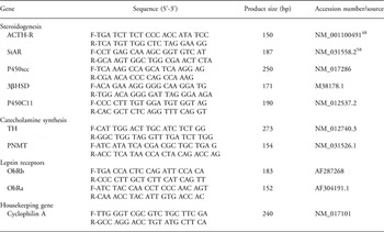
Total RNA was extracted from one whole adrenal gland of each adult male offspring using the Qiagen All-Prep mini Kit (Qiagen, Clifton Hill, Australia). The isolated RNA was quantified by a Nanodrop spectrophotometer (ND-1000, software version 3.1.2, BioLab Ltd) and assessed for integrity on a 1% agarose gel. Total RNA (5 μg) was reverse transcribed at 37°C for 60 min using M-MLV reverse transcriptase (Promega, Madison, WI, USA) and random primers (Invitrogen, Mulgrave, Australia) according to the manufacturer’s instructions. Gene-specific primers for rat ACTH-R, StAR, P450scc, 3βHSD, P450C21, P450C11, TH, PNMT and Cyclophilin A were designed using Primer3 software version 0.4.0Reference Rozen and Skaletsky44 or have been used previously on rat tissue (Table 1). Cyclophilin A was used as an internal control. External standards were generated from pooled adrenal cDNA and 10-fold serial dilutions were made in RNAse-free water (1–105-fold dilutions). Quantitative PCR was performed in 15 μl reaction volumes using the Sybr Green (Invitrogen, Mulgrave, Australia) system using an ABI PRISM 7900HT Sequence Detection System (Perkin-Elmer Applied Biosystems, Foster City, CA, USA) with 10 μM primer concentrations. The PCR cycling conditions were 50°C for 2 min, 95°C for 2 min, 40 cycles of denaturation at 95°C for 10 s, annealing at 60°C for 1 min, followed by the dissociation curve analysis between 90°C, 60°C and 99°C. Dissociation curve analysis showed a single PCR product confirmed to be of the correct size and sequence by gel electrophoresis and sequence analysis (data not shown). Standards, unknowns and non-template controls were included in each assay and measured in triplicate. Expression levels of the genes of interest were expressed relative to Cyclophilin A.
Primer sequences for quantitative PCR

Immunofluorescent localization of PNMT and ObRb protein
Immunofluorescent staining colocalizing ObRb and PNMT was performed on 7 μm paraffin-embedded sagittal adrenal sections. Before labeling, sections were treated with Sudan Black B and ultraviolet (UV) light to reduce autofluorescence.Reference Viegas, Martins, Seco and do Carmo45 Briefly, sections were irradiated with UV light (emitting light in the 253–400 nm range) at room temperature for 2 h. Sections were transferred to phosphate buffered saline (PBS) and then deparaffinized in xylene and rehydrated in descending alcohol series. Slides were treated with 10 mM sodium citrate buffer for antigen retrieval and then placed in 0.1% Sudan Black B (Sigma Chemical Co.) in 70% ethanol for 20 min at room temperature, then washed 3 × 5 min in PBS with 0.02% Tween-20 (Sigma Chemical Co.). Sections were blocked using 10% normal goat serum for 20 min, followed by blocking with avidin/biotin using a blocking kit as per the manufacturer’s protocol (SP-2001, Vector Laboratories, Burlingame, CA, USA). Dual immunofluorescent labeling of ObRb (labeled with fluorescein avidin DCS) and PNMT (labeled with Texas Red©) was performed using a commercial fluorescent avidin kit (A-1100, Vector Laboratories, Burlingame, CA, USA) as per the manufacturer’s protocol using a rabbit anti-ObRb antibody (1:200; 250739, Abbiotec, San Diego, CA, USA), a rabbit anti-PNMT antibody (1:300; AB110, Chemicon, Temecula, CA, USA) and a biotinylated anti-rabbit IgG secondary antibody (BA-1000, Vector Laboratories, Burlingame, CA, USA). Negative controls were included in the experiments: both the ObRb and PNMT primary antibodies were substituted with antibody dilution buffer for the primary negative controls and the secondary anti-rabbit IgG antibody was substituted with antibody dilution buffer for the secondary negative control. Slides were visualized on a ZEISS LSM 510 META upright confocal microscope (AXIOPLAN 2 Imaging microscope, Germany).
Statistical analyses
Statistical analyses were performed using JMP 8 (SAS Institute Inc., Cary, NC, USA). Data were analyzed for normality and equal variances. Data that were of non-normal distribution were log transformed to normalize and data that could not be normalized were analyzed by non-parametric tests. Effects of maternal diet, postweaning diet and their interactions were determined by two-way ANOVA with maternal diet and postnatal diet as factors, with Tukey’s post hoc test for multiple comparisons where appropriate. Relationships between leptin concentrations and steroidogenic and catecholamine-synthesizing enzyme expression levels were evaluated by Pearson Product correlation analysis. Level of significance was set at P = 0.05. Data are presented as means ± s.e.m., with each litter representing an n of one.
Results
Postnatal growth and caloric intake
Body weights and caloric intakes have been previously published.Reference Howie, Sloboda, Kama and Vickers10 Briefly, by postnatal day 150, offspring of MHF and PLHF dams were significantly heavier than CON offspring on both chow and hf diets.Reference Howie, Sloboda, Kama and Vickers10 PLHF offspring had increased caloric intake compared to both CON and MHF offspring from days 30 to 60, after which caloric intakes were not significantly different between any of the treatment groups.Reference Howie, Sloboda, Kama and Vickers10
Adrenal weights
Adrenal weights (unadjusted) were similar between maternal dietary groups but were increased by postweaning hf nutrition (CON-con 60 ± 2 mg, CON-hf 66 ± 2, MHF-con 61 ± 2, MHF-hf 71 ± 4, PLHF-con 62 ± 3, PLHF-hf 66 ± 2, P < 0.005 for postweaning hf diet). When expressed relative to body weight, adrenal weights were not altered by maternal diet but were reduced in postweaning HF offspring (data not shown).
Plasma corticosterone, HCY and leptin concentrations
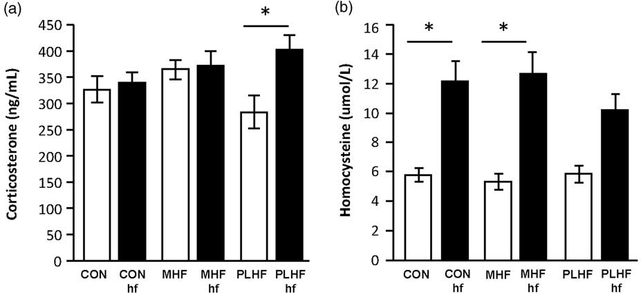
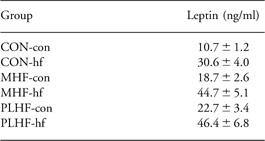
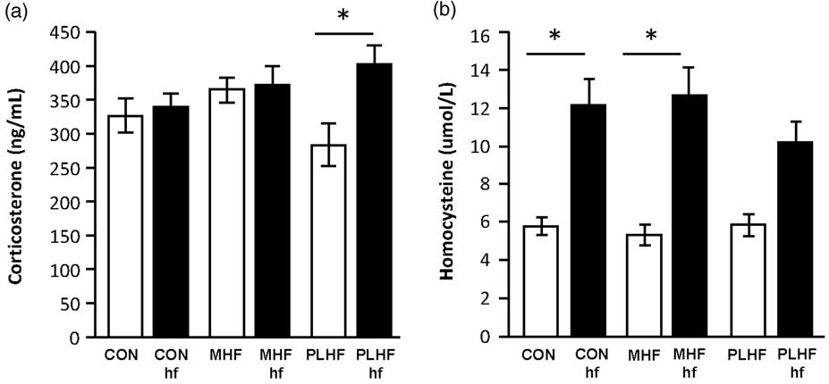
There was no effect of maternal diet on plasma corticosterone concentrations, but levels were higher overall in postweaning hf compared to postweaning con offspring (P = 0.02), due to significantly increased corticosterone concentrations in PLHF-hf offspring compared to PLHF-con (P = 0.05; Fig. 1a). There was a significant interaction between maternal and postweaning diets (Tukey, P = 0.05). Plasma HCY concentrations were unaffected by maternal diet, but were increased in postweaning hf-fed offspring compared to postweaning con (P < 0.0001), in which concentrations were increased in CON-hf and MHF-hf compared to CON-con and MHF-con offspring (Fig. 1b). There was no interaction between maternal and postweaning diets on plasma HCY concentrations. Plasma leptin and insulin levels have been previously reported.Reference Howie, Sloboda, Kama and Vickers10 Overall, both maternal (P < 0.0001) and postweaning (P < 0.0001) HF diets significantly increased offspring circulating leptin concentrations compared to controls, although no significant interaction between maternal and postweaning diets on offspring leptin levels was observed (Table 2).
Circulating plasma corticosterone and homocysteine (HCY) concentrations in male rats at P160. (a) Plasma corticosterone concentrations: No effect of maternal diet; P = 0.02 for postnatal high fat diet effect; P = 0.05 for interaction between maternal and postnatal high fat diets. (b) Plasma HCY concentrations: No effect of maternal diet; P < 0.0001 for postnatal high fat diet; no significant interactions. Data are mean ± s.e.m. *denotes differences after Tukey’s post hoc test.
Mean plasma leptin concentrations in adult male rats
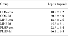
Data are mean ± s.e.m. P < 0.0001 for maternal HF diet effect (PLHF and MHF v. CON); P < 0.0001 for postweaning HF diet effect; no significant interactions.
Adrenal steroidogenic enzyme expression levels
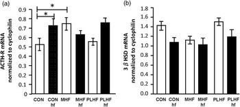
ACTH-R mRNA expression levels were significantly elevated in MHF-con compared to CON-con offspring (Tukey, P = 0.03; Fig. 2a) and were higher overall in postweaning hf compared to postweaning con offspring (P = 0.03), but this was only significant in CON offspring (Tukey, P = 0.03; Fig. 2a). Circulating leptin concentrations correlated positively with ACTH-R mRNA expression levels (y = −0.82 + 0.11(0.06), r = 0.22, P = 0.058). 3βHSD mRNA expression levels overall were significantly reduced in MHF compared to PLHF, but not CON, offspring (P = 0.04) and were significantly lower overall in postweaning hf compared to postweaning con offspring (P = 0.006; Fig. 2b). There was no interaction between maternal and postweaning diets on 3βHSD mRNA levels. There was a significant negative correlation between circulating leptin concentrations and 3βHSD mRNA expression levels (y = 1.65−0.14(0.06), r = −0.25, P = 0.03). Overall, StAR mRNA levels were significantly higher in MHF compared to PLHF, but not CON, offspring (P = 0.01), and the increase was only present in postweaning hf groups (CON-con 0.88 ± 0.06, CON-hf 1.07 ± 0.08, MHF-con 1.04 ± 0.12, MHF-hf 1.31 ± 0.11, PLHF-con 0.99 ± 0.01, PLHF-hf 0.79 ± 0.07; Tukey, P = 0.05). There were neither effects of maternal or postweaning diet on P450scc, P450C11, ObRb or ObRa mRNA levels, and nor were there significant correlations between circulating leptin concentrations and mRNA expression levels of these genes (data not shown).
Adrenal cortical steroidogenic enzyme mRNA expression in male rats at P160. (a) ACTH-R mRNA: No effect of maternal diet; P = 0.03 for postnatal high fat diet effect; P = 0.03 for interaction between maternal and postnatal diets. (b) 3βHSD mRNA: P = 0.04 for maternal diet effect (PLHF v. MHF); P = 0.006 for postnatal high fat diet effect; no significant interactions. Data are mean ± s.e.m. *denotes differences after Tukey’s post hoc test.

Adrenal catecholamine-synthesizing enzyme expression levels
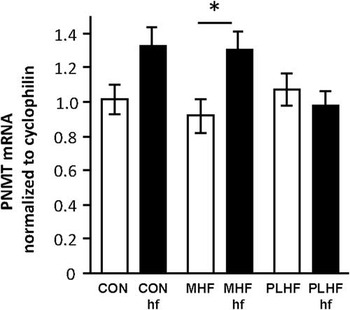
Although PNMT mRNA was unaffected by maternal HF diet, there was an overall increase in expression levels in offspring fed a postweaning hf diet compared to postweaning con (P = 0.01); levels were higher in MHF-hf offspring compared to MHF-con (Tukey, P = 0.04; Fig. 3). There was no effect of maternal or postweaning diet and no interaction between these factors on TH mRNA expression levels (data not shown). There was no significant relationship between circulating leptin concentrations and either PNMT or TH mRNA expression levels (data not shown).
Adrenal medullary PNMT mRNA expression in male rats at P160. PNMT mRNA: No effect of maternal diet; P = 0.01 for postnatal high fat diet effect; P = 0.04 for interaction between maternal and postnatal diets. Data are mean ± s.e.m. *denotes differences after Tukey’s post hoc test.

Localization of ObRb and PNMT protein
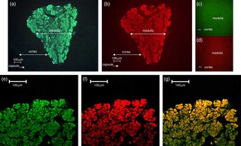
Immunoreactive (ir) ObRb and PNMT were colocalized within discrete regions in the adrenal gland: ObRb was localized to the adrenal medulla but not in the surrounding cortex (Fig. 4a, e). PNMT was localized to the cytoplasm of chromaffin cells in the adrenal medulla, but not in the surrounding cortex (Fig. 4b, f). Double labeling of ir-ObRb and PNMT were present throughout the adrenal medulla, with dense labeling observed in the majority of chromaffin cells (Fig. 4g). Figure 4c and d depict representative negative controls.
Adrenal medullary ObRb and PNMT protein localization in male rats at P160. (a) Immunofluorescent labeling of ObRb at 10× magnification demonstrates labeling in the adrenal medulla with no labeling in the surrounding cortex. (b) Immunofluorescent labeling of PNMT at 10× magnification demonstrates labeling in the adrenal medulla with no labeling in the surrounding cortex. (c, d) Primary negative controls for ObRb (c) and PNMT (d) at 10× magnification. (e) Immunofluorescent labeling of ObRb at 20× magnification demonstrates labeling in the adrenal medulla with no labeling in the surrounding cortex. (f) Immunofluorescent labeling of PNMT at 20× magnification demonstrates labeling in the adrenal medulla with no labeling in the surrounding cortex. (g) Dual immunofluorescent labeling of ObRb and PNMT in the adrenal medulla at 20× magnification demonstrates colocalization of these proteins. Photographs are of the same tissue section viewed by different fluorescence filters.

Methionine and pyridoxine intake in adulthood
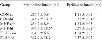
Owing to the changes observed in HCY concentrations, dietary intakes of methionine and pyridoxine, which are associated with HCY metabolism and methyl balance, were determined. Overall, methionine intake was significantly increased in offspring fed a postweaning hf diet compared to postweaning con offspring (P < 0.0001; Table 3). Conversely, the overall pyridoxine intake was significantly reduced in offspring fed a postweaning hf diet compared to postweaning con offspring (P < 0.0001; Table 2).
Mean dietary intakes of methionine and pyridoxine in adult male rats

Data are mean ± s.e.m. *P < 0.0001 for postweaning HF diet effect.
Discussion
We present novel data suggesting that a postweaning HF diet in offspring induced changes in adrenal gene expression levels that are, in part, dependent upon the level of maternal nutrition. We have shown higher corticosterone and HCY concentrations in animals fed a postweaning hf diet, dependent upon the level of maternal nutrition, and higher leptin concentrations following maternal and postweaning HF diets. ACTH-R mRNA expression levels were elevated in MHF-con and CON-hf offspring compared to controls, and there was a positive relationship between circulating leptin levels and adrenal ACTH-R mRNA expression. 3βHSD mRNA expression levels were lower in postweaning hf offspring compared to postweaning con offspring and there was a significant negative relationship between circulating leptin levels and adrenal 3βHSD mRNA expression. Furthermore, PNMT mRNA expression levels were higher with postweaning hf diet and we colocalized ir-PNMT and ir-ObRb to the medulla of the adrenal gland.
Previous studies have shown that changes in maternal nutrition have long-term effects on the function and activity of the HPA axis in offspring and that the timing of the maternal nutritional challenge is critically important.Reference Bloomfield, Oliver and Giannoulias17, Reference Gardner, Van Bon and Dandrea46 The importance of the preconceptional period is clear; short periods of maternal undernutrition immediately before and after conception alter offspring HPA function. The mechanisms underpinning these changes, however, are unclear. In sheep, we have previously shown that a brief period of periconceptional undernutrition results in changes in fetal adrenal P450C17 expressionReference Connor, Bloomfield, Oliver, Harding and Challis47, but whether this has long-term effects on adrenal gene expression is unknown. In this study, offspring born to mothers fed a lifetime of HF nutrition (MHF) demonstrated increased ACTH-R and decreased 3βHSD mRNA levels. These observations are consistent with the notion that balanced nutrition is essential during the preconceptional period. Of note, despite these observed changes in adrenal mRNA levels, maternal diet was not associated with altered basal corticosterone concentrations in MHF offspring. Furthermore, a postweaning HF diet increased ACTH-R and decreased 3βHSD mRNA levels in offspring born to control mothers, and was not associated with changes in basal corticosterone concentrations. We hypothesized that, given our previous observation of hyperleptinemia in offspring born to HF-fed mothersReference Howie, Sloboda, Kama and Vickers10, we would observe changes in corticosterone levels and related changes in adrenal gene expression levels. During adrenal steroidogenesis, leptin acts to inhibit the expression levels of StARReference Cherradi, Capponi, Gaillard and Pralong21 and P450sccReference Hsu, Chang and Chiu22, and consequently, modulation of corticosterone secretion by leptin may have direct influences on GC-dependent processes. We did not observe changes in basal corticosterone levels in our offspring following changes in maternal nutrition. This agrees with previous reports using cultured rat and human adrenal cells, in which leptin treatment did not alter basal GC levels but did inhibit ACTH-induced GC secretion.Reference Pralong, Roduit and Waeber26, Reference Salzmann, Otis and Long48 Reduced expression of StAR protein and reduced GC production by leptin have been shown only under stimulated conditions.Reference Cherradi, Capponi, Gaillard and Pralong21 Our data are consistent with these observations and suggest that basal adrenal function is maintained in offspring exposed to HF nutrition as adults despite changes in ACTH-R and 3βHSD. We recognize, however, that a maternal HF diet may result in differential adrenal responses to secondary stressful challenges in offspring and this should be investigated further. Intriguingly, a postweaning HF diet did induce an increase in basal corticosterone concentrations in PLHF offspring. These changes, however, were not associated with changes in adrenal gene expression levels in these animals, but could be a reflection of altered adrenal sensitivity or central drivers of HPA function. We acknowledge that to conclusively determine if there is primary programming of adrenal function on obesity risk in adults, we must investigate the function of the HPA axis at earlier times during the life course before the onset of the obese phenotype.
Our study presents data suggesting that postweaning HF nutrition alters adrenal medullary gene expression in a manner that is dependent upon maternal nutritional history and we speculate that leptin plays a role in this process. In the medulla, leptin stimulates THReference Shibuya, Utsunomiya and Toyohira23, Reference Trevenzoli, Valle and Machado24, Reference Utsunomiya, Yanagihara and Tachikawa49 and increases synthesis of epinephrine from norepinephrine, mediated by PNMT.Reference Kelner and Pollard31 Although neither maternal nor postnatal HF nutrition altered TH mRNA expression levels, postnatal HF nutrition increased PNMT mRNA, in which levels were higher in MHF-hf compared to MON-con offspring. Neonatal leptin treatment in rodents results in adult hyperleptinemia, increased body weight and TH mRNA levels.Reference Trevenzoli, Valle and Machado24 However, unlike the work of Trevenzoli and colleagues, our model did not result in hyperleptinemia at birth, but in fact resulted in low neonatal leptin levels.Reference Howie, Sloboda, Kama and Vickers10 Whether this low neonatal leptin during the stress hyporesponsive period in the rat influences mRNA levels of key adrenal medullary genes is unknown. It has been suggested that leptin may play a direct role in regulating medullary cell function, increasing epinephrine synthesis and secretionReference Cao, Considine and Lynn28, but the mechanisms are not fully known. Shibuya et al.Reference Shibuya, Utsunomiya and Toyohira23, Reference Shibuya, Utsunomiya and Toyohira51 showed that incubation of cultured bovine medullary cells with leptin resulted in increased activity of mitogen-activated protein kinases (MAPKs), which are known to phosphorylate and activate TH.Reference Haycock, Ahn, Cobb and Krebs50 This was associated with increased catecholamine synthesis from tyrosine, which was blocked by a selective MAPK kinase inhibitor, suggesting that leptin acts, in part, through an MAPK-dependent pathway to stimulate TH.Reference Shibuya, Utsunomiya and Toyohira51 Although we were not able to measure circulating catecholamines in this study, we have confirmed the colocalization of immunoreactive ObRb and PNMT in chromaffin cells, strengthening the hypothesis that leptin is directly implicated in catecholamine synthesis or secretion. Further studies are required to determine whether this colocalization has functional significance on circulating catecholamine levels and the intracellular mechanisms through which leptin may alter PNMT activity and expression.
Our observation that offspring fed a postweaning HF diet demonstrated elevated plasma HCY levels is consistent with the known association between HCY, obesity and cardiovascular diseaseReference Zhu, Huang, Li and Neubauer35, Reference Boushey, Beresford, Omenn and Motulsky52 and may be associated with our observed reduction in pyridoxine intake.Reference Selhub, Jacques, Wilson, Rush and Rosenberg39 These data may be suggestive of the reduced metabolism of HCY to cysteine by the transsulfuration pathway. The HCY metabolic pathway and its cofactors have been implicated in catecholamine synthesis. The conversion of norepinephrine to epinephrine by the methyl transferase PNMT is dependent upon the availability of the cofactor SAM (the major biological methylating agent) and the subsequent metabolism of HCY to cysteine is influenced by the availability of pyridoxine.Reference Franken, Boers, Blom, Trijibels and Kloppenborg38 Pyridoxine is also a cofactor required to convert L-DOPA to dopamine, a critical step before conversion of norepinephrine to epinephrine.Reference Siow and Dakshinamurti53 The observed increase in dietary methionine intakes in offspring fed a postweaning HF diet may contribute to the increased availability of SAM as a methyl donor for PNMT activity and have downstream effects on catecholamine synthesis, although further studies are needed to confirm this. Recent studies utilizing low-protein models have speculated on the role of dietary methionine as a driver for altered HCY metabolism and subsequent programming of physiologic processes, but have produced conflicting conclusions.Reference Rees54–Reference Engeham, Haase and Langley-Evans56
We recognize that the use of whole adrenal tissue may have masked any discernable changes specific to either the cortex or the medulla, had they been processed and examined separately. However, given that we have demonstrated robust changes in some medullary- and cortical-specific enzymes, we suggest that our methods have not limited our results. Nevertheless, the previously reported differential expression analysis of ObRb and ObRa mRNA in the adrenal cortex and medulla by Cao et al.Reference Cao, Considine and Lynn28 and Malendowicz et al.Reference Malendowicz, Neri and Markowska57 provide strong evidence for examining these regions independently and will be considered for future independent studies.
In summary, we have shown that a postweaning HF diet results in changes in the levels of specific steroidogenic and medullary genes in adult offspring that are dependent upon the nutritional background of the mother. Despite our previous observation that hyperleptinemia is common in offspring of HF mothers, we did not observe consistent changes across nutritional treatment groups as previously reported for adrenal steroidogenic enzyme expression. We speculate that in our model under basal conditions, the primary effects of hyperleptinemia may be directed toward increasing medullary catecholamine synthesis, rather than inhibiting cortical steroidogenic capacity. The possibility of changes in cortical steroidogenic function and corticosterone output under stimulated conditions in our model cannot be ruled out, and may provide an important and relevant approach for future investigations.
Acknowledgments
This work was supported by the National Research Centre for Growth and Development and the Health Research Council of New Zealand. The authors would like to acknowledge Graham Howie for his involvement in the animal work.
Statement of Interest
None.