Introduction
Precise tumour staging is a fundamental prerequisite of head and neck cancer management. The precision of such staging has a direct impact upon tumour prognosis and therapeutic strategy.
Recent radiological advances, such as multi-channel magnetic resonance scanning with multiple phased-array coils, have enabled whole-body magnetic resonance imaging (MRI) without spatial resolution compromise. Head and neck cancer often presents as a systemic disease, and whole-body imaging techniques are increasingly being used. Whole-body MRI scanners, with a field strength of 3 T, have recently been given clinical approval. Image resolution can therefore be improved, with consequent higher sensitivity. A high signal-to-noise ratio reduces the overall examination time, while also enabling highly resolved isotropic three-dimensional data sets, thus optimising the diagnosis of pathology in anatomically complex areas such as the head, neck and skull base.Reference Schmidt, Reiser and Baur-Melnyk1
Another powerful imaging modality is positron emission tomography – computed tomography (PET-CT), which offers a broad approach to cancer diagnosis, with superb anatomical delineation integrating functional, metabolic and molecular information about the neoplasm. This modality offers high-resolution, three-dimensional, quantitative imaging. It also has high sensitivity and a high negative predictive value in the visualisation of residual or recurrent disease following radiotherapy.Reference Garcia and Flamen2
In comparisons of these two imaging modalities, whole-body MRI has been shown to be at least as accurate as CT in the detection of distant metastases.Reference Chen, Zhu, Hou, Wang, Jiang and Feng3 Furthermore, whole-body MRI has shown improved visualisation of distant metastatic disease, compared with fluoro-deoxy-glucose (FDG) PET, in lung cancer patients.Reference Ohno, Nogami, Takenaka, Yoshikawa, Yoshimura and Kotani4 For initial staging, PET-CT is more accurate than whole-body MRI in the definition of tumour (T) and node (N) stages.Reference Yi, Shin and Lee5, Reference Schmidt, Baur-Melnyk and Herzog6
There is a relative paucity of data comparing the use of whole-body MRI and PET-CT for head and neck cancer staging. This comparison was the focus of the current prospective, blinded study.
Materials and methods
This prospective study included 13 cases of primary tumour and two cases of recurrent cancer of the head and neck. It was conducted over a six-month period from January to July 2009.
Institution ethical approval was obtained.
Two consultant radiologists blinded to the scanned patients' identities assessed the scan images in two sittings. All scans were staged in accordance with the American Joint Committee on Cancer tumour–node–metastasis (TNM) classification.7
All pathology reporting was undertaken by consultant pathologists, and discussed at our head and neck multidisciplinary team conference.
The study aimed to analyse the TNM staging concordance and/or discordance between the two radiological modalities.
In order to test the above hypotheses, several statistical techniques were used. The chi-square test was used to assess whether there was a significant difference between the number of agreements and the number of disagreements for each new variable, at a 5 per cent significance level. If the test was significant, frequency analysis (using a bar chart) was conducted to determine whether the compared staging modalities differed or not (a difference was concluded if the frequency of ‘disagree, discordance’ was greater than that of ‘agree, concordance’).Reference Schmidt, Reiser and Baur-Melnyk1
Results and analysis
Fifteen consecutive head and neck cancer patients (11 men and four women; mean age 59 years; age range 19 to 81 years) underwent PET-CT and whole-body MRI scans for pre-therapeutic evaluation. The tumour sites involved were the salivary gland, larynx and pharynx.
All scans were conducted on a 1.5 Tesla scanner.
Tumour staging
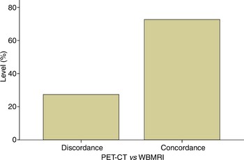
Analysis of results for both imaging modalities indicated that the sample proportions were significantly different from the hypothesised values of 0.5 and 0.5 (p < 0.05). Figure 1 indicates that results for the two modalities were significantly similar as regards T staging. In this regard, the percentage concordance was 74 per cent.
Concordance and discordance for positron emission tomography – computed tomography (PET-CT) versus whole-body magnetic resonance imaging (WBMRI), for tumour staging.

Node staging
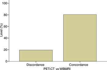
Analysis of results for both modalities indicated that the sample proportions were significantly different from the hypothesised values of 0.5 and 0.5 (p < 0.05). Figure 2 indicates that results for the two modalities were significantly similar as regards N staging. In this regard, the percentage concordance was 80 per cent.
Concordance and discordance for positron emission tomography – computed tomography (PET-CT) versus whole-body magnetic resonance imaging (WBMRI), for node staging.

Metastasis staging
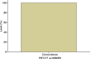
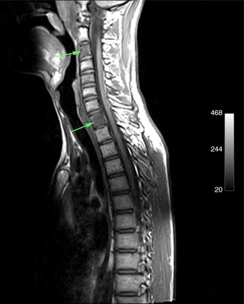
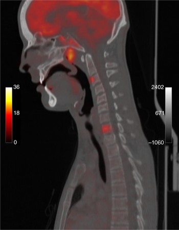
We noted 100 per cent concordance between both modalities for the detection of metastases (Figure 3). However, superior visualisation of metastases was noted for whole-body MRI (Figure 4) compared with PET-CT (Figure 5).
Concordance for positron emission tomography – computed tomography (PET-CT) versus whole-body magnetic resonance imaging (WBMRI), for metastasis staging.

Sagittal whole-body magnetic resonance imaging scan of a patient with nasopharyngeal primary cancer with spinal metastases (arrows).

Sagittal positron emission tomography – computed tomography scan of the same patient shown in Figure 4.

Tumour recurrence staging
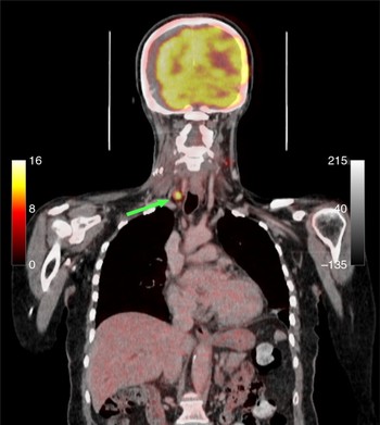
There were two cases of head and neck cancer recurrence. Concordance between the two imaging modalities was noted in the first case. However, in the second case, the relevant nodal metastatic deposit was detected on PET-CT (Figure 6) but was not visible on whole-body MRI.
Coronal positron emission tomography – computed tomography scan of a patient with paratracheal nodal recurrent disease (arrow).

Whole-body MRI scanning took on average 20 minutes to complete, as opposed to 2 hours for PET-CT.
Discussion
The clinical applicability of 2-deoxy-2-(F-18) fluoro-d-glucose positron emission tomography in head and neck squamous cell carcinoma is well documented. It has greater sensitivity than specificity, and false positives (due to infection or inflammation) continue to be problematic. One PET-CT scanning session takes approximately two hours, and results in a large radiation exposure (17–25 mSv). Whole-body MRI is an appealing diagnostic modality, given its absence of ionising radiation, fast acquisition of high soft tissue contrast and good spatial resolution.
In Ireland, the current cost of a PET-CT scan is approximately €2500–3000. Whole-body MRI has a cost advantage, at approximately €500 per scan.
There are few comparative studies assessing the use of these two imaging modalities in head and neck cancer. A study of lung cancer staging involving 165 patients has been reported: for tumour staging, an accuracy of 86 per cent was found for whole-body MRI, compared with 82 per cent for PET-CT.Reference Yi, Shin and Lee5
Detection of lymph node metastases is the undoubted advantage of PET-CT scanning, based as it is on cell metabolism. Studies comparing whole-body MRI with PET-CT for N staging report diagnostic accuracies of 91–97 per cent for PET-CT and 79–82 per cent for whole-body MRI.Reference Schmidt, Baur-Melnyk and Herzog6, Reference Balogova, Perie, Kerrou, Grahek, Montravers and Angleard8
Whole-body MRI is an accurate diagnostic technique, and may be considered at least as effective as FDG-PET for M staging. Indeed, some reported findings indicate that whole-body MRI has advantages for the detection of bony metastases. In the current study, we report data and give examples (e.g. Figure 4 compared with Figure 5) of superior visualisation of tumour metastases with whole-body MRI.
• Precise tumour staging is a fundamental pre-requisite of head and neck cancer management, and has a direct impact upon tumour prognosis and therapeutic strategies
• This prospective, blinded trial investigated the concordance between positron emission tomography – computer tomography and whole-body magnetic resonance imaging (MRI) used for the staging of head and neck cancer
• A discordance was found between these imaging modalities
• Whole-body MRI is an emerging staging modality which enables superior visualisation of metastatic disease, without exposure to ionising radiation
In a study of 98 patients, Antoch et al. correctly estimated the overall TNM staging in 77 per cent of PET-CT scans, compared with 54 per cent of whole-body MRI scans. These authors also found whole-body MRI to be more accurate in the detection of bone and liver metastases.Reference Antoch, Vogt and Freudenberg9 In our study, we noted 74 per cent concordance in final TNM staging, comparing these two modalities.
Positron emission tomography – computed tomography has poor sensitivity in the first two to three months after therapy. This is because the tumour load increases with time. It must also be remembered that not all tissues with high metabolic activity are malignant, and that, conversely, not all tissues with minimal activity are benign. Correct clinical application allows PET-CT to exclude residual disease in most patients, particularly those with lymphadenopathy, who would otherwise require neck dissection.
A prospective trial is required to confirm that lack of 18F-FDG uptake excludes tumour viability with a high level of certainty.Reference Ong, Schoder, Lee, Patel, Carlson and Fury10
In the future, we aim to report results for a larger patient cohort, to further compare PET-CT and whole-body MRI for the staging of head and neck cancer.
Conclusion
We report a prospective, blinded trial assessing concordance between the results of PET-CT and whole-body MRI for staging head and neck cancer. We observed advantages and disadvantages for both imaging modalities. Concordance levels were 74 per cent for T stage, 80 per cent for N stage and 100 per cent for M stage. Whole-body MRI continues to reveal superior metastatic visualisation. With its lack of ionising radiation and rapid image acquisition, there is a definite role for whole-body MRI, particularly for evaluation of early and long term tumour response. However, its spatial resolution is not as good as that of focussed surface coil MRI scanning, and lesions smaller than 2 mm are therefore difficult to assess. Positron emission tomography – computed tomography remains an excellent diagnostic modality, albeit with limitations of high radiation exposure, slow image acquisition and reduced patient throughput. Both imaging modalities provide complementary, synergistic information for staging head and neck cancer.