Hypoplastic left heart syndrome represents 2–3% of CHD cases.Reference Gobergs, Salputra and Lubaua 1 Ebstein’s anomaly represents <1% of CHD.Reference Luxford, Arora and Ayer 2 The combination of hypoplastic left heart syndrome with Ebstein’s anomaly is very rare in CHD, and the management of this type of anomaly is difficult as the prognosis is poor.Reference Stamm, Anderson and Ho 3 – Reference Castaneda-Zuniga, Nath, Moller and Edwards 6 We reported a case of hypoplastic left heart syndrome with Ebstein tricuspid valve using prenatal fetal imaging, and post-natal two-dimensional and three-dimensional echocardiographic imaging of this lesion.
Case presentation
A 37-year-old patient presented at 19 weeks and 3 days of gestation for a fetal echocardiogram as a referral from an outside institution for suspected fetal cardiac anomalies. A fetal echocardiogram was performed and it demonstrated a moderately hypoplastic left ventricle, severely hypoplastic mitral valve, a severely hypoplastic aortic valve, a dysplastic tricuspid valve, and significant left ventricular endocardial fibrosis.
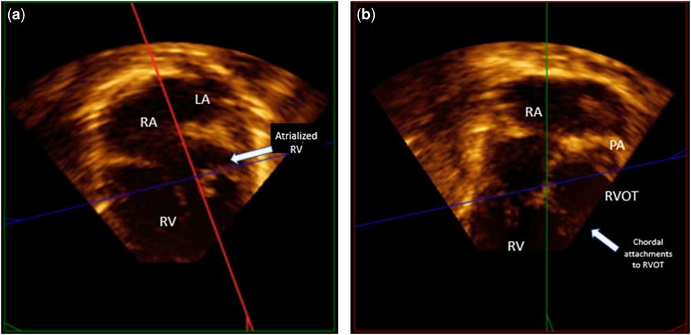
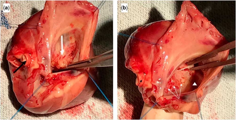
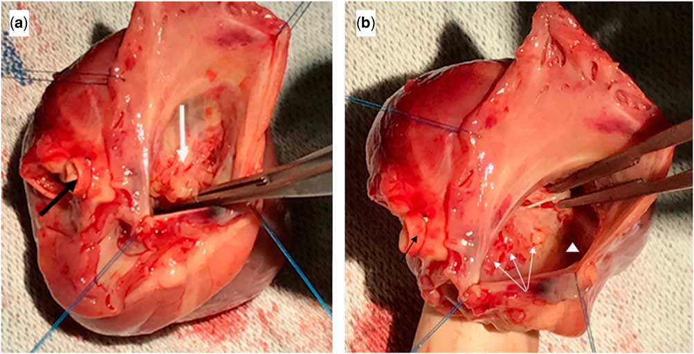
The infant was born by Caesarean delivery at 40 weeks of gestation. A complete transthoracic echocardiogram confirmed a moderately hypoplastic left ventricle, severely hypoplastic mitral valve with minimal antegrade mitral valve flow, an atretic aortic valve, and a dysplastic tricuspid valve with moderate tricuspid valve insufficiency with concern for Ebstein’s anomaly (Supplementary Fig 1). A three-dimensional echocardiogram was then performed using the X7 transthoracic transducer with full-volume four-beat acquisition. After cropping, we demonstrated the Ebstein tricuspid valve from above (Supplementary video 1). The three-dimensional imaging was an additive component to the diagnosis of the tricuspid valve morphology in this complex patient. Using multi-plane reconstruction, we could demonstrate the chordal attachment of the anterior leaflet of the tricuspid valve to the right ventricular outflow tract (Fig 1). This patient did not qualify for a staged surgical palliation as the single-ventricle palliation with the addition of tricuspid valve repair would become very high risk. The patient was listed and received a heart transplantation at 55 days old. The explanted heart demonstrated a displaced tricuspid valve septal leaflet and anteriorly displaced anterior tricuspid valve leaflet (Fig 2).
( a ) The four-chamber view demonstrating the large section of atrialised right ventricle. ( b ) Right ventricular inflow/outflow view demonstrating the chordal attachments of the Ebstein valve to the right ventricular outflow tract. LA=left atrium; PA=pulmonary artery; RA=right atrium; RV=right ventricle, RVOT=right ventricular outflow tract.

( a ) Hypoplastic left heart syndrome (HLHS) with Ebstein’s anomaly: black arrow demonstrates transected hypoplastic ascending aorta, and white arrow demonstrates sail-like anterior leaflet with severely dysplastic leading edge. ( b ) HLHS with Ebstein’s anomaly: black arrow demonstrates the transected hypoplastic ascending aorta, white arrows demonstrate dysplastic septal leaflet with apical displacement, and white triangle is the atrialised portion of right ventricle.
Discussion
Hypoplastic left heart syndrome with Ebstein’s anomaly is a rare congenital heart lesion that has poor prognosis.Reference Hammel, Danford, Spicer and Kutty 5 Hammel et al reported a case with hypoplastic left heart syndrome with Ebstein’s anomaly with severe tricuspid regurgitation who did not survive. Tricuspid valve regurgitation is a poor prognostic factor that leads to poor outcomes in a single-stage palliation.Reference Kutty, Graney and Khoo 7 The Ebstein valve would not be suited to function as a systemic atrioventricular valve. Three-dimensional echocardiographic imaging complemented our two-dimensional imaging by providing better delineation of the tricuspid valve function with chordal attachment of the papillary muscle to the right ventricular outflow tract. Multi-plane reconstruction using the three-dimensional data set was helpful in delineating the anatomy well and in understanding the anatomy in different cut planes.Reference Bharucha, Anderson, Lim and Vettukattil 8 Multi-plane reconstruction allows for quick interrogation of the anatomy of the chordae tendinae and papillary muscles from one single three-dimensional data set, and provides cut planes that would not have been obtained from the traditional two-dimensional imaging. This allows for understanding the anatomy well in three dimensions and assists in communicating this anatomy to the surgeon.Reference Acar, Abadir and Roux 9 The explanted heart anatomy corresponded well to the three-dimensional images and multi-plane reconstruction.
Conclusion
We report a three-dimensional evaluation of a patient with hypoplastic left heart syndrome and Ebstein’s anomaly with significant tricuspid valve regurgitation. Owing to the severity of the tricuspid valve dysfunction, the patient was listed and received a heart transplantation.
Acknowledgements
None.
Financial Support
This research received no specific grant from any funding agency or from commercial, or not-for-profit sectors.
Conflicts of Interest
None.
Supplementary material
To view supplementary material for this article, please visit https://doi.org/10.1017/S1047951118000367