introduction
The protozoan parasite Toxoplasma gondii is the aetiological agent of toxoplasmosis, a worldwide infection affecting 500 million to 1 billion people (Tenter et al. Reference Tenter, Heckeroth and Weiss2000). Members of the Felidae family are considered the definitive hosts for toxoplasmosis, but intermediate hosts are the most affected by toxoplasmosis (Dubey, Reference Dubey2010). In immunocompromised individuals, Toxoplasma is an important opportunistic pathogen due to fulminate re-emergence of chronic infections (Luft et al. Reference Luft, Brooks, Conley, McCabe and Remington1984). Acute infections acquired during pregnancy are also capable of causing severe birth defects, including hydrocephaly, calcification, neurological defects and chorioretinitis, which may be recurrent (Wong and Remington, Reference Wong and Remington1994). Recent studies have shown that an infection with T. gondii may affect behaviours of hosts (Gatkowska et al. Reference Gatkowska, Wieczorek, Dziadek, Dzitko and Dlugonska2012) and may be a causative or contributory factor for certain psychiatric disorders, sterility and cancers (Thomas et al. Reference Thomas, Thomas, Salmon, Lewis and Smith2004; Yuan et al. Reference Yuan, Gao, Liu, Xia, Liu, Liu and Hu2007; Li et al. Reference Li, Cui, Zhao, Dai, Zong, Zuo, Chen, Jin, Gao and Liu2011).
Prophylaxis and treatment of T. gondii infection is often by combination chemotherapy with pyrimethamine and sulfadiazine, or in cases of sulfa intolerance, pyrimethamine is combined with clindamycin. Several studies have shown that trimethoprim/sulfamethoxazole is effective as primary prophylaxis of toxoplasmosis (Grossman and Remington, Reference Grossman and Remington1979; Derouin and Chastany, Reference Derouin and Chastany1989; Carr et al. Reference Carr, Tindall, Brew, Marriott, Harkness, Penny and Cooper1992). However, generally these drugs are poorly tolerated, presenting severe side effects, and cannot act against chronic T. gondii infections. In addition, resistance to some of these drugs has been noted (Dannemann et al. Reference Dannemann, McCutchan, Israelski, Antoniskis, Leport, Luft, Nussbaum, Clumeck, Morlat, Chiu, Vilde, Orellana, Feigal, Bartok, Heseltine, Leedom and Remington1992; Aspinall et al. Reference Aspinall, Joynson, Guy, Hyde and Sims2002; Baatz et al. Reference Baatz, Mirshahi, Puchta, Gumbel and Hattenbach2006). Added to the fact that there is no effective vaccine available for the prevention of T. gondii infection in humans (Vermeulen, Reference Vermeulen1998) the development of effective drugs with low toxicity, associative therapies, or treatments of support, which may enhance the effectiveness or reduce the adverse effects of traditional chemotherapy, are necessary to treat toxoplasmosis.
In this sense some protocols have been developed, such as the utilization of supplementation with selenium compounds. The potential health-promoting activities of dietary selenium consumption at supranutritional levels (above 55−70 μg per day) have been widely documented (Boosalis, Reference Boosalis2008; Hoffmann and Berry, Reference Hoffmann and Berry2008; Rayman, Reference Rayman2008). The most evident biological effects of selenium include enhanced immune response and antiviral effects, protection of thyroid, cognitive and reproductive functions, reduced risk of cancer and coronary heart disease (Fleming et al. Reference Fleming, Ghose and Harrison2001; Lu and Jiang, Reference Lu and Jiang2005; Rayman, Reference Rayman2005; Naithani, Reference Naithani2008). Based on these properties, we hypothesized if a supplementation with selenium compounds could improve the therapeutic efficacy.
Therefore, the aim of this study was to assess the effect of sulfamethoxazole/trimethoprim supplemented with diphenyl diselenide (DPDS) and sodium selenite (SSe) in oxidative damage and inflammatory response, in mice infected with T. gondii (RH strain).
MATERIALS AND METHODS
Experimental animals
Eighty-four adult male BALB/c mice (Mus musculus), average 60 days old and with weight varying from 22 to 24 grams were used in our study. They were kept in an experimental room with controlled temperature and humidity (25 °C; 70% RH), fed a commercial ration, and with water ad libitum. The animals were submitted to a period of 7 days of adaptation. The procedure was approved by the Animal Welfare Committee of Universidade Federal de Santa Maria, number 73/2012.
Toxoplasma gondii strain and inoculum preparation
This experiment used a strain of T. gondii (RH, isolated by Sabin 1941), kept in liquid nitrogen in laboratory. First, one mouse was inoculated intraperitoneally with tachyzoites of T. gondii (0·20 mL, containing 1·32×107 tachyzoites mL−1). Four days later, peritoneal fluid containing the parasite was collected and inoculated into other mice (at same dose and parasite concentration). This procedure was repeated three times, in order to reactivate the parasite virulence; the last inoculation was carried out in 5 mice with the aim of obtaining a large number of T. gondii tachyzoites for the mice infection.
Reagents
Diphenyl diselenide (Ph2Se2) (99·9%) and sodium selenite (Na2O3Se) (99%) were purchased from SIGMA (St. Louis, MO, USA). Sulfamethoxazole/trimethoprim (ST) (Bactrim® F suspension), available as a commercial product, was purchased from ROCHE (Rio de Janeiro, RJ, Brazil). SSe was diluted in physiological solution (0·9%) resulting in a final concentration of 1·67%. DPDS was dissolved in dimethyl sulfoxide (DMSO, Sigma-Aldrich, USA) at final concentration of 0·1%.
Experimental design
The 84 animals were divided in seven groups (A, B, C, D, E, F, G with 12 animals in each group). These groups were divided again in two subgroups (A1 and A2, B1 and B2, C1 and C2, D1 and D2, E1 and E2, F1 and F2, G1 and G2 – with 6 animals/each group). All infected mice received, intraperitoneally, a volume of 0·5 mL of peritoneal fluid, containing 1·2×107 tachyzoites. 0·5 mL saline was administered in group A, by the same method. Treatment with ST was performed twice a day (0·5 mg kg−1), orally; from day 1 to 20 p.i. SSe (intramuscularly) and DPDS (subcutaneously) were administered on the first day p.i. at 1 mg kg−1 and 5 μmol kg−1, respectively.
The treatments were carried as follows: Groups A and B served as negative (uninfected) and positive (infected) controls, respectively; in group C, the animals were infected with T. gondii and treated with ST; group D comprised mice infected with T. gondii and supplemented with SSe; in group E the mice were infected with T. gondii and received DPDS; group F comprised animals infected with T. gondii and treated with combination ST and SSe; and finally, the mice of group G were infected with T. gondii and treated with the combination of ST and DPDS.
On day 4 p.i. (subgroups A1, B1, C1, F1 and G1) and day 20 p.i. (subgroups A2, C2, F2 and G2) the sample collections were performed. Animals of groups D1, D2, E1 and E2 died before day 4 p.i., and mice from group B2 died before day 20 p.i. In both periods, the surviving animals were anaesthetized in a chamber with isoflurane and submitted to cervical dislocation, for posterior collection of blood by cardiac puncture (1·0 mL) and liver tissue. Fragments of livers were homogenized in 10 mmol Tris-HCl buffer pH 7·2, and then centrifuged at 5000 g for 10 min. The supernatant was collected for analysis of TBARS, AOPP and GR. Samples were stored in tubes and frozen at −80 °C until analysed. All the procedures described above were performed at refrigerated temperature (4 °C).
TBARS and AOPP
All the evaluations were performed in liver tissue (Mordue et al. Reference Mordue, Monroy, La Regina, Dinarello and Sibley2001).
TBARS
Lipid peroxidation (TBARS levels) was measured as described by Ohkawa et al. (Reference Ohkawa, Ohishi and Yagi1979). Briefly, the reaction mixture contained 200 μL of liver homogenate or standard malondialdehyde (0·03 mm; MDA), 200 uL of 8·1% sodium dodecylsulphate (SDS), 750 uL of acetic acid solution (2·5 m HCl, pH 3·5) and 750 uL of 0·8% thiobarbituric acid (TBA). Since sucrose interferes in the TBARS assay, a portion of the liver was weighed, homogenized at a proportion of 1 g of tissue to 10 mL of buffer Tris/HCl 50 mm pH 7·4 plus 10% of SDS. The mixtures were heated at 95 °C for 90 min. After centrifugation at 1700 g for 5 min, the absorbance was measured at 532 nm. TBARS tissue levels were expressed as nmol MDA per mg of protein.
AOPP
The advanced oxidation protein products (AOPP) were measured as the oxidation of iodide anion to diatomic iodine by AOPP (Witko-Sarsat et al. Reference Witko-Sarsat, Friedlander, Nguyen Khoa, Capeillere-Blandin, Nguyen, Canteloup, Dayer, Jungers, Drueke and Descamps-Latscha1998). Briefly, the technique consists in treating 100 μL of tissue homogenates in PBS (1 mL) with 50 μL of potassium iodide 1·16 m followed by 100 μL of acetic acid. The absorbance of the reaction mixture was immediately read at 340 nm. Results of AOPP levels were expressed in μmol L−1 protein.
Glutathione reductase (GR)
GR activity in homogenized liver was assessed by the automated technique using a Cobas Mira® analyser (Hermes et al. Reference Hermes, Hausen, Sangoi, Almeida, Carvalho, Gomes and Moresco2013), with results expressed as U/g protein. Briefly, NADPH (3·6 mm) and GSSG (10 mm) were diluted in 0·1 m monobasic potassium phosphate buffer pH 7·6 containing 6·3 mm of EDTA. GR was prepared in this buffer (10, 40, 80, 120 and 200 U L−1) for the development of the calibration curve in triplicate. GR activity was measured according to the method described by Mannervik and Carlberg (Reference Mannervik and Carlberg1985), with some modifications in the Cobas Mira® automated analyser (Roche Diagnostics, Basel, Switzerland) as follows: 160 μL of monobasic potassium phosphate buffer and 20 μL of oxidized glutathione (GSSG) were pipetted. Then, 20 μL of the sample (homogenate of liver tissue) was pipetted into the reaction cuvette after 25 s. The reaction starts with the addition of 20 μL of NADPH, 50 s after the addition of the sample.
Cytokines
Cytokines in serum samples were assessed by ELISA using commercial kits for mouse INF-γ and IL-10 (eBioscience®, San Diego, USA), according to the manufacturer's instructions. Briefly, microplates were sensitized with the primary antibody at room temperature (RT) for 30 min. Then, the serum sample was added and incubated (37 °C, for 30 min). After washing, the secondary antibody conjugated with peroxidase was added and incubated. The presence and concentration of the cytokines were determined by the intensity of the colour measured by spectrometry in a micro ELISA reader.
Statistical analysis
The data were submitted to analysis of variance followed by the Student t-test (P<0·05), performed using SAS statistical package (SAS Institute, Cary, NC, USA) with a significance level of 5%.
RESULTS
Course of infection after treatment
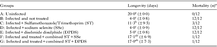
Results on longevity and mortality using treatment with Bactrim® F, sodium selenite and diphenyl diselenide in mice experimentally infected with T. gondii are presented in Table 1. Mice treated only with selenium compounds (groups D and E) were not able to control the disease, dying within 5 days after infection as happened with the positive control (group B). The treatment only with chemotherapy (group C) or in combination with selenium compounds (groups F and G) controlled the disease, preventing mortality in most of the animals tested, and thus prolonging longevity.
Mean and standard deviation of longevity and mortality using treatment with Bactrim® F, sodium selenite and diphenyl diselenide in mice experimentally infected with Toxoplasma gondii
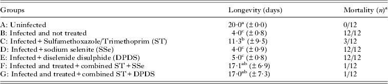
Note: Results are the mean±standard deviation. Results followed by the same letters in the same column do not differ significantly in Student t-test. Total of animals/group = 12, 6 of them evaluated at each period.
a Mice dead during the study.
TBARS and AOPP
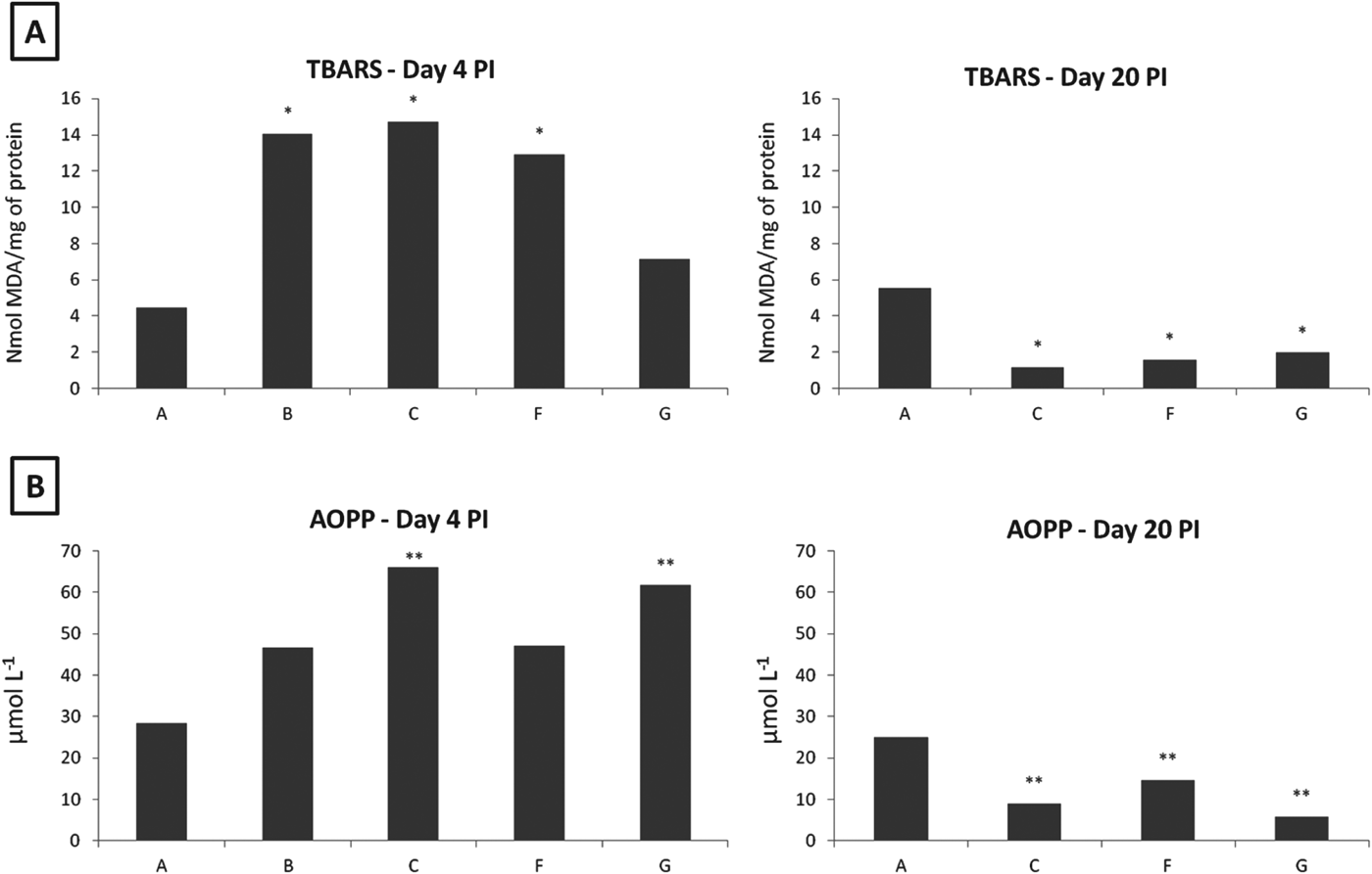
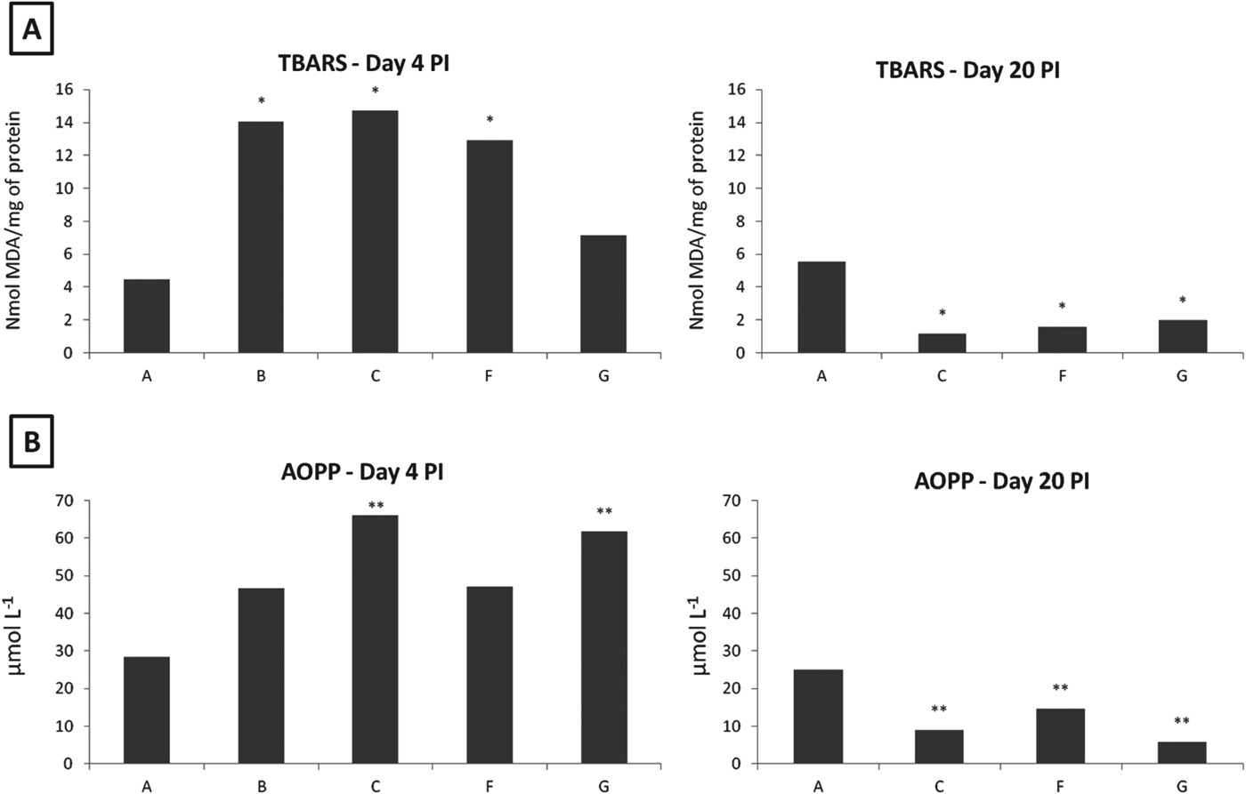
Biomarkers of lipid peroxidation and protein oxidation in liver tissue were clearly altered in mice experimentally infected with T. gondii. Figure 1A shows that TBARS levels significantly increased (P<0·05) in groups B, C, D, E and F on day 4 p.i., while they were reduced (P<0·05) in groups C, F and G on day 20 p.i., when these data were compared with the control group. Levels of AOPP (Fig. 1B) significantly increased (P<0·001) in groups C and G on day 4 p.i., and were reduced in groups C, F and G (P<0·001) on day 20 p.i. when compared with group A.
Levels of TBARS (1A) and AOPP (1B) in liver homogenate of mice experimentally infected with Toxoplasma gondii, RH strain, on day 4 and 20 p.i., when compared with non-infected animals (*P<0·05; **P<0·001). Total of animals/group = 12, 6 of them evaluated at each period. Obs. Groups D and E (days 4 and 20 p.i.), and B (day 20 p.i.) are not presented in the figure, because all the mice died before these dates.
Glutathione reductase
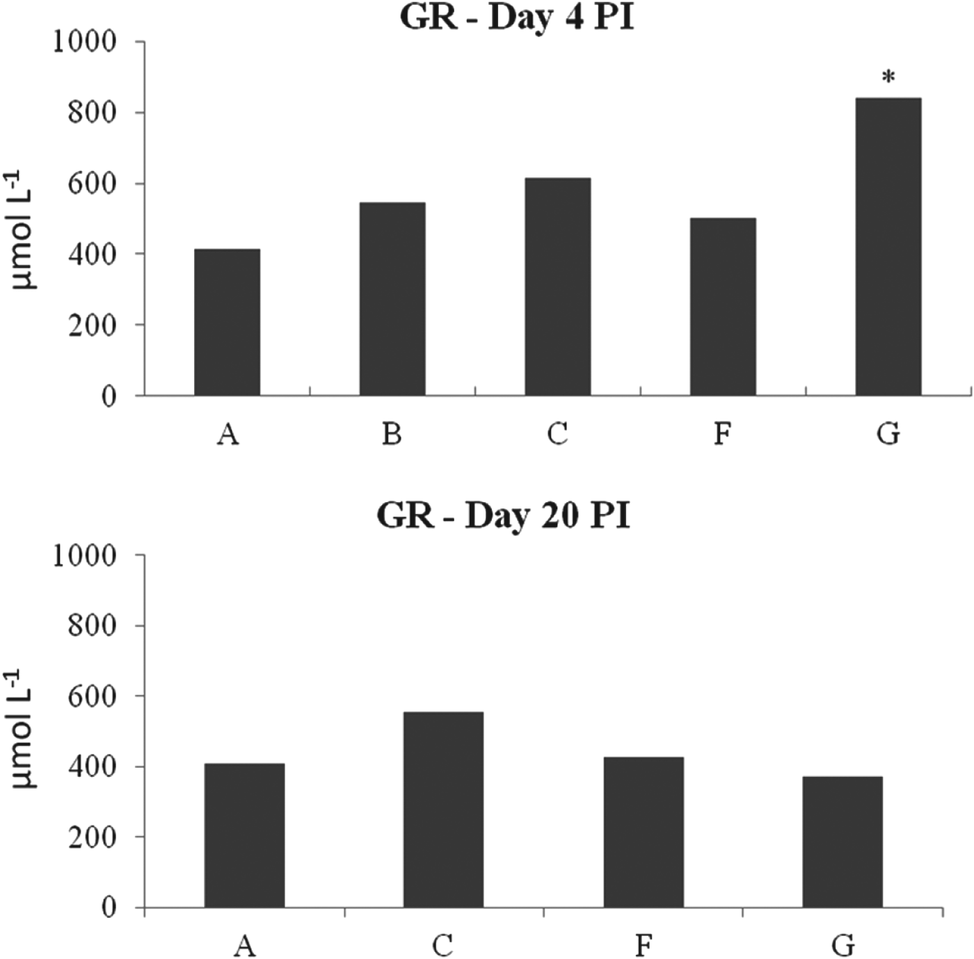
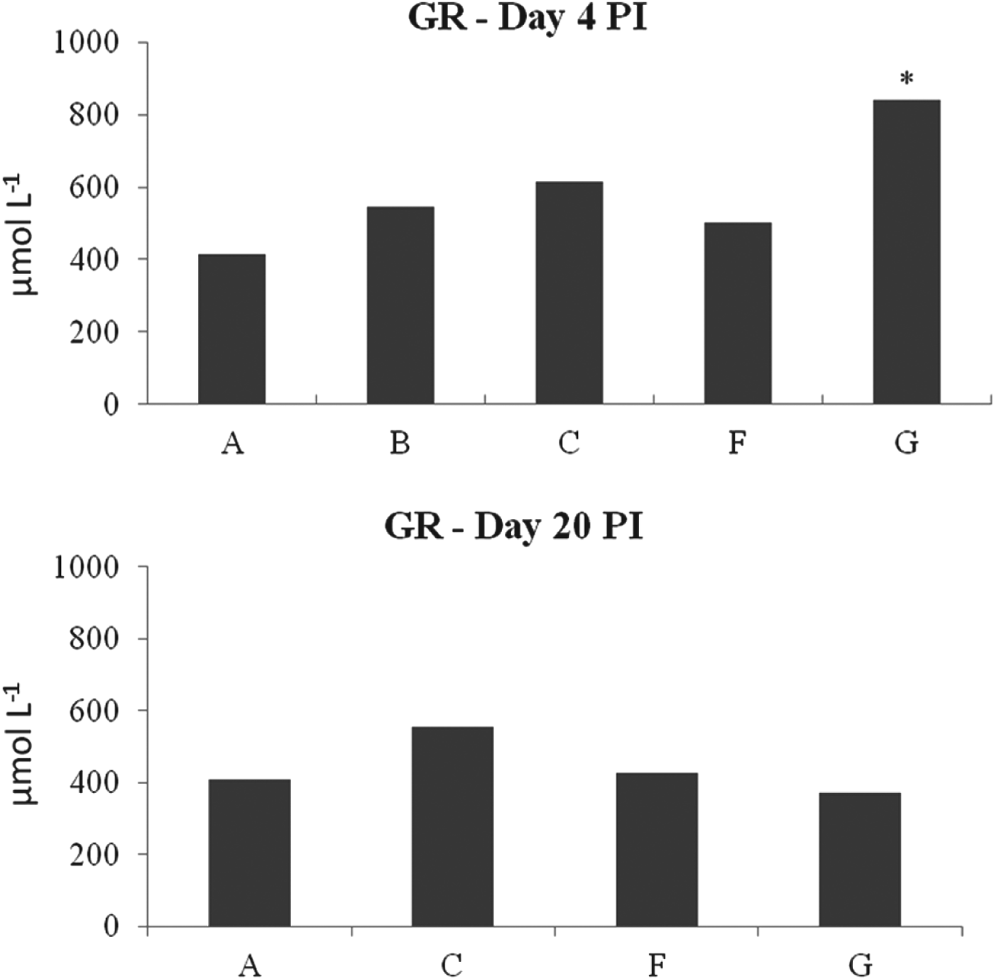
GR activity was significantly (P<0·01) increased on day 4 p.i. in group G when it was compared with the control group, while on day 20 p.i. no statistical differences were observed. Data are shown in Fig. 2.
Levels of glutathione reductase (GR) in liver of mice experimentally infected with Toxoplasma gondii, RH strain, on days 4 and 20 p.i., when compared with non-infected animals (*P<0·05). Total of animals/group = 12, 6 of them evaluated at each period. Obs. Groups D and E (days 4 and 20 p.i.), and B (day 20 p.i.) were not presented in the figure, because all the mice died before these dates.
Cytokines
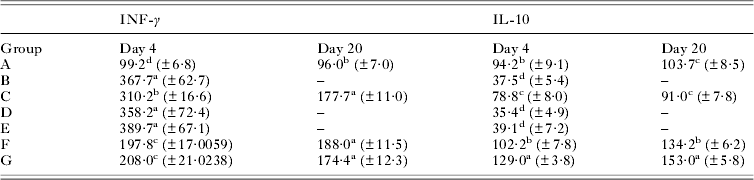
Levels of cytokines are shown in Table 2. It was possible to observe statistical differences in both periods. Levels of INF-γ were significantly increased (P<0·001) in both periods, day 4 p.i. (groups B, C, D, E, F and G) and 20 p.i. (groups C, F and G), when compared with the untreated/control group. Levels of IL-10 were significantly reduced (P<0·001) on day 4 p.i. in groups B, D and E while at the same period it was increased (P<0·001) in group G when compared with group A. On day 20 p.i. the IL-10 had its levels increased (P<0·001) in groups F and G, when compared with the untreated/control group.
Levels of INF-γ and IL-10 in mice experimentally infected with Toxoplasma gondii on days 4 and 20 post infection
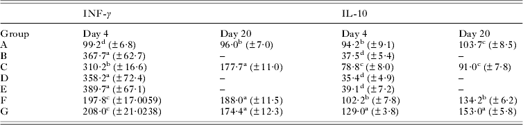
Note: Results are the mean±standard deviation. Results followed by the same letters in the same column do not differ significantly in Student t-test. Total of animals/group = 12, 6 of them evaluated at each period.
DISCUSSION
Our experimental design led the mice to develop an acute toxoplasmosis, and due to the high virulence of the RH strain of T. gondii, some interesting patterns in the evaluated parameters were observed. TBARS levels (Fig. 1A) significantly increased on day 4 p.i. in groups B, C, D, E and F when compared with group A (uninfected) and group G. This situation was expected, especially for groups B and C, because of the parasitic infection, associated with the fact that these groups did not receive antioxidant therapy. We expected a reduction in TBARS levels in groups where the chemotherapy was combined with SSe and DPDS, since the antioxidant activity of selenium and its inorganic forms are well documented (Mugesh et al. Reference Mugesh, du Mont and Sies2001; Rossato et al. Reference Rossato, Ketzer, Centuriao, Silva, Ludtke, Zeni, Braga, Rubin and Rocha2002; Brenneisen et al. Reference Brenneisen, Steinbrenner and Sies2005). Interestingly, TBARS levels were statistically reduced only in group G, where the combination of ST with DPDS was used. DPDS has been shown to be even more active as a glutathione peroxidase (GPx) mimetic (Meotti et al. Reference Meotti, Borges, Zeni, Rocha and Nogueira2003; Nogueira et al. Reference Nogueira, Meotti, Curte, Pilissao, Zeni and Rocha2003). Therefore, the maintenance of TBARS levels in group G (day 4 p.i.), was strongly linked to the inclusion of DPDS together with the chemotherapy. This hypothesis is corroborated by the increased activity of glutathione reductase observed on day 4 p.i. GR is a key enzyme because it regenerates reduced glutathione (GSH) from oxidized glutathione (GSSG) in the presence of NADPH, maintaining millimolar concentrations of GSH, the most abundant reductant enzyme in human cells (Kamerbeek et al. Reference Kamerbeek, van Zwieten, de Boer, Morren, Vuil, Bannink, Lincke, Dolman, Becker, Schirmer, Gromer and Roos2007). Thus, with the increased activity of GR, an enzymatic detoxification was started, maintaining TBARS at levels almost the same as the uninfected group on day 4 p.i.
TBARS levels on day 20 p.i. were unexpectedly reduced in groups C, F and G, when compared with group A. These results might be explained by the cytochrome P450 enzymatic system (CYPs). CYPs is in charge of the oxidative metabolism of hundreds of drugs (Cooper et al. Reference Cooper, Levin, Narasimhulu and Rosenthal1965; Nebert and Dalton, Reference Nebert and Dalton2006; Guengerich, Reference Guengerich2008). Among the various CYPs enzymes, the 2C8 and 2C9 isoforms have particular relevance in patients under trimethoprim–sulfamethoxazole (ST) therapy, because the 2C8 isoform is inhibited by trimethoprim, while the C9 isoform is inhibited by sulfamethoxazole (Wen et al. Reference Wen, Wang, Backman, Laitila and Neuvonen2002). Additionally, cytochrome P450-2E1 (CYP2E1) is among the most active CYPs in producing reactive oxygen species (ROS); therefore, its over-expression is associated with several cellular biomarkers of oxidative stress, as well as with decreased cellular viability, as a result of both necrosis and apoptosis (Wu and Cederbaum, Reference Wu and Cederbaum2001; Thum and Borlak, Reference Thum and Borlak2002; Schattenberg et al. Reference Schattenberg, Wang, Rigoli, Koop and Czaja2004; Butura et al. Reference Butura, Nilsson, Morgan, Morgan, French, Johansson, Schuppe-Koistinen and Ingelman-Sundberg2009). Lu et al. (Reference Lu, Ma, Zhang, Bao, Dong, Lian, Huang and Zhang2012) reported that CYP2E1 knockout mice significantly ameliorated their oxidative stress status by decreasing H2O2 and MDA, added to an increased level of GSH. Therefore, TBARS results on day 20 p.i. probably were outcomes of an inhibition of CYPs by prolonged use of ST. However, knowing that glutathione reductase regenerates GSH (Kamerbeek et al. Reference Kamerbeek, van Zwieten, de Boer, Morren, Vuil, Bannink, Lincke, Dolman, Becker, Schirmer, Gromer and Roos2007) added to the fact that a depletion in CYP2E1 is able to increase GSH levels, the results observed on day 20 p.i., for GR, might explain the GR levels being statistically equal in groups C, F and G, when compared with group A.
Quite similar results of TBARS were observed in both periods for AOPP (Fig. 1B), differing on day 4 p.i., where levels of AOPP were statistically increased in groups C and G when compared with groups A and F. The exquisite vulnerability of proteins to ROS is well documented (Davies, Reference Davies1987; Davies and Goldberg, Reference Davies and Goldberg1987; Heinecke et al. Reference Heinecke, Li, Daehnke and Goldstein1993; Dean et al. Reference Dean, Fu, Stocker and Davies1997), therefore this increase in groups C and G probably was a result of the interaction between the parasitic infection, leading to an increased ROS production (van Gisbergen et al. Reference van Gisbergen, Geijtenbeek and van Kooyk2005). The interesting finding in the assessment of this parameter was the inversion in the protective action of antioxidants, since for AOPP, the protective effect was provided mostly by the association of ST with SSe (group F), instead of by ST associated with DPDS (group G), as occurred for TBARS. A possible explanation of this situation lies on two aspects; first, the oxidation pathway; and second, the chemical classification and distribution of these compounds. Regarding the first aspect, it is important to remember that reactive carbonyl compounds (RCCs), formed endogenously during lipid peroxidation and the glycoxidation of carbohydrates, are precursors of advanced glycation end products (AGEs) and advanced lipid peroxidation end products (ALEs), which form cross-links on tissular proteins (carbonyl stress), accumulating during ageing and in chronic diseases (Dalle-Donne et al. Reference Dalle-Donne, Giustarini, Colombo, Rossi and Milzani2003; Smit and Lutgers, Reference Smit and Lutgers2004). Carbonyl stress induces progressively protein dysfunction and damage in all tissues, leading to increased levels of AOPP, with pathological consequences, such as inflammation and apoptosis, contributing to the progression of diseases (Dalle-Donne et al. Reference Dalle-Donne, Giustarini, Colombo, Rossi and Milzani2003; Petersen and Doorn, Reference Petersen and Doorn2004). In this sense, it is possible to place AOPP as an immediate and direct consequence of lipid peroxidation and glycoxidation of carbohydrates. In relation to selenium compounds distribution, or our second possible explanation, it is important to mention the highly lipophilic property of diphenyl diselenide. Prigol et al. (Reference Prigol, Schumacher, Nogueira and Zeni2009) demonstrated that DPDS exhibits a concentration-time profile characterized by an early peak, followed by a rapid distribution from blood to the central nervous system, where it exerts its pharmacological and toxicological effects. On the other hand, Davidson-York et al. (Reference Davidson-York, Galey, Blanchard and Gardner1999) observed that when pigs received repeated oral toxic doses of SSe, the biological half-life of SSe was on average 12 days. Additionally, the organic forms of Se, such as DPDS, have higher bioavailability, as well as better antioxidant properties, than inorganic forms (Mahan et al. Reference Mahan, Cline and Richert1999; Mahmoud and Edens, Reference Mahmoud and Edens2003), represented in this experiment by SSe. Therefore, the protective effect on AOPP production, generated by the association of ST plus SSe, might have been due to the characteristics of SSe (less bioavailability), in comparison with the highly lipophilic and absorption characteristics of DPDS. Thus, with DPDS being more lipophilic and with a fast absorption, we expected a satisfactory action on preventing generation of lipid peroxidation, while an antioxidant with less bioavailability (SSe) and with longer half-life, may prevent a late protein peroxidation. AOPP findings on day 20 p.i. were quite similar to the TBARS assessment, i.e. reduced in all groups when compared with group A. Knowing that both biomarkers are mainly generated by oxidative stress processes, probably the same mechanism was in charge of the reduction of AOPP on day 20 p.i.
Based on the discussion of the changes observed in these two biomarkers of injury cell, it was possible to assume that an inflammatory response was generated. Several studies have shown the great importance of T cells in resistance against T. gondii (Denkers and Gazzinelli, Reference Denkers and Gazzinelli1998). A hallmark of T. gondii infection is the induction of a strong Th1-mediated immunity, especially with production of IFN-γ (Denkers and Gazzinelli, Reference Denkers and Gazzinelli1998). Therefore, cytokines, such as IFN-γ and TNF-α, are important for controlling tachyzoite replication during both acute and chronic phases of infection (Suzuki et al. Reference Suzuki, Conley and Remington1989; Gazzinelli et al. Reference Gazzinelli, Hakim, Hieny, Shearer and Sher1991, Reference Gazzinelli, Xu, Hieny, Cheever and Sher1992, Johnson, Reference Johnson1992; Scharton-Kersten et al. Reference Scharton-Kersten, Wynn, Denkers, Bala, Showe, Grunvald, Hieny, Gazzinelli and Sher1996). In contrast, interleukin-10 (IL-10) appears to be crucial at the initial phase of infection, and less important during chronic toxoplasmosis (Gazzinelli et al. Reference Gazzinelli, Wysocka, Hayashi, Denkers, Hieny, Caspar, Trinchieri and Sher1994, Reference Gazzinelli, Wysocka, Hieny, Scharton-Kersten, Cheever, Kuhn, Muller, Trinchieri and Sher1996). Our results highlighted some important alterations in the levels of the cytokines assessed in animals under ST single treatment, or ST combined with DPDS/SSe. It was possible to observe that IFN-γ statistically increased in both instances, in all the infected groups, when compared with group A, demonstrating an expected response against the parasite. However, the most interesting finding, on IFN-γ, is when its production is compared among groups C, F and G, or the infected groups. Mordue et al. (Reference Mordue, Monroy, La Regina, Dinarello and Sibley2001), when comparing the response of immunocompetent outbreed mice infected by different strains of T. gondii, found that infection with the RH strain was characterized by its rapid ability to reach tissue burdens associated with lethality. These authors additionally reported that the lethal infections were associated with over-induction of inflammatory cytokines, rather than an insufficient immune response. In our study the levels of IFN-γ were statistically reduced in groups F and G, when compared with group C on day 4 p.i., demonstrating the influence of SSe and DPDS when combined with ST. These findings strongly support the protective action of both antioxidants in acute infection with T. gondii, preventing excessive tissue damage during the initial phase of the infection. The levels of IL-10 also support this theory, since IL-10 plays a key role in the regulation of many functions of the immune system by inhibiting the proliferation of B and T lymphocytes (Arababadi et al. Reference Arababadi, Mosavi, Khorramdelazad, Yaghini, Zarandi, Araste, Pourali, Nekhei and Kennedy2010). Comparing the results of INF-γ and IL-10, on day 4 p.i., it is possible to observe an inverse role, since in group C, IL-10 presented its lower level, while IFN-γ had its highest level. In groups F and G, the levels of IL-10 were statistically increased when compared with group C, contrasting with the statistical reduced levels of IFN-γ on these groups. Therefore, the association of DPDS with SSe generated a compensatory and protective mechanism, since they provided a balance between pro- and anti-inflammatory responses, preventing extra tissue damage. Probably on day 20 p.i., the tissue levels of both antioxidants were lower (as discussed above), causing the maintenance of IFN-γ levels at the same levels among groups C, F and G.
Therefore, our results highlight that inorganic (sodium selenite) and organic (diphenyl diselenide) forms of selenium associated with sulfamethoxazole/trimethoprim were able to reduce lipid peroxidation and protein oxidation, mainly during the acute phase of the experimental infection, as well as providing an immunological balance between the production of pro- and anti-inflammatory cytokines. Our results show that perhaps, through this association, it is possible to reduce the oxidative stress and exacerbated immune response in toxoplasmosis.