In this issue of the journal, Rogers et alReference Rogers, Klein, James and FitzGerald
1
from Cincinnati describe the results of a questionnaire circulated among the centres in the United States of America providing training in paediatric cardiology. The responses were salutary and of particular interest to those of us who continue to research and teach using specimens retained subsequent to autopsies of those who died with congenitally malformed hearts. Responses were received from the directors of training in just over half the centres polled, along with responses from 66 of the current fellows in training. It is encouraging to note that almost nine-tenths of those responding indicated that autopsy specimens continued to be used for the purposes of training. It was less encouraging to learn that only one-third of the directors considered that their trainees had “adequate abilities to apply segmental anatomy, identify associated cardiac lesions, or communicate complex CHD”. Therefore, to overcome these shortcomings, it was suggested that structured teaching, protected time, and the availability of specimens were likely to improve the current training. Almost three-quarters of the directors indicated that they would support the development of an online curriculum to provide the required information on the morphology of the congenitally malformed heart. I know that Lindsey Rogers is already actively addressing the need to provide such a curriculum. To achieve this goal, she will need help, along with support from many of those who currently curate the archives containing the autopsy specimens.
In their discussion, the authors highlight the current lack of consensus for describing various lesions that make up CHD. They also identify the steps already in hand to remedy this deficiency, specifically the efforts of the International Nomenclature Committee for Pediatric and Congenital Heart Disease. This committee was established subsequent to a meeting held at the World Congress in Toronto in 2001. The initial task undertaken by the committee was to merge the coding systems that had been developed separately by the North American and European Paediatric Cardiac Surgeons, on the one hand, and the Association for European Paediatric Cardiology, on the other hand, at the same time incorporating the systems already in use in Boston and Canada. These efforts were successful in producing the unified code now available on the website maintained by the International Nomenclature Society and in providing cross-mapping between the individual systems. More importantly, the International Committee has now been tasked with providing codes that will form the 11th iteration of the International Classification of Disease (ICD-11). Not only has the committee produced a listing of just over 300 terms for this purpose, but definitions have been debated and agreed. Lindsey is herself now a member of the committee, which has convened an archiving group tasked with providing images that illustrate the phenotypic features of all the lesions listed within ICD-11.
The existence and content of this website should hopefully provide the basis for the online curriculum proposed by Rogers et al. It is axiomatic that, if fellows understand all the phenotypes now contained within ICD-11, they will be more than capable of diagnosing the various lesions likely to confront them throughout their career. Rogers et al point out that their questionnaire was circulated only among the centres in the United States of America. It is highly likely that the findings are just as relevant for those providing training throughout the rest of the World. I know from personal experience that not all those training in the United Kingdom have access to archives of congenitally malformed hearts. Steps are in hand to unify the cataloguing of the several archives that do exist, using the newly created list for ICD-11 for this purpose. It is hoped that such cooperation between centres, particularly those holding archives, with become international. Already the Idriss archive held at Lurie Children’s Hospital in Chicago and the Van Mierop archive maintained at University of Florida in Gainesville have been catalogued in a manner that will permit coding using ICD-11, this being the product of the hard work of Diane Spicer, who is also the archivist of the Working Group of the International Nomenclature Committee. Such international cooperation is likely to be needed if we are to achieve the admirable goals now established by Rogers et al.
It is also intriguing to me personally to note that the authors comment “Utilizing a common language to describe cardiac morphology is imperative to standardizing communication”. The questionnaire has served to identify the problems that continue to exist because of the use of “Van Praaghian” as opposed to “Andersonian” terminologies. They then draw attention to the recent “dictionary” produced by David Ezon et al2 in the hope of finding middle ground. In reality, the differences between the systems are not as great as often perceived. When my colleagues and I proposed expanding the initial segmental approachReference Van Praagh
3
to include detailed analysis of the junctions, producing the modification that we called sequential segmental analysis,Reference Shinebourne, Macartney and Anderson
4
we had no idea it would produce a counter modification by Van Praagh himself, in which he promoted the notion of “alignments” for the feature we had termed “connections”.Reference Van Praagh
5
It took some time before we became aware that the major reason for this divergence of description was our own misinterpretation of the initial definitions for “concordance” and “discordance”. We had presumed that, when Van PraaghReference Van Praagh
3
introduced the notion of the atrioventricular concordance and discordance, he had meant that the atrial chambers were joined in the usual or opposite manner to the underlying ventricles. We had not appreciated that, in fact, Van Praagh had used segmental harmony or disharmony as the basis of concordance as opposed to discordance. Thus, when using the segmental approach, any patient with {S,D,*} or {I,L,*} has atrioventricular concordance, irrespective of whether there is double-inlet ventricle, tricuspid atresia, or even, on exceedingly rare connections, when the atrial chambers are joined to morphologically appropriate ventricles. Therefore, to circumvent this problem in description, it is now our policy to describe the arrangements at the atrioventricular junctions in terms of concordant or discordant atrioventricular connections, emphasising that it is the cavities of the chambers that are joined together.Reference Anderson and Ho
6
In this way, we distinguish explicitly between the alternative connections of double-inlet ventricle, the mixed connections found in the setting of hearts with isomeric atrial appendages, and situations in which one or other of the normal atrioventricular connections is absent. It is the latter arrangement that, at least to our eyes, points to the ongoing difficulty in using “alignments” to describe the specific morphology of the atrioventricular junctions. Thus, in “classical” tricuspid atresia, the right atrium is normally aligned with the morphologically right ventricle, but there is no communication between their cavities because of the absence of the right atrioventricular connection (Fig 1).
Figure 1
The image is a “four-chamber” section of a heart from a patient with “classical” tricuspid atresia. The cavity of the right atrium is aligned with the hypoplastic cavity of the right ventricle, but has no direct union with it because of the absence of the right atrioventricular connection. The arrow is through the “dimple” in the floor of the right atrium, showing that this is in potential connection with the cavity of the left ventricle. The image shows that “alignments” cannot be considered directly analogous to the feature described in terms of “connections”. AV=atrioventricular.
Rogers et al, as a result of their questionnaire, also discovered that the directors were of the opinion that only one-tenth of the fellows were capable of “describing the embryological origin of cardiac structures”, whereas only one-fifth of the fellows themselves considered that they possessed this capability. This is hardly surprising, as teaching cardiac development is even more fragmented compared with cardiac morphology, and lacks the foundation now provided for morphology by the efforts of the International Nomenclature Committee. It is, perhaps, paradoxical that for the larger part of my own career I shunned the temptation to stray into discussions of cardiac development. At one stage, indeed, along with my very good friend and colleague Anton Becker, we produced a provocative review, suggesting that embryology was a hindrance rather than a help in understanding congenital cardiac morphology.Reference Becker and Anderson
7
At that time, there were good reasons for this statement, as the evidence underscoring the extant concepts of development tended to come from the armchair rather than the workbench. All that has changed in the decades leading to the turn of the century and in the new millennium. We now have much better knowledge of the details of cardiac embryology. This, in turn, provides new evidence regarding the ongoing real differences in interpretation, as opposed to description, concerning the way that Van Praagh and I view the congenitally malformed heart. As is pointed out in the Ezon “dictionary”,Reference Ezon, Goldberg and Kyle
2
Van Praagh still maintains that the small chamber found in the setting of double-inlet left ventricle is no more than an infundibulum, whereas my colleagues and myself, for several years, have argued that it is an incomplete right ventricle.Reference Jacobs and Anderson
8
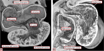
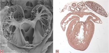
The evidence now available from examination of the developing heart supports our own contention. At the early stage of embryogenesis, the default option for the developing heart is double inlet to the developing left ventricle and double outlet from the developing right ventricle. At this early stage, however, it is clear that the apical component of the right ventricle has already ballooned from the outlet component of the ventricular loop even before the ventricle achieves its inlet component (Fig 2). Evidence from genetically modified mice then shows unequivocally that it is possible for the embryos to develop with isomeric right or left atrial appendages (Fig 3), giving the lie to the suggestion that isomerism is no more than a “useful mnemonic”.Reference Ezon, Goldberg and Kyle
2
It is, however, only the appendages that are truly isomeric. Those of us using the morphology of the appendages to identify atrial arrangement recognise fully the need to describe in a straightforward manner all the remaining components of the heart.Reference Loomba, Hlavacek, Spicer and Anderson
9
This, after all, is the basis of segmental analysis.
Figure 2
The images are from an episcopic data set prepared from a mouse embryo sacrificed at the 11th day of development. (
a
) A “four-chamber” section, revealing that, at this early stage, the atrial chambers are connected through the atrioventricular (AV) canal to the developing left ventricle, which is ballooning from the inlet part of the ventricular loop. The apical component of the right ventricle is beginning to balloon from the outlet component of the loop. (
b
) Oblique cut through the developing right-sided chambers. The cavity of the right atrium is already aligned relative to the developing right ventricle, but at this stage the cavities are not connected one to the other (white dashed line). Already, nonetheless, it is possible to recognise the developing apical component of the right ventricle, which supports the entirety of the developing outflow tract.
Figure 3
The images show isomeric atrial appendages from mouse hearts, induced in (
a
) by knocking out the Pitx2 gene and in (
b
) by knocking out the Lefty1 gene.
Rogers et al, therefore, are to be congratulated for highlighting the current deficiencies in providing a standard curriculum for congenital cardiac morphology. The steps they suggest for remedying this deficiency are to be applauded. It is difficult to imagine that anyone will take exception to their proposals. The need now is to implement their suggestions; one of the comments listed in Table 4 is particularly pertinent in this regard – namely “the cardiac morphologists in our field are aging and there are few of them”. I am more than aware of the validity of the first part of this sentence. There is also some truth to the second part. The authors have already themselves pointed to one way of overcoming this problem, when emphasising the value to be gained from three-dimensional reconstruction and printing of images obtained during clinical investigations, particularly CT. The quality of the reconstructions is now such that, in several instances, the printed models are providing the “gold standard” for the anatomy of the congenitally malformed heart. This is the more so, as the morphological features can be demonstrated with suitable sectioning to replicate the images produced echocardiographically in the clinic. In future, therefore, the interested paediatric cardiologists, surgeons, and radiologists will become their own morphologists. In the meantime, there are sufficient younger morphologists already on the scene who, together with the surviving dinosaurs, will hopefully help provide the standardised curriculum proposed by Rogers et al.







 You have Access
In this issue of the journal, Rogers et alReference Rogers, Klein, James and FitzGerald 1 from Cincinnati describe the results of a questionnaire circulated among the centres in the United States of America providing training in paediatric cardiology. The responses were salutary and of particular interest to those of us who continue to research and teach using specimens retained subsequent to autopsies of those who died with congenitally malformed hearts. Responses were received from the directors of training in just over half the centres polled, along with responses from 66 of the current fellows in training. It is encouraging to note that almost nine-tenths of those responding indicated that autopsy specimens continued to be used for the purposes of training. It was less encouraging to learn that only one-third of the directors considered that their trainees had “adequate abilities to apply segmental anatomy, identify associated cardiac lesions, or communicate complex CHD”. Therefore, to overcome these shortcomings, it was suggested that structured teaching, protected time, and the availability of specimens were likely to improve the current training. Almost three-quarters of the directors indicated that they would support the development of an online curriculum to provide the required information on the morphology of the congenitally malformed heart. I know that Lindsey Rogers is already actively addressing the need to provide such a curriculum. To achieve this goal, she will need help, along with support from many of those who currently curate the archives containing the autopsy specimens.
In their discussion, the authors highlight the current lack of consensus for describing various lesions that make up CHD. They also identify the steps already in hand to remedy this deficiency, specifically the efforts of the International Nomenclature Committee for Pediatric and Congenital Heart Disease. This committee was established subsequent to a meeting held at the World Congress in Toronto in 2001. The initial task undertaken by the committee was to merge the coding systems that had been developed separately by the North American and European Paediatric Cardiac Surgeons, on the one hand, and the Association for European Paediatric Cardiology, on the other hand, at the same time incorporating the systems already in use in Boston and Canada. These efforts were successful in producing the unified code now available on the website maintained by the International Nomenclature Society and in providing cross-mapping between the individual systems. More importantly, the International Committee has now been tasked with providing codes that will form the 11th iteration of the International Classification of Disease (ICD-11). Not only has the committee produced a listing of just over 300 terms for this purpose, but definitions have been debated and agreed. Lindsey is herself now a member of the committee, which has convened an archiving group tasked with providing images that illustrate the phenotypic features of all the lesions listed within ICD-11.
The existence and content of this website should hopefully provide the basis for the online curriculum proposed by Rogers et al. It is axiomatic that, if fellows understand all the phenotypes now contained within ICD-11, they will be more than capable of diagnosing the various lesions likely to confront them throughout their career. Rogers et al point out that their questionnaire was circulated only among the centres in the United States of America. It is highly likely that the findings are just as relevant for those providing training throughout the rest of the World. I know from personal experience that not all those training in the United Kingdom have access to archives of congenitally malformed hearts. Steps are in hand to unify the cataloguing of the several archives that do exist, using the newly created list for ICD-11 for this purpose. It is hoped that such cooperation between centres, particularly those holding archives, with become international. Already the Idriss archive held at Lurie Children’s Hospital in Chicago and the Van Mierop archive maintained at University of Florida in Gainesville have been catalogued in a manner that will permit coding using ICD-11, this being the product of the hard work of Diane Spicer, who is also the archivist of the Working Group of the International Nomenclature Committee. Such international cooperation is likely to be needed if we are to achieve the admirable goals now established by Rogers et al.
It is also intriguing to me personally to note that the authors comment “Utilizing a common language to describe cardiac morphology is imperative to standardizing communication”. The questionnaire has served to identify the problems that continue to exist because of the use of “Van Praaghian” as opposed to “Andersonian” terminologies. They then draw attention to the recent “dictionary” produced by David Ezon et al2 in the hope of finding middle ground. In reality, the differences between the systems are not as great as often perceived. When my colleagues and I proposed expanding the initial segmental approachReference Van Praagh 3 to include detailed analysis of the junctions, producing the modification that we called sequential segmental analysis,Reference Shinebourne, Macartney and Anderson 4 we had no idea it would produce a counter modification by Van Praagh himself, in which he promoted the notion of “alignments” for the feature we had termed “connections”.Reference Van Praagh 5 It took some time before we became aware that the major reason for this divergence of description was our own misinterpretation of the initial definitions for “concordance” and “discordance”. We had presumed that, when Van PraaghReference Van Praagh 3 introduced the notion of the atrioventricular concordance and discordance, he had meant that the atrial chambers were joined in the usual or opposite manner to the underlying ventricles. We had not appreciated that, in fact, Van Praagh had used segmental harmony or disharmony as the basis of concordance as opposed to discordance. Thus, when using the segmental approach, any patient with {S,D,*} or {I,L,*} has atrioventricular concordance, irrespective of whether there is double-inlet ventricle, tricuspid atresia, or even, on exceedingly rare connections, when the atrial chambers are joined to morphologically appropriate ventricles. Therefore, to circumvent this problem in description, it is now our policy to describe the arrangements at the atrioventricular junctions in terms of concordant or discordant atrioventricular connections, emphasising that it is the cavities of the chambers that are joined together.Reference Anderson and Ho 6 In this way, we distinguish explicitly between the alternative connections of double-inlet ventricle, the mixed connections found in the setting of hearts with isomeric atrial appendages, and situations in which one or other of the normal atrioventricular connections is absent. It is the latter arrangement that, at least to our eyes, points to the ongoing difficulty in using “alignments” to describe the specific morphology of the atrioventricular junctions. Thus, in “classical” tricuspid atresia, the right atrium is normally aligned with the morphologically right ventricle, but there is no communication between their cavities because of the absence of the right atrioventricular connection (Fig 1).
The image is a “four-chamber” section of a heart from a patient with “classical” tricuspid atresia. The cavity of the right atrium is aligned with the hypoplastic cavity of the right ventricle, but has no direct union with it because of the absence of the right atrioventricular connection. The arrow is through the “dimple” in the floor of the right atrium, showing that this is in potential connection with the cavity of the left ventricle. The image shows that “alignments” cannot be considered directly analogous to the feature described in terms of “connections”. AV=atrioventricular.
Rogers et al, as a result of their questionnaire, also discovered that the directors were of the opinion that only one-tenth of the fellows were capable of “describing the embryological origin of cardiac structures”, whereas only one-fifth of the fellows themselves considered that they possessed this capability. This is hardly surprising, as teaching cardiac development is even more fragmented compared with cardiac morphology, and lacks the foundation now provided for morphology by the efforts of the International Nomenclature Committee. It is, perhaps, paradoxical that for the larger part of my own career I shunned the temptation to stray into discussions of cardiac development. At one stage, indeed, along with my very good friend and colleague Anton Becker, we produced a provocative review, suggesting that embryology was a hindrance rather than a help in understanding congenital cardiac morphology.Reference Becker and Anderson 7 At that time, there were good reasons for this statement, as the evidence underscoring the extant concepts of development tended to come from the armchair rather than the workbench. All that has changed in the decades leading to the turn of the century and in the new millennium. We now have much better knowledge of the details of cardiac embryology. This, in turn, provides new evidence regarding the ongoing real differences in interpretation, as opposed to description, concerning the way that Van Praagh and I view the congenitally malformed heart. As is pointed out in the Ezon “dictionary”,Reference Ezon, Goldberg and Kyle 2 Van Praagh still maintains that the small chamber found in the setting of double-inlet left ventricle is no more than an infundibulum, whereas my colleagues and myself, for several years, have argued that it is an incomplete right ventricle.Reference Jacobs and Anderson 8 The evidence now available from examination of the developing heart supports our own contention. At the early stage of embryogenesis, the default option for the developing heart is double inlet to the developing left ventricle and double outlet from the developing right ventricle. At this early stage, however, it is clear that the apical component of the right ventricle has already ballooned from the outlet component of the ventricular loop even before the ventricle achieves its inlet component (Fig 2). Evidence from genetically modified mice then shows unequivocally that it is possible for the embryos to develop with isomeric right or left atrial appendages (Fig 3), giving the lie to the suggestion that isomerism is no more than a “useful mnemonic”.Reference Ezon, Goldberg and Kyle 2 It is, however, only the appendages that are truly isomeric. Those of us using the morphology of the appendages to identify atrial arrangement recognise fully the need to describe in a straightforward manner all the remaining components of the heart.Reference Loomba, Hlavacek, Spicer and Anderson 9 This, after all, is the basis of segmental analysis.
The images are from an episcopic data set prepared from a mouse embryo sacrificed at the 11th day of development. ( a ) A “four-chamber” section, revealing that, at this early stage, the atrial chambers are connected through the atrioventricular (AV) canal to the developing left ventricle, which is ballooning from the inlet part of the ventricular loop. The apical component of the right ventricle is beginning to balloon from the outlet component of the loop. ( b ) Oblique cut through the developing right-sided chambers. The cavity of the right atrium is already aligned relative to the developing right ventricle, but at this stage the cavities are not connected one to the other (white dashed line). Already, nonetheless, it is possible to recognise the developing apical component of the right ventricle, which supports the entirety of the developing outflow tract.
The images show isomeric atrial appendages from mouse hearts, induced in ( a ) by knocking out the Pitx2 gene and in ( b ) by knocking out the Lefty1 gene.
Rogers et al, therefore, are to be congratulated for highlighting the current deficiencies in providing a standard curriculum for congenital cardiac morphology. The steps they suggest for remedying this deficiency are to be applauded. It is difficult to imagine that anyone will take exception to their proposals. The need now is to implement their suggestions; one of the comments listed in Table 4 is particularly pertinent in this regard – namely “the cardiac morphologists in our field are aging and there are few of them”. I am more than aware of the validity of the first part of this sentence. There is also some truth to the second part. The authors have already themselves pointed to one way of overcoming this problem, when emphasising the value to be gained from three-dimensional reconstruction and printing of images obtained during clinical investigations, particularly CT. The quality of the reconstructions is now such that, in several instances, the printed models are providing the “gold standard” for the anatomy of the congenitally malformed heart. This is the more so, as the morphological features can be demonstrated with suitable sectioning to replicate the images produced echocardiographically in the clinic. In future, therefore, the interested paediatric cardiologists, surgeons, and radiologists will become their own morphologists. In the meantime, there are sufficient younger morphologists already on the scene who, together with the surviving dinosaurs, will hopefully help provide the standardised curriculum proposed by Rogers et al.
Acknowledgements
I am indebted to my colleagues Professors Nigel Brown of St George’s Medical University, London, and Chikara Meno, of Kyushu University, Japan, who provided the original material for our previous collaborations and from which I prepared Figure 3.
Financial Support
This research received no specific grant from any funding agency, commercial or not-for-profit sectors.
Conflicts of Interest
None.