INTRODUCTION
Trichomonas vaginalis is an amitochondriated parasitic protozoan responsible for the sexually transmitted disease trichomonosis (World Health Organization, 2007). In eukaryotes, glycolysis consists of 10 sequential steps that convert 1 molecule of glucose into 2 molecules of pyruvate with a total gain of 2 ATP molecules. In anaerobic organisms glycolysis may be subject to strong selective pressure that can be manifested in the use of more efficient enzymes or as gene duplication events necessary for the robustness of this metabolic pathway (Slamovits and Keeling, Reference Slamovits and Keeling2006). For instance, it is suggested that gene duplication in Saccharomyces cerevisiae provides an advantage for fermentative growth (Conant and Wolfe, Reference Conant and Wolfe2007, Reference Conant and Wolfe2008) and that the response of a duplicated gene depends on growth conditions (DeLuna et al. Reference DeLuna, Springer, Kirschner and Kishony2010). Glycolysis in T. vaginalis is a mosaic of enzymes from bacterial and eukaryotic origins. Although the evolutionary origin of pyruvate phosphate dikinase (Slamovits and Keeling, Reference Slamovits and Keeling2006), glyceraldehyde-3-phosphate dehydrogenase, glucosephosphate isomerase (Henze et al. Reference Henze, Horner, Suguri, Moore, Sanchez, Muller and Embley2001), and fructose-1,6-bisphosphate aldolases (Sanchez et al. Reference Sanchez, Horner, Moore, Henze, Embley and Muller2002) has been elucidated, to date the only glycolytic enzymes biochemically characterized from T. vaginalis are a glucokinase that replaces the classical hexokinase (Mertens and Muller, Reference Mertens and Muller1990) and a pyruvate kinase (Mertens et al. Reference Mertens, Van Schaftingen and Muller1992). Other glycolytic enzymes like α-enolase (Mundodi et al. Reference Mundodi, Kucknoor and Alderete2008) and glyceraldehyde-3-phosphate dehydrogenase (Lama et al. Reference Lama, Kucknoor, Mundodi and Alderete2009) have been characterized as moonlighting proteins involved in the host-parasite interplay as virulence factors (Carlton et al. Reference Carlton, Hirt, Silva, Delcher, Schatz, Zhao, Wortman, Bidwell, Alsmark, Besteiro, Sicheritz-Ponten, Noel, Dacks, Foster, Simillion, Van de Peer, Miranda-Saavedra, Barton, Westrop, Muller, Dessi, Fiori, Ren, Paulsen, Zhang, Bastida-Corcuera, Simoes-Barbosa, Brown, Hayes, Mukherjee, Okumura, Schneider, Smith, Vanacova, Villalvazo, Haas, Pertea, Feldblyum, Utterback, Shu, Osoegawa, de Jong, Hrdy, Horvathova, Zubacova, Dolezal, Malik, Logsdon, Henze, Gupta, Wang, Dunne, Upcroft, Upcroft, White, Salzberg, Tang, Chiu, Lee, Embley, Coombs, Mottram, Tachezy, Fraser-Liggett and Johnson2007).
Triosephosphate isomerase (TIM) catalyses the isomerization between glyceraldehyde-3-phosphate and dihydroxyacetone phosphate (Blacklow et al. Reference Blacklow, Raines, Lim, Zamore and Knowles1988). The catalytic activities of TIMs from Giardia lambia, Leishmania mexicana, Trypanosoma cruzi, Entamoeba histolytica and Plasmodium falciparum are almost completely abolished by thiol-reactive compounds whereas the catalytic activity of TIM from Homo sapiens is unaltered (Velanker et al. Reference Velanker, Ray, Gokhale, Suma, Balaram, Balaram and Murthy1997; Gao et al. Reference Gao, Garza-Ramos, Saavedra-Lira, Cabrera, De Gomez-Puyou, Perez-Montfort and Gomez-Puyou1998; Rodriguez-Romero et al. Reference Rodriguez-Romero, Hernandez-Santoyo, del Pozo Yauner, Kornhauser and Fernandez-Velasco2002; Enriquez-Flores et al. Reference Enriquez-Flores, Rodriguez-Romero, Hernandez-Alcantara, Oria-Hernandez, Gutierrez-Castrellon, Perez-Hernandez, de la Mora-de la, Castillo-Villanueva, Garcia-Torres, Mendez, Gomez-Manzo, Torres-Arroyo, Lopez-Velazquez and Reyes-Vivas2011). This differential in inactivation profiles is due to subtle structural differences between TIMs. Thus, although the overall barrel structure of TIM is conserved among all species, TIM is a potential drug target against pathogens that heavily depend on glycolysis for energy, like apicomplexans, trypanosomatides and amitochondriates.
In the majority of organisms TIM is present as a single gene. The only reported exceptions are plants, teleost fish, Klebsiella pneumoniae and Sinorhizobium meliloti (Feierabend et al. Reference Feierabend, Kurzok and Schmidt1990; Merritt and Quattro, Reference Merritt and Quattro2001; Zheng et al. Reference Zheng, Sun, van den Heuvel and Zeng2006; Poysti and Oresnik, Reference Poysti and Oresnik2007). Gene duplication protects against harmful mutations, because if a duplicated gene is lost the other copy might be used. Furthermore a gene duplication that evolved point mutations can regulate the function of the organism in several growth conditions. For instance, neurally expressed TIM from the teleost fish has accumulated mutations to negatively charged amino acids and TIMs from S. meliloti have differential transcriptional regulation (Merritt and Quattro, Reference Merritt and Quattro2001; Poysti and Oresnik, Reference Poysti and Oresnik2007).
MATERIALS AND METHODS
Database mining and genome analysis of expressed sequence tags (EST)
The search and in silico studies were performed using the database TRICHDB (www.trichdb.org). Possible initiator elements (Inr) and the sequences required for polyadenylation were searched based on the comparison of the previously reported consensus sequences (Liston and Johnson, Reference Liston and Johnson1999; Espinosa et al. Reference Espinosa, Hernandez, Lopez-Griego and Lopez-Villasenor2002). These motifs were confirmed by analysing the reported ESTs obtained from parasites grown in different culture conditions, in the same database, by alignments of their genomic sequences with the DNAMAN program. Amino acid sequences of amitochondriated and human TIMs were aligned using ClustalW.
Structural models of TvTIM1 and TvTIM2
The structural models of TvTIM1 and TvTIM2 were constructed with the Molecular Operating Environment (MOE) program using the coordinates of the crystal structure of TIM from the beetleTenebreio molitor (PDB ID 219E) as a template (Knobeloch et al. Reference Knobeloch, Schmidt, Scheerer, Krauss, Wessner, Scholz, Kuttner, von Rintelen, Wessel and Hohne2010). Briefly, the amino acid sequence of TvTIM1 and TvTIM2 were aligned with the amino acid sequence of the template molecule and 50 initial homology models were created. The final models were energy-minimized and the stereochemistry of the final models was assessed by Ramachandran plot analysis.
Parasite growth
Parasites of T. vaginalis clinical isolate CNCD147 (Leon-Sicairos et al. Reference Leon-Sicairos, Leon-Felix and Arroyo2004) were cultured in trypticase–yeast extract–maltose (TYM) medium (Diamond, Reference Diamond1957) supplemented with 10% heat-inactivated horse serum (TYM-HS) and incubated at 37 °C for 24 h. The parasites were maintained in in vitro culture for up to 2 weeks. To obtain iron-depleted or iron-rich conditions parasites were grown in TYM-HS medium containing 20 μ m iron supplemented with 100 μ m of 2-2′ dipyridyl (Sigma) for iron depletion or 250 μ m ammonium ferrous sulfate (JT Baker) for iron enrichment, 24 h before parasite culture started (Alvarez-Sanchez et al. Reference Alvarez-Sanchez, Solano-Gonzalez, Yanez-Gomez and Arroyo2007).
RT-PCR assays
Total RNA from parasites grown at 0, 20, or 250 μ m iron was extracted with TRIzol following the manufacturer's recommendations. To obtain cDNA, 5 μg of total RNA was treated with DNase I and reverse transcribed using an oligo dT primer. For PCR amplification, 500 ng of cDNA, EASY-A polymerase (Stratagene) and gene-specific primers designed based on the reported sequences in TRICHDB for tpi1 (TVAG_497370) and tpi2 (TVAG_096350) genes were used. For tpi1 amplification the RT-PCR primers were 5′-tac att ctt tgt cgg agg c-3′ (forward) and 5′-aat gtt gat gaa acc tgg-3′ (reverse). For tpi2 the RT-PCR primers were 5′-aca ttc ttc gtc ggt ggt-3′ (forward) and 5′-aat gtt gat gaa gcc tgg-3′ (reverse). The 3′ end of the forward primers is different between tpi1 and tpi2 (bold). DNA polymerases are inefficient in amplifying from a 3′ mismatch, thus the amplifications should be specific for each RT-PCR product. The amplification program consisted of 25 cycles of denaturation at 94 °C for 1 min, annealing at 55 °C for 30 sec and extension at 72 °C for 1 min. A 112 bp fragment of β-tubulin of T. vaginalis was amplified as an internal control (Leon-Sicairos et al. Reference Leon-Sicairos, Leon-Felix and Arroyo2004). RT-PCR products were cloned into a pGEM-T plasmid and the identity of the 5 independent amplificons was corroborated by automated DNA sequencing.
Antibody production
A New Zealand male rabbit was intramuscularly immunized using 300 μg of recombinant protein TvTIM2 homogenized with TiterMax adjuvant (1∶1, v/v). Five weeks after immunization serum was obtained (anti-TvTIM2). The pre-immune serum (PI) was obtained before the immunization schedule began and served as a negative control in all experiments with antibodies such as Western blots (WB) and indirect immunofluorescence.
Western blot analysis
12% SDS-polyacrylamide gel electrophoresis containing approximately 2 μg of TvTIM1 or TvTIM2 recombinant proteins or 50 μg of total parasite extracts of T. vaginalis grown in distinct iron conditions were transferred onto nitrocellulose membranes (NC). NC membranes were blocked with 5% skim milk in PBS/0·05% Tween 20 (PBS-T) for 18 h at 4 °C, and incubated with the anti-TvTIM2 antibody (1∶1000 for recombinant TvTIM2, or 1∶5000 for trichomonad total protein extracts) or with pre-immune (PI) serum (1∶1000) for 18 h at 4 °C. NC membranes were washed 5 times with PBS-T and incubated with goat anti-rabbit IgG conjugated to peroxidase (1∶3000) for 2 h at room temperature (RT). The membranes were washed 5 times with PBS-T and developed with 4-chloro-1-naphthol. Anti-α-tubulin antibody (1∶3000) was used as a loading control for WB assays of total extracts of parasites grown under different iron conditions.
Indirect immunofluorescence assay
Parasites grown on cover slips for 18 h at 37 °C were fixed with paraformaldehyde at 4% in PBS for 1 h at 37 °C and washed 3 times with PBS. Samples were treated with 50 mm ammonium chloride, washed 10 times with PBS, permeabilized with 0·2% Triton X-100 for 2 min at room temperature (RT), washed 5 times with PBS, and blocked with 1% fetal bovine serum and 0·2 M glycine for 15 min and 1 h at RT, respectively. The cover slips were washed 3 times with PBS and incubated with the anti-TvTIM2 antibody (1∶500) and PI serum (1∶500) for 18 h at 4 °C, washed 5 times with PBS, incubated with FITC-conjugated anti-rabbit IgG secondary antibody (1:100) for 1 h at RT, washed 5 times with PBS, and mounted with DAPI-Vectashield solution. The parasites were analysed by confocal microscopy.
Cloning of TvTIM1 and TvTIM2
tpi1 and tpi2 ORFs were amplified from genomic DNA of T. vaginalis. For tpi1 the PCR primers were 5′- cac acc cgt cat atg aga aca ttc ttt gtc gga g -3′ (forward) and 5′- aac cgt cgg atc ctt act tag agt gga cat tgg agt tga caa tgt tga aac ctg g -3′ (reverse). For tpi2 the PCR primers were 5′- cac acc cgt cat atg aga aca ttc ttc gtc ggt g -3′ (forward) and 5′- aac cgt cgg atc ctt act tgg agt gga cat tgg agt tga caa tgt tga tga agc cgg c -3′ (reverse). The identity of the 3′ base is different between the reverse oligonucleotides. These primers contained restriction sites for NdeI and Bam HI restriction endonucleases (the recognition sequences are underlined). Four positive clones of each TvTIM were confirmed by automated DNA sequencing. Site-directed mutagenesis of the conserved Lys11 to Ala was carried out using the Quick-Change kit.
Expression and purification of TvTIM1 and TvTIM2
E. coli BL21 (DE3) Star Rossetta II was transformed with pET19:TvTIM1 or pET19:TvTIM2 plasmids and plated onto an agar plate supplemented with 100 μg/ml ampicillin. A single colony was used to grow a 100 ml overnight LB culture supplemented with 100 μg/ml ampicillin, which was used to inoculate a 2 L culture. This culture was grown at 37 °C until it reached an OD600 of 0·8 and was then induced by 1 mm IPTG. The induced cell culture was centrifuged and the pellet was re-suspended in 40 ml of lysis buffer (20 mm Tris-HCl, pH 8·0, 50 mm NaCl). Bacteria were lysed by sonication and centrifuged at 10 000 g for 30 min at 4 °C. The supernatant was passed through a 5 ml Ni Sepharose High Performance Column equilibrated with lysis buffer. Three sequential washes with 10 ml of lysis buffer, 20 ml of lysis buffer with 20 mm imidazole, and 20 ml of lysis buffer with 40 mm imidazole were performed. TvTIM1 or TvTIM2 were eluted with 5 ml of lysis buffer with 500 mm imidazole. The eluate was immediately dialysed against 1 L of 20 mm Tris-HCl, pH 8·0, 50 mm NaCl, 5 mm DTT, 2 mm EDTA for 12 h. For kinetic assays the protein samples were extensively dialysed against 0·1 M triethanolamine (TEA) buffer, pH 7·4, 50 mm NaCl. The histidine tags of TvTIM1 and TvTIM2 were cleaved by the addition of PreScission Protease using 0·1 mg of protease per 10 mg of purified protein. Histidine-tag removal was carried out by gel filtration.
In vivo complementation studies
Calcium-competent E. coli VR101 cells in which a kanamycin cassette replaces the endogenous tpi gene (Saab-Rincon et al. Reference Saab-Rincon, Juarez, Osuna, Sanchez and Soberon2001) were transformed with plasmids pET19:TvTIM1, pET19:TvTIM2, pTvTIM1-Lys11Ala, and pTvTIM2-Lys11Ala. The in vivo complementation studies were conducted as previously reported (Saab-Rincon et al. Reference Saab-Rincon, Juarez, Osuna, Sanchez and Soberon2001).
Oligomeric state of TvTIMs
TvTIM1 and TvTIM2 oligomeric state was estimated by size exclusion chromatography. Molecular weight standard and recombinant proteins were passed through a Superdex 75 column previously equilibrated with 20 mm Tris-HCl, 50 mm NaCl buffer at pH 8·0. Samples of 500 μl of TvTIM1 and TvTIM2 were injected at 5 mg/ml.
Circular Dichroism (CD) spectroscopy
CD measurements were performed in a JASCO J-815 spectropolarimeter (Jasco Inc., Easton, MD) equipped with a PFD-425S Peltier-type cell holder for temperature control and magnetic stirring. CD spectra were recorded at a concentration of 0·6 mg/ml from 200 to 250 nm at 0·5-nm intervals with a spectral bandwidth of 1 nm and 4 s integration time, using 0·10 cm path-length cells. Ellipticities are reported as mean residue ellipticity [θ], (deg cm2 dmol−1).
Kinetic constants
Enzymatic activity was determined by a coupled assay with α-glycerolphosphate dehydrogenase (α-GDH), following NADH absorbance changes at 340 nm (Rozacky et al. Reference Rozacky, Sawyer, Barton and Gracy1971). The reaction was carried out in a buffer containing 100 mm triethanolamine, 10 mm EDTA, 0·2 mm NADH, 1 mm glyceraldehyde-3-phosphate, and 0·9 units α-glycerolphosphate dehydrogenase (pH 7·4) (Sigma-Aldrich). The reaction was started by the addition of 5 ng of either TvTIM1 or TvTIM2.
Analysis of duplicated TIM genes in all genomes
All genomes reported with a duplicated TIM gene (KO number: K01803) by the KEGG ORTHOLOGY (KO) Database (Wixon and Kell, Reference Wixon and Kell2000; Kanehisa et al. Reference Kanehisa, Goto, Sato, Furumichi and Tanabe2008) were selected for analysis of its corresponding TIM protein sequences. The alignments of all TIM protein sequences were performed with Clustal W (Thompson et al. Reference Thompson, Higgins and Gibson1994). To compute the distance matrix and phylogenetic analyses of protein sequences, we used Phylip 3·67 software (Felsenstein, Reference Felsenstein1993), which implements the Neighbor-Joining method and the UPGMA method of clustering. The program FigTree was used for viewing and editing the Phylogenetic Tree.
RESULTS
Trichomonas vaginalis contains two open reading frames (ORF) for the enzyme triosephosphate isomerase
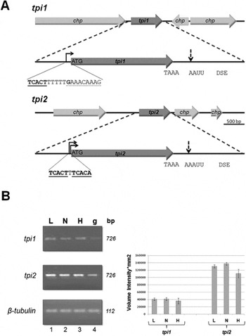
A BLAST search using the amino acid sequence of TIM from S. cerevisiae in the TrichDB (http://TrichDB.org) revealed 2 ORFs for the enzyme triosephosphate isomerase in the genome of T. vaginalis (Aurrecoechea et al. Reference Aurrecoechea, Brestelli, Brunk, Carlton, Dommer, Fischer, Gajria, Gao, Gingle, Grant, Harb, Heiges, Innamorato, Iodice, Kissinger, Kraemer, Li, Miller, Morrison, Nayak, Pennington, Pinney, Roos, Ross, Stoeckert, Sullivan, Treatman and Wang2009). These ORFs are located in different loci and encode proteins of 254 amino acids (Fig. 1A). Both proteins share 98·4% and 97·6% identity at their amino acid and nucleotide sequences, respectively. Thus, these TIMs only differ in the character of 4 amino acids and 18 nucleotides (Fig. S1, online version only). The ORF located in locus vg97417.m00128 is referred to as tpi1 and its gene product as TvTIM1 and the ORF located in locus vg83581.m00073 is named tpi2 and its gene product TvTIM2 throughout this work. The tpi1 gene is located in a locus in which 3 ORFs are oriented in the same strand and 1 ORF is oriented in the opposite strand (Fig. 1A). This is in contrast to the genomic organization of tpi2 that is located in a locus in which all four ORFs are oriented in the same strand (Fig. 1A). An in silico analysis of the 5′ UTR reveals that tpi1 contains a typical transcription initiation site (Inr) located -19 nt of the initial ATG. An EST analysis indicates that the G (Fig. 1A) located at -9 nt is the putative Inr. tpi2 contains 2 Inr sequences located at -5 nt and -11 nt of the initial ATG. Both genes have a TAA stop codon that included the polyadenylation signal in it, and an AAUU and polyT down stream elements (DSE) corresponding to putative polyadenylation regulatory sequences (Fig. 1A).
Genomic organization and expression of TvTIMs. (A) In silico analysis of tpi genes showing the physical map the 2 tpi genes of Trichomonas vaginalis. The arrows represent predicted open reading frames indicating transcription directionality. tpi 1 is flanked by conserved hypothetical proteins (CHP) oriented in 2 polarities. The putative Inr site and the transcription start site (G) are indicated. tpi2 contains 2 putative Inr sequences (underlined). Both genes contain a putative polyadenylation site (TAAA), a cleavage site (AAUU) and the down stream element (DSE). (B) Expression profile of tpi genes. Semi-quantitative RT-PCR of tpi1, tpi2 and β-tubulin using mRNA from T. vaginalis CNCD147, grown in low (L), normal (N), and high (H) iron concentrations and genomic DNA (g). The graph shows the average of 3 independent RT-PCRs assays corrected with tubulin expression.

An EST analysis in TrichDB revealed that tpi1 contains 55 ESTs, whereas tpi2 contains 896 ESTs, indicating the in vivo over-expression of tpi2 in most growth conditions (Fig. S2, online version only). In order to determine the expression level for each tpi gene, it was necessary to design oligonucleotides that selectively amplify each gene. The latter can be a problem in this case since both genes are 97·6% identical at the nucleotide level. Thus, to selectively RT-PCR amplify each gene, the 3′ end of the reverse primer was ‘specific’. In the case of tpi1, its 3′ end contained a cytidine, while in tpi2, its 3′ end contained a thymidine. The selective amplification is due to the fact that replicative DNA polymerases are not able to extend a mispaired primer-terminus (Huang et al. Reference Huang, Arnheim and Goodman1992). We also sequenced 5 independent clones of each tpi RT-PCR product and all of them were specific (data not shown).
A multitude of genes in T. vaginalis are regulated by iron, differentially affecting its cytopathogenicity including cytoadherence. Because several glycolytic enzymes of T. vaginalis function as adhesins, we tested the expression levels of tpi genes under distinct iron concentrations by RT-PCR assays (Fig. 1B). RT-PCR analysis of the expression levels of tpi1 and tpi2 indicate that the expression of these genes is constitutive. However, tpi2 is over-expressed approximately 3·3-fold as compared to tpi1 in the 3 iron conditions tested (Fig. 1B). These data correlate with the relative EST abundance observed in TrichDB for tpi2 (Fig. S2, online version only).
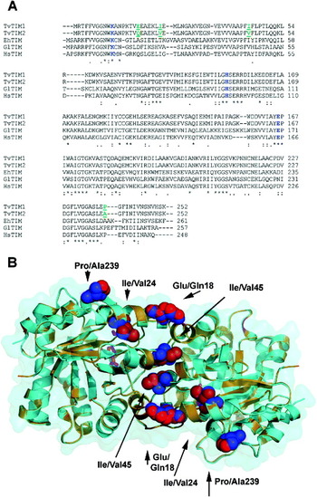
As illustrated in a multi-amino sequence alignment of TIMs from amitochondriate protozoa (G. lambia and E. histolytica) and H. sapiens, both TvTIM1 and TvTIM2 proteins contain the conserved Lys11, His94 and Glu166 catalytic residues (Fig. 2A). A homology model using the TIM crystal structure from T. molitor indicates that both TvTIM1 and TvTIM2 display the archetypical (β/α)8 fold (Fig. 2B). The localization of the 4 amino acid substitutions in this homology model indicates that these residues are in α-helix 1 (Asp/Asn 18,Glu/Gln 18 and Ile/Val 24), α-helix 2 (Ile/Val 45) and α-helix 8 (Pro/Ala 239). Residues Asp/Asn 18, Glu/Gln 18 and Pro/Ala 239 are solvent exposed, whereas Ile/Val 24 is part of the monomeric hydrophobic core, and residue Ile/Val 45 is part of the dimer interface.
Primary and predicted tertiary structure of TvTIMs. (A) Amino acid sequence alignment of TvTIMs with TIMs from Entamoeba histolytica, Giardia lamblia, and Homo sapiens. The conserved catalytic residues (Lys11, His 94, Glu 166) are coloured in blue. The four amino acid differences between TvTIM1 and TvTIM2 are green coloured. (B) Homology model of TvTIMs showing the archetypical (β/α)8 barrel fold. Ribbon representation of dimeric TvTIMs in which the conserved catalytic residues are in a stick representation and are magenta coloured. The differential residues between TvTIM1 and TvTIM2 are shown as spheres and are coloured red and blue respectively.

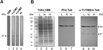
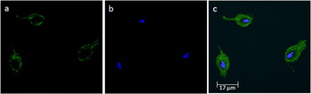
We carried out a WB assay using the anti-TvTIM2 polyclonal antibody against TvTIM1 and TvTIM2 recombinant proteins. As expected, the anti-TvTIM2 polyclonal antibody also recognized TvTIM1 (Fig. 3A, lane 2), thus this antibody can be used to test the localization of both enzymes in the parasite. Total extracts of T. vaginalis grown in low, normal and high iron conditions (Fig. 3B, lanes 2, 3 and 4) blotted onto NC membranes were tested against PI serum (Fig. 3B, lanes 5–7) or against anti-TvTIM2 antiserum (Fig. 3B, lanes 8–10). A single protein band of approximately 27 kDa was detected using the anti-TvTIM2 antiserum (Fig. 3B, lanes 8–10) and, as expected, no reactivity was observed with the PI serum (Fig. 3A, lanes 5–7). As a loading control, the blots were treated with an anti-tubulin antibody (Fig. 3B, lanes 5–10), showing reactivity with a single 50 kDa band. The uniformity between the loading control and the expression levels of TvTIMs indicated that no differences in the amount of TvTIMs is observed regarding the iron concentration, suggesting that neither TvTIM1 or TvTIM2 are post-transcriptionally regulated by iron. Immunofluorescence assays using permeabilized parasites revealed that TvTIMs are cytoplasmic proteins in T. vaginalis (Fig. 4) as previously shown for TIMs of amitochondriated organisms (Reyes-Vivas et al. Reference Reyes-Vivas, Diaz, Peon, Mendoza-Hernandez, Hernandez-Alcantara, De la Mora-De la, Enriquez-Flores, Dominguez-Ramirez and Lopez-Velazquez2007).
Protein expression profiles of TvTIMs. (A) WB analysis with the anti-TvTIM2 antibody. Lane 1, Coomassie brilliant blue (CBB) recombinant TvTIM2; lane 2, recombinant TvTIM1 blotted onto NC membrane and reacted with the anti-TvTIM2 antiserum; lane 3, recombinant TvTIM2 blotted onto NC membrane and reacted with the anti-TvTIM2 antiserum; lane 4, recombinant TvTIM2 blotted onto NC membrane and reacted with the PI serum. (B) WB on total trichomonad extracts (50 μg per lane) from parasites grown in different iron conditions. 12% SDS-PAGE stained with Coomassie brilliant blue of total extracts of Trichomonas vaginalis grown in low (L), normal (N), and high (H) iron conditions (lanes 2–4). WB of these extracts incubated with an anti-tubulin antibody and PI serum (PI) (lanes 5–7), or with an anti-tubulin antibody used as a loading control and the anti-TvTIM2 serum (lanes 8–10).

Cellular localization of TvTIMs. Indirect immunofluorescence analysis of permeabilized parasites incubated with the anti-TvTIM2 antibody. (a) Parasites treated with anti-TvTIM2 antibody (1:1000) and secondary antibody conjugated with FITC. (b) DAPI (diamidino-2-phenylindole) stained. (c) Merge analysis.

TvTIMs are able to complement a tpi E. coli deletion strain
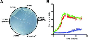
In order to test the enzymatic properties of TvTIM1 and TvTIM2, both ORFs were selectively amplified from genomic DNA of T. vaginalis using a set of specific oligonucleotides. E. coli lacking its endogenous TIM was used to complement a tpi E. coli deletion strain (tpi−) by TvTIMs (Saab-Rincon et al. Reference Saab-Rincon, Juarez, Osuna, Sanchez and Soberon2001). As observed in Fig. 5A, both TvTIM1 and TvTIM2 were able to complement a tpi− E. coli strain; however, the catalytically incompetent Lys11Ala mutants and an empty pET19 vector were unable to complement it. Furthermore, we analysed and monitored the growth of 3 independent colonies for each construct in liquid minimal medium at 30 °C. No significant difference in growth rate between E. coli deletion strains complemented with TvTIM1 or TvTIM2 was observed, indicating that both enzymes may share similar catalytic properties (Fig. 5B). TvTIM1 and TvTIM2 were also able to complement the E. coli tpi- strain grown on minimal media using lactate or glycerol as a unique carbon source (data not shown).
In vivo complementation of TvTIMs. (A) Minimal medium plates showing complementation of E. coli tpi − by TvTIMs. E. coli grown on minimal medium agar plates containing casaaminoacids, 0·1% glucose and 1 mm IPTG at 30 °C. (B) Growth curves of E. coli- tpi− by TvTIMs. The growth rates of E. coli tpi − that contained a plasmid encoding TvTIM1 and TvTIM2 are shown in green and red, respectively. E. coli tpi− transformed by plasmids containing the Lys11Ala substitution start to growth after 24 h (blue trace). The data represent the average of 3 independent cell cultures.

Heterologously expressed TvTIM1 and TvTIM2 are homodimers with similar secondary structure assessment and kinetic properties
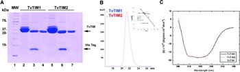
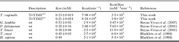
Heterologous expression of recombinant TvTIMs resulted in soluble proteins at physiological salt concentrations (150 mm NaCl). After a single purification step, both recombinant proteins were approximately 90% pure as indicated by the presence of a single protein band of 27 kDa by SDS-polyacryalmide gel electrophoresis (SDS-PAGE) (Fig. 6A, lanes 2 and 5). The histidine tag was removed by addition of PreScission Protease and each proteolytically cleaved enzyme was subjected to gel filtration chromatography; the collected TvTIMs were more than 95% pure as tested by SDS-PAGE gels stained with Coomassie brilliant blue (Fig. 6A, lanes 3, 4 and 6,7). The oligomeric state of TvTIMs was estimated by gel filtration and commercial molecular-mass standards at a protein concentration of 5 mg/ml. Both TvTIM1 and TvTIM2 were retained as a protein with a Stokes radius that corresponds to a globular protein of 52 kDa (Fig. 6B). This indicates that, like all TIM proteins studied to date, with the exception to TIMs from archaebacteria, TvTIMs are dimers. Furthermore, the CD spectra of TvTIMs superimpose with the spectra from S. cerevisiae´s TIM indicating that the content of α–helix and β-sheets is conserved among these three proteins. In addition, the CD spectra validate the (β/α)8–barrel fold observed in the molecular modelling for TvTIM1 and TvTIM2 (Fig. 6C). The kinetic characterization of TvTIMs was measured in the forward direction using glyceraldehyde-3-phosphate as substrate. Both enzymes followed Michaelis–Menten kinetics and exhibited a specific activity of 2367 U/mg for TvTIM1 and 2437 U/mg for TvTIM2. The catalytic efficiency of both TvTIMs is in the same order of magnitude as that reported for TIMs from Plasmodium falciparum, Trypanosoma cruzi, and T. brucei (Table 1). In order to assure that the catalytic activity was not due to the endogenous triosephosphate isomerase from E. coli BL21 (DE3) Star/Rossetta II strain, the Lys11Ala mutants of TvTIM1 and TvTIM2 were purified following the same protocol and no detectable activity was observed even at 100-fold of enzyme concentration with respect to wild type TvTIMs (data not shown).
Biophysical characterization of heterologous expressed TvTIMs. (A) SDS-PAGE analysis of the recombinant TvTIMs. Lane 1, molecular weight markers (MW); lane 2, IMAC purified TvTIM1, lane 3, proteolytically cleaved TvTIM1; lane 4, TvTIM1 after size-exclusion chromatography; lane 5, IMAC purified TvTIM2, lane 6, proteolytically cleaved TvTIM2; lane 7, TvTIM2 after size-exclusion chromatography. (B) Oligomeric determination of TvTIMs by gel filtration. Gel filtration chromatogram showing the retention time of TvTIMs in a Superdex-75 column. The molecular weight was determined by the elution volume relative to elution volume of standards with different molecular weight (inset). (C) CD spectra of TvTIMs in comparison to TIM from S. cerevisiae.

Steady-state kinetic parameters of recombinant TIMs

Analysis of duplicated TIM genes in different genomes
In order to ascertain the evolutionary origin of TvTIMs, we analysed all duplicated TIMs from eucaryotes and compared their evolutionary relationships (Fig. S3, online version only). We found 130 genomes with a duplicated tpi gene according to the Kegg Orthology (KO) pathway annotations for the enzyme triosephosphate isomerase (Table S1, online version only). To elucidate the evolutionary origin of TvTIMs, we performed a phylogenetic analysis with duplicated TIMs and found that TvTIMs are located in the same branch that duplicated TIMs from fungi (Fig. S4, online version only). A phylogenetic analysis indicates that within protists, only T. vaginalis and Paramecium tetraurelia contain 2 similar TIM genes in their genome (98% and 89% amino acid sequence identity, respectively) (Table S2, online version only). Only 7 organisms were found (including T. vaginalis) with duplicated TIMs with an amino acid sequence identity higher than 60%, suggesting that maintaining 2 very similar copies of a TIM gene is a particular event in T. vaginalis and in a few other organisms (Table S2). Thus, TIM duplication may be related to massive gene duplication events that the genome of T. vaginalis genome has passed during its evolution. Interestingly, the higher sequence conservation between the two tpi genes, which is a unique characteristic of the T. vaginalis (Table S2), can reflect a higher selective pressure by the original function of the TIM gene (related to retain a gene dosage) or to an event of very recent duplication. Without selective pressure imposed by the original activity on the new TIM copy, this copy can accumulate mutations until it is converted to a pseudogene or it can be selected for a different task in a process of neofunctionalization. Finally, the evolutionary relationships of all duplicated TIM genes showed that between all organisms reported by KO Database with duplicated TIM genes (Fig. S3), only Z. mays presents 2 very similar copies of a tpi gene with more than 98% of sequence identity (Table S2).
DISCUSSION
The 160Mb genome of T. vaginalis is unusually large for a protozoan parasite (Cui et al. Reference Cui, Smith and Samuelson2007). T. vaginalis contains a large number or duplicated genes, like transmembrane cyclases, family B2 DNA polymerases, RNA helicases, adenylate cyclases, N-acetylneuraminidase lyases and integrases (Carlton et al. Reference Carlton, Hirt, Silva, Delcher, Schatz, Zhao, Wortman, Bidwell, Alsmark, Besteiro, Sicheritz-Ponten, Noel, Dacks, Foster, Simillion, Van de Peer, Miranda-Saavedra, Barton, Westrop, Muller, Dessi, Fiori, Ren, Paulsen, Zhang, Bastida-Corcuera, Simoes-Barbosa, Brown, Hayes, Mukherjee, Okumura, Schneider, Smith, Vanacova, Villalvazo, Haas, Pertea, Feldblyum, Utterback, Shu, Osoegawa, de Jong, Hrdy, Horvathova, Zubacova, Dolezal, Malik, Logsdon, Henze, Gupta, Wang, Dunne, Upcroft, Upcroft, White, Salzberg, Tang, Chiu, Lee, Embley, Coombs, Mottram, Tachezy, Fraser-Liggett and Johnson2007; Singh et al. Reference Singh, Singh, Singh, Gautam, Farmer, Lodhi and Wadhwa2009; Cui et al. Reference Cui, Das, Smith and Samuelson2010). It has been hypothesized the massive gene duplication in Trichomonas is associated with a change in life style from an enteric to an urogenital environment (Singh et al. Reference Singh, Singh, Singh, Gautam, Farmer, Lodhi and Wadhwa2009). An example of the latter is gene duplication of metabolic enzymes, as the genome of T. vaginalis contains at least 20 enolases and several pyruvate:ferredoxin oxidoreductases. These metabolic duplicated genes have been involved in the host-parasite interaction (Mundodi et al. Reference Mundodi, Kucknoor and Alderete2008; Meza-Cervantez et al. Reference Meza-Cervantez, González-Robles, Cárdenas-Guerra, Ortega-López, Saavedra, Pineda and Arroyo2011). Herein we report that T. vaginalis contains 2 tpi ORFs in its genome. These tpi genes have a different chromosomal organization and are expressed at different mRNA levels. Interestingly, among the organisms that encode TIM as a duplicated gene only T. vaginalis presents a high sequence amino acid identity between both copies, suggesting that this is a recent gene duplication event. Specifically TvTIMs only differ in the character of 4 amino acids and all 4 substitutions are to conserved amino acids, and therefore it is expected that both TIM structures are practically identical. As TIM is an essential enzyme in amitochondriate protists, this scenario can be exploited to design drugs that inhibit the two enzymes of T. vaginalis. TvTIMs share only 54% amino acid sequence identity with TIM from H. sapiens, which opens the possibility that the structural differences between them can be used for rational drug design. The archetypical rationale for drugs against protozoan parasites is based on exposed cysteines; however, in our structural model of TvTIMs, no exposed cysteine was observed, indicating that specific drugs targeting the cysteine corresponding to Cys14 of other protozoans would be inefficient against TvTIMs (Reyes-Vivas et al. Reference Reyes-Vivas, Hernandez-Alcantara, Lopez-Velazquez, Cabrera, Perez-Montfort, de Gomez-Puyou and Gomez-Puyou2001). However, the dimer interface between parasitic and human TIMs has been exploited to design drugs that are species specific, indicating the possibility that the dimer interface of TvTIMs is a potential drug design target (Olivares-Illana et al. Reference Olivares-Illana, Perez-Montfort, Lopez-Calahorra, Costas, Rodriguez-Romero, Tuena de Gomez-Puyou and Gomez Puyou2006, Reference Olivares-Illana, Rodriguez-Romero, Becker, Berzunza, Garcia, Perez-Montfort, Cabrera, Lopez-Calahorra, de Gomez-Puyou and Gomez-Puyou2007).
tpi1 and tpi2 are transcribed in vivo and their transcription has no dependence on iron concentration. Interestingly, tpi2 contains 2 putative transcription start sites and is expressed approximately 3·3-fold more abundantly than tpi1. Confocal fluorescence microscopy indicates that TvTIMs, like TIMs from other amitochondriates, are located in the cytoplasm (Reyes-Vivas et al. Reference Reyes-Vivas, Diaz, Peon, Mendoza-Hernandez, Hernandez-Alcantara, De la Mora-De la, Enriquez-Flores, Dominguez-Ramirez and Lopez-Velazquez2007).
Recombinantly expressed TvTIMs exist as dimers in gel filtration, as is the case for TIMs from most organisms with the exception of Pyroccoccus woesei and Thermoproteus tenax (Kohlhoff et al. Reference Kohlhoff, Dahm and Hensel1996; Bell et al. Reference Bell, Russell, Kohlhoff, Hensel, Danson, Hough and Taylor1998). Recombinantly expressed TvTIMs display similar catalytic rate constants in comparison to TIMs from other organisms, indicating that the amino acid changes between them do not alter their catalytic function. The high amino acid and nucleotide similarity between TvTIMs suggest that the original TvTIM gene was subject to gene duplication. Interestingly, the distribution of nucleotide and amino acid changes show that those changes are not uniformly distributed along the two genes. Instead these changes are mainly concentrated in the first 240 pb of each gene or in the first 80 amino acids of the protein. This section includes 14 of the18 nucleotides and 3 of the 4 amino acid changes between both genes, suggesting a possible selective pressure. It is known that gene duplication implies an energetic cost. Thus, the question is how can the presence of 2 tpi genes in T. vaginalis be explained?
The bacterium S. meliloti contains 2 tpi genes; one involved in erythritol catabolism and the other in gluconeogenesis (Poysti and Oresnik, Reference Poysti and Oresnik2007). We speculate that one possibility for T. vaginalis tpi to be duplicated is to increase gene dosage in the parasite. This may be inferred from the similar catalytic efficiency (3·5 and 3·8 × 105 mM/min) of both TvTIMs which is in the range of the catalytic efficiency of other TIMs. Thus, gene duplication of triosephosphate isomerase may contribute to the robustness of glycolysis in T. vaginalis. It is relevant to point out that TIMs from other organisms act as adhesion molecules and that other metabolic enzymes from T. vaginalis like, malic enzyme, succinyl CoA synthetase and pyruvate:ferredoxin oxidoreductase are cell-adhesion molecules (Alderete et al. Reference Alderete, O'Brien, Arroyo, Engbring, Musatovova, Lopez, Lauriano and Nguyen1995; Mundodi et al. Reference Mundodi, Kucknoor and Alderete2008; Lama et al. Reference Lama, Kucknoor, Mundodi and Alderete2009; Meza-Cervantez et al. Reference Meza-Cervantez, González-Robles, Cárdenas-Guerra, Ortega-López, Saavedra, Pineda and Arroyo2011).
Thus, it is possible that the recent duplication event of TvTIMs represents a process in which one of these enzymes is diverging towards moonlighting activity. Supporting this notion, the dimer stability of TvTIM2 with respect to TvTIM1 differs by 15 kJ mol−1 (Lara et al. manuscript in preparation). Interestingly, the character of residue 45 at the dimer interface is isoleucine for TvTIM1 and valine for TvTIM2. It is possible that TvTIM2 duplicated from TvTIM1 and that the loss of the methyl group at the dimer interface maybe an evolutive event in which TvTIM2 may function as a monomeric adhesion molecule. Although speculative, our results evidence the necessity of further studies in order to fully understand why the high sequence conservation between tpi genes is exclusively maintained in a few organisms and the nature of this duplication event in T. vaginalis.
ACKNOWLEDGEMENTS
We thank Corina Díaz Quezada and Leticia Ávila González for their technical assistance and Dr Cei Abreu-Goodger for critical reading of the manuscript.
FINANCIAL SUPPORT
This work was supported by grants ICYTDF-6968 to C.G.B.C. and ICYTDF-299 and 219 to R.A.; CONACYT-162123 to R.A. H.S.L. and P.E.H. received fellowships from CONACYT. E.E.F.A. was a recipient of a Fellowship from ICYTDF-299 grant.