Introduction
Echinococcus granulosus infection leads to cystic echinococcosis (CE) caused by the metacestode (larval) stage of the parasite. The metacestode stage of the parasite consists of a fluid-filled cavity, surrounded by a germinal layer which, in turn, is supported by an acellular laminated layer and a fibrous adventitial layer derived from the host. Within the intermediate host, protoscoleces develop by clonal expansion. Detailed studies on the characterization, expression and stage-specific expression of the 14-3-3 gene in E. granulosus and E. multilocularis have been reported by several researchers (Siles-Lucas et al., Reference Siles-Lucas, Felleisen, Hemphill, Wilson and Gottstein1998, Reference Siles-Lucas, Nunes and Zaha2001, Reference Siles-Lucas, Merli and Gottstein2008; Nunes et al., Reference Nunes, Zaha, Gottstein, Müller and Siles-Lucas2004). The deduced amino acid sequence of E14-3-3.2 (zeta type) showed 88% identity to the E14-3-3.1 isoform (zeta type) and 52% identity to a third Echinococcus isoform (E14-3-3.3) (epsilon type). These different isoforms are orthologous, thus each individual isoform is more highly conserved among species than within a single species (Rosenquist et al., Reference Rosenquist, Sehnke, Ferl, Sommarin and Larsson2000). This indicates a selective pressure on isoform specific functions, although the specific involvement of the different 14-3-3 isoforms in specialized cellular processes remains unclear. The 14-3-3 proteins play an important role in morphological changes in the life cycle of the parasite. The stage-specific expression of this chaperone protein appears to be crucial as an immunodominant antigen (Siles-Lucas & Gottstein, Reference Siles-Lucas and Gottstein2003). This study was undertaken to evaluate the relative expression of the vital 14-3-3 protein in different morphotypes of cysts collected from naturally infected buffaloes.
Materials and methods
Collection of cysts
Larval stages of the parasite were collected from buffalo from a slaughterhouse under the jurisdiction of the Kolkata Municipality Corporation, West Bengal, India. The cysts were grouped into six different morphotypes as follows. Cysts with a white germinal layer with protoscoleces and clear cystic fluid were designated as S1. Cysts with transparent germinal layer without protoscoleces and clear cystic fluid were termed S2. A designation of S3 was given to those cysts that contained protoscoleces, germinal layer and turbid cystic fluid. Cysts with a white germinal layer and clear cystic fluid without protoscoleces were categorized as S4. Those with slightly yellowish and thickened germinal layer, clear fluid without protoscoleces were termed S5. And finally, cysts with no protoscoleces and calcified and turbid cystic content were designated as S6. Ten samples of S1 cysts were collected while eight cysts each were collected from the remaining S2, S3, S4, S5 and S6 categories.
Isolation of protoscoleces and the germinal layer
Protoscoleces and the germinal layer from both the fertile and the sterile cysts were isolated, rinsed in sterilized phosphate-buffered saline (PBS) (pH 7.2) three times and stored until further use (Eckert et al., Reference Eckert, Gemmell, Meslin and Pawlowski2001).
DNA extraction
Genomic DNA was isolated from protoscoleces and the germinal layer using the B10 101® system GNOME® (Qbiogene/MP Biomedicals, Irvine, California, USA) according to the protocol provided within the kit. The same kit and methodology were used for both sterile and fertile cysts. The genomic DNA was dissolved in 25 μl Tris–EDTA (TE) buffer (pH 8.0).
RNA extraction and cDNA preparation
Total RNA was isolated from fresh protoscoleces and the germinal layers using TRI Reagent® (Sigma, St Louis, Missouri, USA). Each sample was incubated for 45 min at 37°C with RQ1 RNase-free DNase (Promega, Madison, Wisconsin, USA) and heat inactivated at 65°C for 10 min using DNase-stop buffer (Promega). Total RNA was quantified after determination of optical density at 260 nm. A total of 3 μg of RNA was used for cDNA preparation for each isolate. The cDNA was synthesized from each RNA sample using high-capacity cDNA reverse transcription kit (Applied Biosystems, Foster City, California, USA). cDNA samples were kept at − 70°C until further use.
Primer design and semi-quantitative RT-PCR analysis
Primers were used to amplify actin II (housekeeping gene) and 14-3-3 protein using Primer Premier Version 5 (PREMIER Biosoft International, Palo Alto, California, USA). All primers were designed from available actin II sequence information (accession number L07774) and 14-3-3 protein (accession number AY 265907). The designs of primers were:
Actin II forward: 5′-CGAGCAGGAAATGATAACG-3′
Actin II reverse: 5′-GAA CAG GGC TTC AGG ACA-3′
14-3-3 gene forward: 5′-ATG TCT TCT CTC AGT AAG CGC G- 3′
14-3-3 gene reverse: 5′-CTT TTC AAG TTG CGC TCA GTT C-3′.
Polymerase chain reaction (PCR) was performed using GeneAmp® PCR System 9700 (Applied Biosystems) in a final 25 μl volume containing 200 μm of each deoxynucleoside triphosphate (dNTP) (Genei, India), 10 × PCR buffer, 2.5 mm MgCl2, 10 pmol of each gene-specific primer and 1 unit of Taq DNA polymerase (Fermentas, Glen Burnie, Maryland, USA) and an equal volume of cDNA was used as template for each isolate in reaction buffer. Briefly, the PCR process comprised an initial denaturing step (95°C for 5 min.); followed by 35 cycles of 95°C for 45 s (denaturation), annealing at 56°C (Eg14-3-3) or 61°C (actin II) for 45 s and elongation at 72°C for 90 s; and a final extension step (72°C for 10 min). The specificity and size of the amplified product was assessed by electrophoresis in 2% (w/v) Tris acetate–EDTA agarose gel stained with ethidium bromide. The amplified product was quantified directly using GelQuant software version 2.7 (DNR Bio-Imaging Systems Ltd., Israel). Relative quantification of expression of Eg14-3-3 was normalized against actin II (housekeeping gene or internal control) in each isolate. The transcription levels of the selected 14-3-3 gene were compared with the housekeeping gene and relative quantification was determined as a ratio.
Statistical analysis
Data were expressed as mean ± standard error (SE) and evaluated for their statistical significance of difference with analysis of variance (ANOVA). P values of less than 0.05 were considered to be statistically significant.
Analysis of E. granulosus genotype
Genotyping of all isolates was accomplished by mitochondrial adenosine triphosphatase (ATPase) subunit 6 (ATP6) sequence, as previously described (Gudewar et al., Reference Gudewar, Pan, Bera, Das, Konar, Rao, Tiwari and Bhattacharya2009).
In silico analysis
DNA sequences were aligned with other reported 14-3-3 isoforms of E. granulosus available in a public database using CLUSTAL W analysis by MEGA 4 (Tamura et al., Reference Tamura, Dudley, Nei and Kumar2007). The predicted amino acid sequence of 14-3-3 was deduced on the basis of genetic codes available at the National Center for Biotechnology Information (www.ncbi.nlm.nih.gov). Clustering of the 14-3-3 isoforms was done by the unweighted pair group method with arithmetic mean (UPGMA) using the above-mentioned programme (Tamura et al., Reference Tamura, Dudley, Nei and Kumar2007).
Results and discussion
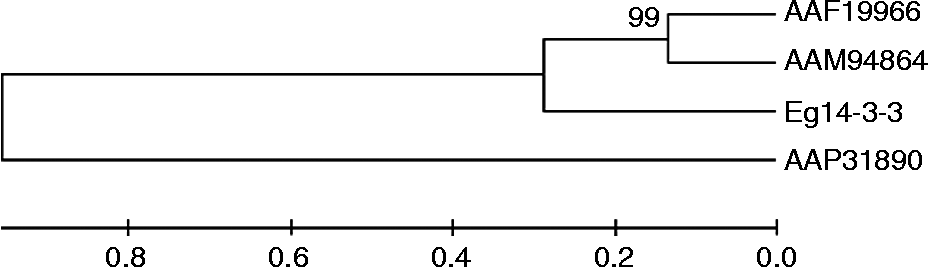
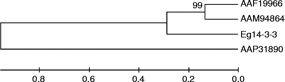
The sizes of amplified products of the actin II and 14-3-3 genes were 291 bp (accession number FJ 897573) and 295 bp (accession number FJ 897574), respectively. Genotyping of all the isolates revealed that they belonged to a common sheep strain (G1) based on analysis of the ATP6 gene. A phylogenetic tree constructed on the basis of 98 predicted amino acid sequences revealed that this partial fragment of the 14-3-3 gene was closely clustered with isoforms 1 and 2 (zeta types) (fig. 1).
Phylogenetic analysis from predicted amino acid sequence information of 14-3-3 protein of Indian isolates of Echinococcus granulosus constructed using unweighted pair group method with arithmetic mean (UPGMA). Numbers at nodes represent percentage occurrence of clades in 1000 bootstrap replications of data. AAF19966 (amino acid sequence of accession no. AF207964), AAM94864 (amino acid sequence of accession no. AF529419), AAP31890 (amino acid sequence of accession no. AY265907), Eg14-3-3 (accession no. FJ897574). AAF19966, AAM94864, reported isoforms 1 and 2 (zeta type); AAP31890, reported isoform 3 (epsilon type).
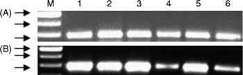
Gene expression levels of the 14-3-3 protein were quantitatively evaluated in relation to the housekeeping molecule and directly compared for the six different categories of cysts. On the basis of relative quantification, the expression of transcripts of the 14-3-3 gene was higher in S1 cysts followed in order by S3, S5, S2, S4 and S6 cysts (fig. 2 and table 1). The relative expression of this protein was higher in fertile cysts (cysts with protoscoleces) when compared to cysts without protoscoleces (sterile cysts). This finding suggests that the expression of the 14-3-3 gene plays an important role in the regulation of proliferative performance of the parasite during its development. The protoscoleces develop from the germinal layer of the parasite due to clonal expansion of the cells. The 14-3-3 protein is ubiquitous in eukaryotic organisms and has been described as a key molecule in the process of cell differentiation and proliferation (Aitken et al., Reference Aitken, Collinge, van Heusden, Isobe, Roseboom, Rosenfeld and Soll1992). Moreover, uniform expression of the actin gene confirmed the viability of the harvested isolates in this study. Beta-actin is purported to be expressed constitutively in normal living cells (Naora & Naora, Reference Naora and Naora1995). During comparative analysis of expression of the 14-3-3 gene in E. granulosus and E. multilocularis (Siles-Lucas et al., Reference Siles-Lucas, Nunes and Zaha2001), the native 14-3-3 protein could not be detected in the germinal layer of E. granulosus. However, the present investigation confirmed the expression of transcripts of the 14-3-3 protein in the germinal layer of both the viable and sterile cysts of E. granulosus. Significant evidence exists in the literature regarding the diverse functions of the 14-3-3 protein, including signal transduction, cell cycle control, stress response and malignant transformation (Siles-Lucas & Gottstein, Reference Siles-Lucas and Gottstein2003). Therefore, based on the presence of the 14-3-3 protein in the germinal layer of sterile cysts, we can postulate that this protein plays an important role in the development and survival of sterile cysts within the intermediate host.
(A) Expression of actin II mRNA (internal control) in different cystic stages. (B) Expression of 14-3-3 gene mRNA in different cystic stages. Lanes: M, 100 bp DNA ladder (500 bp, 400 bp and 300 bp fragments from the bottom); 1–6, S1, S2, S3, S4, S5 and S6, respectively.

Relative expression of the 14-3-3 gene in different cystic stages.

Significant differences at aP < 0.001 and bP < 0.05.
Acknowledgements
The authors are grateful to the Department of Biotechnology, Government of India for financial support and to the Director, Indian Veterinary Research Institute for provision of infrastructural facilities.