Implications
Surgical castration of male pigs is commonly performed in European farms to avoid unappreciated sexual odors (boar taint) during cooking. Nevertheless, this method is criticized for welfare reasons. One alternative is rearing entire male pigs, provided that the problem of boar taint is solved. A second option is immunocastration that allows boar taint to be reduced. Because testicular hormones exert immunoregulatory functions, the influence of castration and immunocastration on immunity and health was evaluated using blood markers. Overall, the lack of clear negative functional consequences supports the generalization of these new practices.
Introduction
Sex steroids play a significant role in the regulation of the immune function (Bouman et al., Reference Bouman, Heineman and Faas2005) and hence influence the resistance to disease. Surgical castration performed on rodents during adulthood reverses the age-related thymic atrophy and restores the thymic production of lymphocytes (Sutherland et al., Reference Sutherland, Goldberg, Hammett, Uldrich, Berzins, Heng, Blazar, Millar, Malin, Chidgey and Boyd2005). This leads, in secondary lymphoid organs, to an increase in the frequency of both CD8+ and CD4+ naïve T cells, in the T CD4/T CD8 ratio and in the capacity of proliferation of T cells, restoring a juvenile phenotype (Sutherland et al., Reference Sutherland, Goldberg, Hammett, Uldrich, Berzins, Heng, Blazar, Millar, Malin, Chidgey and Boyd2005). Surgical castration of adult rodents also increases the production of B cells in the bone marrow, leading to an increased number of B cells in the spleen and a subsequent splenic enlargement (Viselli et al., Reference Viselli, Stanziale, Shults, Kovacs and Olsen1995). A study carried out on monkeys, using a gonadotropin-releasing hormone (GnRH) antagonist to suppress the neonatal surge of testosterone, showed that neonatal androgens have a long-term influence on the immune development that could be different from the effect of post-pubertal androgens (Gould et al., Reference Gould, Akinbami and Mann1998). Neonatal treatment with an antagonist of GnRH induced a diminution in white blood cells and more so in T- and B-cell numbers during both infancy and adulthood. Very few data describe the effects of testicular hormone deprivation in pigs on blood T cells. We showed that neonatal surgical castration decreased the number of blood lymphocytes at 4 and 5 months, as well as the thymus weight at 5 months, suggesting that castration might affect T-cell production (Merlot et al., Reference Merlot, Thomas and Prunier2013). However, in this study, functional tests involving T cells such as the specific response to vaccination against KLH and the mitogen-induced proliferation of peripheral blood cells were unaltered.
Sex steroid levels vary along the pubertal development. A first peak of androgen secretion occurs during the first few weeks after birth (Colenbrander et al., Reference Colenbrander, Dejong and Wensing1978). Thereafter, not only testosterone but also estradiol levels progressively increase during pubertal development of male pigs, from about 2 until 6 months of age (Allrich et al., Reference Allrich, Christenson, Ford and Zimmerman1982; Brunius et al., Reference Brunius, Zamaratskaia, Andersson, Chen, Norrby, Madej and Lundstrom2011). Immunocastration is based on vaccination against GnRH resulting in the suppression of testicular activity. The primary immunization is usually carried out around 3 months of age. The second injection is carried out some few weeks later and leads to a rapid drop (within a couple of days) in LH and testosterone (Claus et al., Reference Claus, Lacorn, Danowski, Pearce and Bauer2007). Thus, surgical castration suppresses the gonadal steroids from the neonatal period, whereas GnRH and LH are preserved and should even be increased owing to the lack of negative feedback of the testicular hormones on the hypothalamus and pituitary (Colenbrander et al., Reference Colenbrander, Meijer, Macdonald, Vandewiel, Engel and de Jong1987). Immunocastration allows natural activity of the hypothalamic–pituitary–gonadal (HPG) axis until the second injection of the vaccine, followed by a suppression of both pituitary and testicular hormones. Because of the differences in the timing and nature of the suppressed hormones, the two methods of castration may have different effects on the immune system.
The present experiment was carried out to investigate the consequences of two methods of castration that are used in pigs, surgical castration and immunocastration, on peripheral T cells. We hypothesized that the frequency and function of T cells would be similar in immunocastrated and entire male pigs until the second vaccine injection, whereas they could differ between surgically castrated and entire pigs from 3 months of age. The effects of the different treatments were observed from 3 to 5 months of age, as fattening pigs are usually slaughtered at around 5 months. We determined white blood cell counts, proportions of blood T-cell subsets, proliferation of T lymphocytes, markers relevant to evaluating the health status (total IgG, haptoglobin levels and clinical measures at slaughter), and the immune response to a vaccination against influenza.
Material and methods
The experiment was conducted following French guidelines for animal care and use (http://ethique.ipbs.fr/sdv/charteexpeanimale.pdf) in the experimental unit of INRA – Saint Gilles (35590 France). People involved in the experiment were holders of an animal experiment agreement delivered by the veterinary services of the French Ministry of Agriculture.
Animals, experimental design and sampling procedures
Two experiments were conducted on pigs born from Large White×Landrace sows inseminated with Pietrain semen.
Experiment A involved three experimental groups: 18 entire (E), 16 immunocastrated (IC) and 14 surgically castrated (SC) males. This experiment was run in four repetitions undertaken from December 2010 to July 2011. For each replicate, four litters were selected at farrowing, in which a third of the male piglets were surgically castrated at 5 to 6 days of age following the standard procedure used in pig farming. After weaning at 26 to 28 days of age, animals were always housed with pen-mates of the same gonadal status (entire and surgically castrated). At 68 to 71 days of age (29±5 kg live weight), two to five male pigs were chosen in each of the four litters (depending on the number of male pigs available per litter) and assigned to experimental groups (SC, IC and E) on the basis of their BW and gonadal status to equilibrate, as well as possible, BW and litters of origin among treatments. They were transferred into the experimental unit, where they were housed in groups of two or three pigs from the same treatment. Pigs received ad libitum standard growth diet. Water was provided ad libitum. The primary and the booster immunizations of IC pigs against GnRH (Improvac®; Pfizer, Belgium, SC route) were carried out at 87 to 90 and 115 to 118 days of age. All the pigs were vaccinated against influenza (Gripovac; Merial, Lyon, France) at 129 to 132 and 143 to 146 days of age.
Blood samples were drawn by venipuncture in the jugular vein on EDTA-containing (hormonal assay and white blood cell counts) and heparinized (flow cytometry and lymphocyte proliferation) tubes at 87 to 90, 115 to 118 (before each injection of Improvac) and 150 to 153 days of age. At the second and third blood samplings, samples were also drawn in tubes without anti-coagulant for total immunoglobulin (Ig) G and anti-influenza Ig determinations. Plasma and sera were stored at −20°C until use. At 151 to 154 days of age, pigs were transferred to the slaughterhouse and were euthanized the following day by exsanguination after electronarcosis.
Experiment B was run to increase the number of pigs regarding white blood cell counts and lymphocyte subset analyses, and involved 10 entire male pigs and 10 surgically castrated male pigs originating from six different litters. In each litter, half of the male pigs were neonatally castrated, and at 68 to 71 days of age male piglets were chosen (one SC and one E, or two SC and two E per litter, depending of the number of male pigs available in the litter) and assigned to the E and SC experimental groups. This experiment was conducted from February to July 2012 in one replicate in the same rearing conditions as experiment A, except that animals were not vaccinated against influenza. Blood samples were drawn on EDTA-containing (white blood cell counts) and heparinized (flow cytometric analysis) tubes at 115 to 118 and 150 to 153 days of age.
White blood cell count and immunoassays
Total numbers of leukocytes, lymphocytes and granulocytes were measured with a hematology automatic cell counter (MS-9; MeletSchloesing Laboratories, Osny, France). The coulter allows discriminating particles according to their electric impedance that is proportional to their volume, and has been calibrated for growing pigs by the manufacturer. The lymphocyte and granulocyte counts carried out by the coulter and by conventional microscopy in a veterinary laboratory gave correlations with a R 2 of 0.92 and 0.94, respectively. Total IgG levels were assessed by ELISA (Bethyl E100-104, pig IgG Fc Elisa quantitation; Interchim, Montluçon, France), as described previously (Couret et al., Reference Couret, Jamin, Kuntz-Simon, Prunier and Merlot2009). Anti-influenza Ig levels were assessed by ELISA (Swine influenza Virus Antibody Test Kit – H1N1, IDEXX Europe, Hoofddorp, The Netherlands). The relative level of antibody in the sample was determined by calculating the sample to positive standard ratio of optical densities. Plasma testosterone was measured by the AIA 360 automaton (Tosoh Europe, Tessenderlo, Belgium) using a commercial kit (ST AIA – testosterone pack; Tosoh Europe), and haptoglobin was assessed using a commercial kit (Tridelta Ltd., AbCys s.a, Paris, France).
Proliferation assay and flow cytometry
All products were purchased from SIGMA Aldrich (St Louis, Missouri, USA). Peripheral blood mononuclear cells (PBMC) were isolated from heparinized blood by gradient centrifugation on histopaque 1077. They were adjusted at a concentration of 10×106 cells/ml RPMI 1640 supplemented with 10% fetal bovine serum 1% penicillin/streptomycin and 1% l-glutamine as described previously (Couret et al., Reference Couret, Jamin, Kuntz-Simon, Prunier and Merlot2009). A viability analyzer using trypan-blue exclusion test (Vi-Cell XR; Beckman Coulter, Paris, France) was used to determine cell numbers and viability, which was always higher than 95%.
A total of 0.5×10⁶ cells per well in a final volume of 200 µl were cultured in triplicate in the presence or absence of the T-cell mitogen Concavalin A (ConA 6.25 µg/ml) and, after 48 h of incubation, were pulsed with tritiated thymidine (1 µCi per well) for 24 h. Plates were stored at −20°C, and then filtrated on filter glass paper (MultiScreen Filter Plates; Millipore, Molsheim, France). Radioactivity was counted (counts per minute=c.p.m.) in a microplate scintillation counter (TopCount from Perkin Elmer, Courtaboeuf, France). Mitogenic responsiveness was expressed as a proliferation index: PI=(c.p.m. of stimulated cells)/(c.p.m. of unstimulated cells).
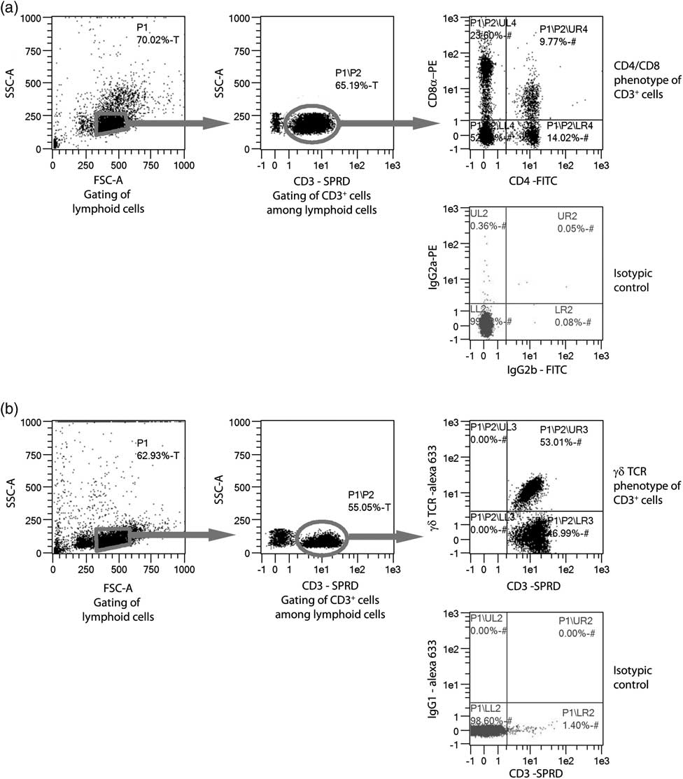
The monoclonal antibodies (mAb) used to detect cell surface markers were SpectralRedTM (SPRD)-conjugated anti-swine CD3 (clone PPT3, IgG1), FITC-conjugated anti-swine CD4 (clone 74-12-4, IgG2b) and PE-conjugated anti-swine CD8α (clone 76-2-1, IgG2a) that were purchased from Southern Biotech (Birmingham, Alabama). Anti-swine γδT-cell receptor (TCR) antibody (clone PGBL22A, IgG1, VMRD; Pullman, WA, USA) was used with an anti-mouse IgG secondary antibody conjugated with Alexa Fluor 633 (Invitrogen, Carlsbad, CA, USA). Appropriate isotype control antibodies were used (Southern Biotech). The direct labeling (CD3/CD4/CD8) was performed on 0.5×10⁶ cells incubated with saturating concentrations of anti CD3, anti CD4 and anti CD8 mAb (1/100 dilutions) in a final volume of 50 µl. The indirect labeling consisted of successive incubations of 1×10⁶ cells with anti γδTCR (1/100 dilution), anti-mouse IgG (1/1000 dilution) and anti-CD3 (1/100 dilution) antibodies in a final volume of 100 µl. All incubations lasted 30 min and were carried out at 4°C. After each staining step, cells were washed twice in 250 µl PBS. Cells were suspended in 400 µl of fluorescence-activated cell sorter buffer before analysis. Analysis of PBMC was performed using a Macs Quant Analyzer (Miltenyi Biotec, Bergisch Gladbach, Germany) with MacsQuant Quantify software. The strategy of gating for the analysis is presented in Figure 1.
Strategy of analysis of lymphocyte subpopulations by flow cytometry. A first gate of lymphoid cells was designed on the forward – side scatter plot (gate P1, 10 000 events acquired). Among lymphoid cells, CD3+ T lymphocytes were selected (gate P2 included in P1). The percentages of CD3+CD4+CD8−, CD3+CD4−CD8+ and CD3+CD4+CD8+ cells (a) and of CD3+ γδTCR+ cells (b) were determined among the CD3+ population.

Clinical measurements at slaughter
In experiment A, the lungs were inspected for pericarditis and pleurisy at slaughter. Pneumonia lesions were scored by summing the score attributed to each of the seven lobes according to the consolidation surface area (from 0 for no lesion to 4 for total pulmonary consolidation), leading to an individual severity score ranging from 0 to 28 (Madec and Kobish, Reference Madec and Kobish1982). Rhinitis was also estimated by summing the shrinking score attributed to each of the four turbinate bones inside the snout (from 0 for intact to 4 for total atrophy) and the distortion score attributed to the nasal wall (from 0 for no deviation to 2 for severe deviation), leading to an individual severity score on a scale from 0 to 18. Stomach lesions were scored between 0 and 7 according to the severity or absence of ulcer (Henry et al., Reference Henry, Dauloudet, Kerisit, Lefeuve, Gaye, Denechaud and Bourdon1970). The testes and spleen were weighed.
Statistical analysis
Analysis of variance was performed using the mixed procedure of SAS (version 8.1, 2000, SAS Institute Inc., Cary, NC, USA). The fixed factors of the analysis were the treatment and replicate. Normality of the data was checked on residuals with the Shapiro–Wilk test. For blood leukocyte counts and cytometry, data from experiments A and B were analyzed together with experiment B declared as a 5th replicate. Because the disequilibrium owing to the lack of IC pigs in experiment B could have biased the analysis, those results were compared with those of an analysis, taking into account only the repetitions from experiment A. The means that were statistically different when pooling experiment A+B were also significant (% CD3+CD4+CD8+ at 5 months of age, blood lymphocyte numbers at 4 months), close to statistical difference (% γδT cells at 5 months) or at least in the same numerical order (blood total leukocyte and lymphocyte counts at 5 months, γδT and CD3+CD4+CD8+% at 4 months) in experiment A alone. Thus, pooling experiments A and B increased the statistical power without inducing any bias. The Bonferroni test was used for post-hoc comparisons when the treatment effect in the ANOVA had a P-value<0.05. Numeric data are presented as least square means±standard errors of the mean (s.e.) calculated by SAS for the lowest group size.
Results
Concentration of testosterone from 3 to 5 months of age
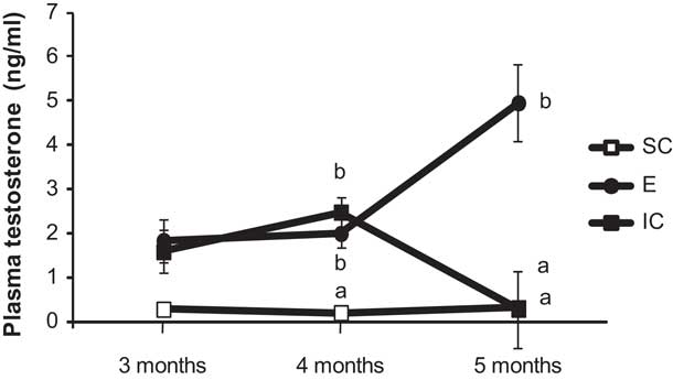
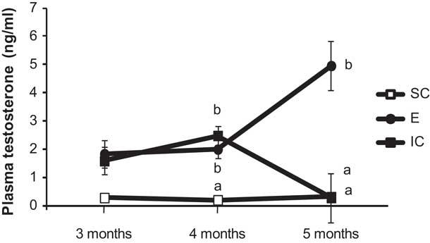
E pigs had higher concentration of testosterone at 4 and 5 months of age than SC pigs (P<0.001 and <0.001, respectively, Figure 2). Until the second vaccination (at 4 months), testosterone concentration was not different in the E and IC pigs (P>0.1) but higher in E than in SC pigs (P<0.001). At 5 months, the concentration in IC pigs fell to levels significantly lower than those of E pigs (P<0.001) and comparable to those of SC pigs (Figure 2).
Levels of testosterone in entire (E, n=18), immunocastrated (IC, n=16 to 18) and surgically castrated (SC, n=14) pigs from experiment A. a,bWithin each age, means with different letters differ with P-values <0.001.
Blood immune measurements
White blood cell counts
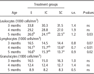
Compared with E male pigs, SC male pigs had a lower number of total leukocytes at 5 months (P=0.03) as well as a lower number of lymphocytes at 4 and 5 months (P<0.01 and P=0.02, respectively) but a similar number of granulocytes at the three ages (Table 1). When the number of cells differed significantly between the SC and E male pigs, it was intermediate in the IC male pigs and never differed significantly from one group or the other.
Numbers of total leukocytes, lymphocytes and granulocytes in entire (E), immunocastrated (IC) and surgically castrated (SC) pigs

Data include the results from experiment A at 3 months of age (E: n=18, IC: n=16 to 18, SC: n=14) and from experiment A+B at 4 and 5 months of age (E: n=28, IC: n=16 to 18, SC: n=24).
ns: P-value of the treatment effect is >0.10.
a,bWithin a row, means with different letters differ at P<0.05.
PBMC subsets and proliferation
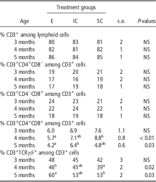
There was no effect of treatment on the percentages of CD3+, CD3+CD4+ and CD3+CD8+ cells (Table 2). E pigs had a percentage of CD3+CD4+CD8+ cells that was lower than in SC pigs at 4 months of age (P<0.01), and lower than in IC pigs at 5 months of age (P=0.03). At 4 and 5 months, E pigs had a higher percentage of TCRγδ + T lymphocytes than the SC male pigs (P=0.02 and 0.03, respectively), whereas the IC pigs had an intermediate percentage not differing from that observed in the E and SC pigs (P>0.1).
T-lymphocyte subsets in entire (E), immunocastrated (IC) and surgically castrated (SC) pigs

Data include the results from experiment A at 3 months of age (E: n=18, IC: n=16 to 18, SC: n=14) and from experiment A+B at 4 and 5 months of age (E: n=28, IC: n=16 to 18, SC: n=24).
ns: P value of the treatment effect is >0.10.
a,bWithin a row, means with different letters differ at P<0.05.
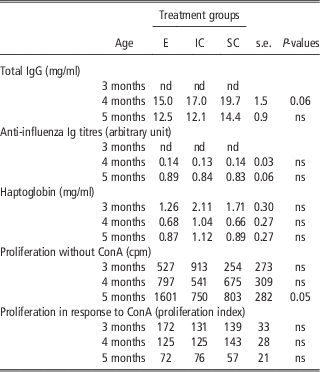
At 5 months, the E male pigs tended to have a higher basal proliferation of T cells than the SC and IC male pigs (P=0.05). T-cell proliferation in response to ConA was not influenced by treatment.
Plasma immunoglobulins and haptoglobin
Treatments did not influence anti-influenza Ig or haptoglobin levels (Table 3). At 4 months of age, gonadal status tended to influence total IgG level (P=0.06), but the effect was no longer present at 5 months (P>0.1).
Blood titres of anti-influenza Ig, concentration of total IgG and haptoglobin and proliferation of peripheral blood mononuclear cells of entire (E, n=18), immunocastrated (IC, n=16 to 18) and surgically castrated (SC, n=14) pigs from experiment A

nd=not determined.
ns: P-value of the treatment effect is >0.10.
Clinical measurements at slaughter
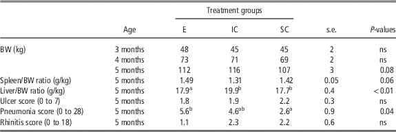
Relative weight of the spleen tended to be influenced by treatment (P=0.06) and that of the liver was higher in IC pigs than in E and SC pigs (P<0.001, Table 4). Lung exams revealed that E pigs had higher scores of pneumonia than SC pigs, but there was no significant difference for stomach and nose lesions.
Body and organ weights and health measures in entire (E, n=18), immunocastrated (IM, n=16 to 18) and surgically castrated (SC, n=14) pigs from experiment A

For stomach ulcer, pneumonia and rhinitis, intact organs received a score of zero.
ns: P-value of the treatment effect is >0.10.
a,bMeans with different letters differ at P<0.05.
Discussion
This experiment showed that the consequences of surgical castration and immunocastration on peripheral immune cells were moderate. Long-term suppression of testicular hormones by surgical castration seemed to have a greater impact than late immunocastration.
Effect of neonatal deprivation of testicular hormones by surgical castration
In our study, SC pigs presented lower numbers of lymphoid cells from 3 to 5 months relatively to entire pigs. Our finding is in agreement with previous studies where castrated male pigs at slaughter age had fewer peripheral lymphoid cells compared with entire male pigs (Odink et al., Reference Odink, Smeets, Visser, Sandman and Snijders1990; Merlot et al., Reference Merlot, Thomas and Prunier2013). The blood lymphoid fraction is composed of T, B and NK cells. As our results showed that surgical castration did not decrease the percentage of T (CD3+) lymphocytes among lymphoid cells, we can hypothesize that the haematopoiesis of T cells, on the one hand, and B plus NK cells on the other was reduced in a similar proportion in SC pigs.
These differences between E and SC pigs in blood cell numbers could result from a direct effect of neonatal and prepubertal androgens on the haematopoiesis of the lymphoid lineage. This would be in contradiction with studies on rodents, showing that both pre- and post-pubertal surgical castration increase the weight of the thymus and spleen and the peripheral pool of both B and T cells during adulthood (Viselli et al., Reference Viselli, Stanziale, Shults, Kovacs and Olsen1995; Ellis et al., Reference Ellis, Moser, Le, Flanigan and Kwon2001; Radojevic et al., Reference Radojevic, Arsenovic-Ranin, Kosec, Pesic, Pilipovic, Perisic, Plecas-Solarovic and Leposavic2007). The divergence between studies on pigs and rodents might result from the fact that the influence of androgens on immune cells depends on the age of the animals at the observational time. They might stimulate the development of the immune system during the neonatal period and infancy, and have an opposite action after puberty. For example, Shioya et al. (Reference Shioya, Inomata, Ninomiya and Nakamura2000) observed that in rats the stimulatory influence of neonatal castration on thymus weight does not appear before 31 days of age, that is, after the theoretical age of puberty. Overall, in the rodent studies (Viselli et al., Reference Viselli, Stanziale, Shults, Kovacs and Olsen1995; Ellis et al., Reference Ellis, Moser, Le, Flanigan and Kwon2001; Radojevic et al., Reference Radojevic, Arsenovic-Ranin, Kosec, Pesic, Pilipovic, Perisic, Plecas-Solarovic and Leposavic2007), the measurements were carried out in sexually mature animals, whereas observations in pigs were carried out during pubertal development. However, another hypothesis to explain the higher number of circulating lymphoid cells in E pigs is that entire pigs are more aggressive than castrates, leading to more injuries when animals are group-housed. These lesions could favor skin microbial infections and thus stimulate the immune system, resulting in an increased number of blood leukocytes. This hypothesis would be in agreement with the tendency for a higher spontaneous proliferation of blood lymphocytes compared with SC pigs, which might also suggest a higher immune stimulation in E than in SC pigs. However, bacterial challenges are usually also associated with increased numbers of neutrophils (Odink et al., Reference Odink, Smeets, Visser, Sandman and Snijders1990), which was not the case in the present experiment. Furthermore, although aggressiveness and skin lesions were not measured in this experiment, our group has recently observed in another study that there was nearly no difference in the frequency of agonistic interactions and the number of skin lesions between groups of E and SC pigs at 4 and 5 months of age, when the pigs are kept in stable social groups (Tallet et al., Reference Tallet, Brillouet, Paulmier, Meunier-Salaun, Bonneau and Prunier2011 and Reference Tallet, Brillouet, Meunier-Salaun, Paulmier, Guerin and Prunier2013), which was the case in the present study. For these two reasons, we defend the hypothesis of a direct effect of androgens on immune cell production.
Surgical castration not only decreased the number of total blood lymphocytes but also modified the frequency of several subsets of T cells. It increased the frequency of the CD3+CD4+CD8+ lymphocyte subset at 4 months. In pigs, these cells are known to be a subset of memory T cells (Pescovitz et al., Reference Pescovitz, Sakopoulos, Gaddy, Husmann and Zuckermann1994; Zuckermann and Husmann, Reference Zuckermann and Husmann1996). The increase in these memory cells unlikely results from a greater immune stimulation of the immune system of surgically castrated male pigs (see the discussion above). Furthermore, this result must be considered cautiously because it could be observed only transiently at 4 but not 3 and 5 months of age.
The frequency of γδT cells was also influenced by treatment. These cells were less frequent in SC compared with E male pigs at 4 and 5 months of age. In humans, testosterone during adulthood is suspected of inhibiting the production of a subset of γδT cells, the Vγ9/Vδ2 T cells (Caccamo et al., Reference Caccamo, Dieli, Wesch, Jomaa and Eberl2006). However, our results suggest that androgens during the neonatal periods and/or infancy might favor the production of γδT cells. To the best of our knowledge, our study is the first to provide results concerning the effect of sex steroids on the production of γδT cells in sexually immature mammals.
The influence of neonatal surgical castration that we observed throughout the present study could be attributed, at least partly, to a long-term effect of the suppression of neonatal androgens (lack of imprinting effect). Indeed, plasma androgens are elevated during the first month of age in pigs (Colenbrander et al., Reference Colenbrander, Dejong and Wensing1978). The surgical castration that was carried out at 5 to 6 days of age shortened this neonatal peak, and may have altered the process of masculinization of the immune system, which has been shown in rodents to be programmed by the neonatal surge of androgens (Leposavic et al., Reference Leposavic, Perisic, Kosec, Arsenovic-Ranin, Radojevic, Stojic-Vukanic and Pilipovic2009). It may also be hypothesized that the influence of surgical castration could be attributed to the suppression of androgens during pubertal development. Indeed, measurements of plasma testosterone from the pigs of experiment A confirmed that concentrations at 3 months of age were much higher in entire than in surgically castrated male pigs and that they increased between 3 and 5 months of age.
From a functional point of view, minor alterations were observed in SC pigs. Basal proliferation of T cells tended to be higher in entire than in castrated pigs at 5 months of age; however, the proliferation index in response to a polyclonal stimulation with Con A was equivalent. The possible effect of neonatal castration on the production of Ig was not clear as total IgG concentrations tended to increase at 4 but not at 5 months, and the titre of specific anti-influenza antibodies measured at 5 months of age did not differ between SC and E pigs. Nevertheless, increases in total IgG levels were observed in castrated pigs at 5 months of age in a previous study (Merlot et al., Reference Merlot, Thomas and Prunier2013). The increase in IgG concentration might result from the absence of testosterone that favors the production of antibodies by B cells (Kanda et al., Reference Kanda, Tsuchida and Tamaki1996). Moreover, in adult rodents, androgen deprivation induces an increase in the pool of B cells (Viselli et al., Reference Viselli, Stanziale, Shults, Kovacs and Olsen1995).
Effect of late deprivation of testicular hormones by immunocastration
We expected different immune effects of immunocastration and surgical castration for at least two reasons. First, deprivation of the testicular hormones did not occur at the same period of sexual development, being effective only after the second injection, that is, after 4 months of age in immunocastrated male pigs. Second, the two methods are very different from the endocrine point of view. Surgical castration suppresses only testicular hormones, whereas immunization against GnRH blocks the action of GnRH and suppresses the secretion of FSH, LH and gonadal steroids (Claus et al., Reference Claus, Lacorn, Danowski, Pearce and Bauer2007). GnRH itself is known to have an immunomodulatory role. Indeed, GnRH is secreted in the thymus (Azad et al., Reference Azad, LaPaglia, Agrawal, Steiner, Uddin, Williams, Lawrence and Emanuele1998), and GnRH receptors are expressed in the thymus and spleen (Jacobson et al., Reference Jacobson, Crofford, Sun and Wilder1998) as well as in blood lymphocytes (Chen et al., Reference Chen, Ganor, Rahimipour, Ben-Aroya, Koch and Levite2002). In particular, GnRH is involved in the homing of T cells to specific organs such as the spleen (Chen et al., Reference Chen, Ganor, Rahimipour, Ben-Aroya, Koch and Levite2002). Therefore, GnRH may affect the immune system independently of sex steroids (Jacobson and Ansari, Reference Jacobson and Ansari2004).
In agreement with our hypothesis, the effect of immunocastration was different from that of surgical castration. Furthermore, this effect was marginal. It reduced basal proliferation of T cells at 5 months of age and enhanced the distribution of blood CD3+CD4+CD8+ memory T cells compared with entire male pigs at 5 months. The increase of CD3+CD4+CD8+ cells in IC pigs 1 month after the second vaccine injection might result from the process of vaccination itself, which is likely to stimulate the production of memory lymphocytes. It is possible that nearly no consequences of immunocastration were detected because the last measures were taken only 1 month after the second vaccine injection. Indeed, it seems that immune alterations after manipulation of the gonadotropic axis in pubertal animals develop in several weeks. For instance, Hirakata et al. (Reference Hirakata, Okumi, Griesemer, Shimizu, Nobori, Tena, Moran, Arn, Boyd, Sachs and Yamada2010) measured the histological modifications of the thymus after suppression of the HPG axis in miniature pigs and showed that thymic rejuvenation appeared 2 months after the beginning of the treatment. Their experiment was conducted on sexually mature pigs (22 and 29 months) and does not prove that such an effect is present in peripubertal animals.
Conclusion
Our data demonstrate that the two methods of castration had different immune consequences. Neonatal surgical castration seemed to influence blood lymphocyte numbers and the frequencies of the different T cell subpopulations until slaughter age, suggesting that immune development and particularly thymic function were altered. Immunocastration at puberty had virtually no effect on the immune system. Overall, no clear functional consequences of either method on commercial pig immune abilities were demonstrated, but more investigations are required to ascertain such a conclusion.
Acknowledgments
The authors thank Françoise Thomas, Raphael Comte, Michel Lefebvre, Rachel Verdet and Sarah Poirot from the PEGASE research unit for their help in the sample analysis, and Josselin Delamarre and Patrice Roger from the experimental unit for their technical assistance. This study was funded by INRA and the French National Research Agency (L’Agence Nationale de la Recherche, ANP-09-BLAN-0083 ANDROPIG). Proliferation plates were kindly processed by Xénoblis (Saint-Grégoire, France).