Introduction
We describe a case of spontaneous cervical oesophageal perforation managed successfully with a conservative approach.
Case report
A 29-year-old man presented with sudden onset of severe pain in his throat, difficulty breathing and a hoarse voice, following an episode of vomiting.
Clinical examination revealed mild stridor but no evidence of respiratory distress.
Initial laboratory test results were normal, except for a neutrophil leukocytosis (leukocyte count 17 × 109/mm3).
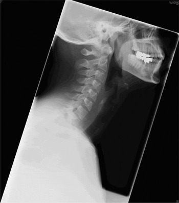
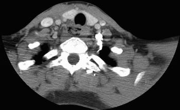
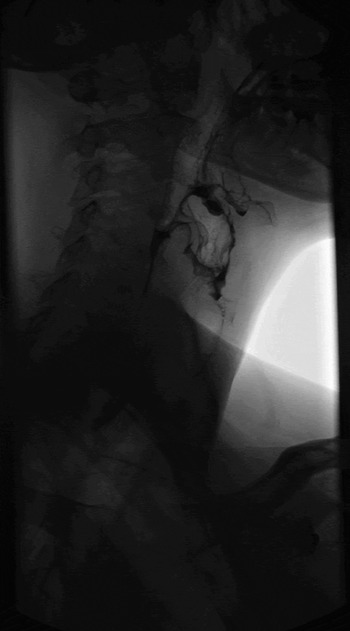
The patient underwent fibre-optic nasendoscopy, which demonstrated a haematoma and oedema in the piriform fossa. Lateral neck radiography revealed air in the retropharyngeal soft tissue at the level of the seventh cervical vertebra (Figure 1). A subsequent computed tomography scan confirmed a 2 cm, loculated, gas-containing collection at the level of the vallecula in the right posterolateral wall, extending to the false vocal folds and communicating between the right parapharyngeal space and the right carotid sheath. There was also evidence of extra-oesophageal gas at the level of the first thoracic vertebra (Figure 2). A contained oesophageal perforation was confirmed by water-soluble contrast swallow (Figure 3).
Lateral neck radiography showing air in the retropharyngeal soft tissue at the level of the seventh cervical vertebra.

Axial, contrast computed tomography scan showing evidence of extra-oesophageal gas at the level of the first thoracic vertebra.

X-ray study using water-soluble contrast swallow, confirming a contained oesophageal leak.

Conservative treatment was adopted, involving nil orally, intravenous antibiotics and nasogastric feeding. A second water-soluble swallow X-ray seven days later showed no evidence of ongoing leakage, and the patient was therefore commenced on oral fluids, eventually converted to a normal diet.
The patient made an uneventful recovery and was discharged 11 days after admission.
Discussion
Based on several articles on the topic, the overall location of oesophageal perforation has been reported as cervical in 27 per cent of cases, intrathoracic in 54 per cent and intra-abdominal in 19 per cent.Reference Khan, Strauss and Mason1 The majority of cervical perforations are iatrogenic secondary to instrumentation.Reference Beggs and Morgan2 Iatrogenic perforation of the cervical oesophagus during endoscopy is a rare but well recognised complication, occurring in elderly patients who have a history of a previous difficult intubation or large cervical osteophytes, and usually involving an inexperienced endoscopist.Reference Das, Sivak and Chak3 Difficult endotracheal intubation due to anatomical problems may also result in trauma to and perforation of the oesophagus, usually posteriorly.Reference Ku, Tong, Ho, Kwan and Hasselt4 Cervical oesophageal perforation has been well documented as an uncommon complication following cervical spinal surgery, secondary to screw migration.Reference Sahjpaul5 Cervical oesophageal perforation has also been described secondary to injury by swallowed sharp foreign bodies or during foreign body removal.Reference Kesler and Buzna6
Some reports have described spontaneous oesophageal perforation precipitated by forcible vomiting, but usually against a partial oesophageal obstruction such as an oesophageal web or in patients with co-morbidities involving ulceration of the gastrointestinal tract.Reference Beggs and Morgan2, Reference Mussack, Landauer, Ladurner, Schieman, Goetzberger and Burchardi7 Cervical oesophageal perforations occurring in young, healthy patients are often secondary to blunt or penetrating trauma to the neck and usually also involve the trachea.Reference Weiman, Walker, Brosnan, Pate and Fabian8
Dr Hermann Boerhaave described the first published case of spontaneous oesophageal rupture in 1724, when he reported the postemetic rupture and subsequent rapid death of Dutch grand admiral Baron Jan van Wessenaer.Reference Scheinin and Wells9 In Boerhaave's syndrome, barotraumatic oesophageal injury is caused by vomiting occurring in an uncoordinated fashion against a closed cricopharyngeus, despite pyloric closure and diaphragmatic contraction. In 90 per cent of cases, this results in full thickness rupture of the left lower oesophagus.Reference Khan, Strauss and Mason1 The classical features of vomiting, chest pain and subcutaneous emphysema, known as Mackler's triad, are often absent and thus the diagnosis is often delayed. Mediastinitis ensues and may lead to sepsis and multi-organ failure. For this reason, the ‘gold standard’ treatment of oesophageal perforation is primary repair within 24 hours. However, management remains controversial, and many advocate a conservative approach with sepsis control by collection drainage.Reference Vogel, Rout, Martin and Abbitt10 Despite advances in surgery and critical care, Boerhaave's syndrome continues to carry high morbidity with mortality rates, of 10–75 per cent.Reference Khan, Strauss and Mason1
Postemetic rupture of an essentially normal oesophagus is extremely rare. We undertook a Medline search of the English language literature and were unable to locate any previously reported cases.
Perforation of the cervical oesophagus carries a lower mortality rate compared with perforation of the thoracic and abdominal oesophagus, due to prevention of spread of infection by the anatomical planes of the neck. For this reason, conservative treatment of cervical oesophageal perforations has been reported to have greater success.Reference Scheinin and Wells9
• Cervical oesophageal perforations account for 27 per cent of all oesophageal perforations
• The majority of cervical perforations are iatrogenic, secondary to instrumentation, or traumatic
• Postemetic rupture of an essentially normal oesophagus is extremely rare; no previously reported cases could be found
• Cervical oesophageal perforations carry a lower mortality rate compared with perforations of the thoracic and abdominal oesophagus, due to prevention of spread of infection by the anatomical planes of the neck
• Cervical perforations are treated conservatively with greater success, compared with oesophageal perforation at other sites
Conclusion
Postemetic rupture of the cervical oesophagus can occur in the absence of any underlying iatrogenic or anatomical cause. Based on our single case experience, this condition can be successfully managed conservatively.