The branched colonies of several hydroids are a suitable substrate for sessile (Hughes, Reference Hughes1975, 1978a,b) and vagile epibiontic organisms (Lagardère & Tardy, Reference Lagardère and Tardy1980; Bavestrello et al., Reference Bavestrello, Cerrano, Cattaneo Vietti, Sarà, Bouillon and Cornelius1996). Generally the hydroid does not represent for these organisms a mere low-competition substratum but several examples of relationships occurring between the host and its epibionts are here described. Lagardère & Tardy (Reference Lagardère and Tardy1980) recorded a complex behaviour of a syllid polychaete that fixes its tubes immediately under the polyps of Tubularia larynx. In this situation, the polychaete eats part of the food captured by the polyps, and is able to defend the host from nudibranch predation. Some species of caprellid amphipods living on Eudendrium are able to use the polyps as traps to collect planktonic prey. When a polyp has caught and engulfed prey, the caprellid opens the polyp mouth by its gnatopods and extracts and eats the prey (Bavestrello et al., Reference Bavestrello, Cerrano, Cattaneo Vietti, Sarà, Bouillon and Cornelius1996). It was observed that the polyps of Eudendrium never eat the harpacticoids living on their colonies (Bavestrello et al., Reference Bavestrello, Cerrano, Cattaneo Vietti, Sarà, Bouillon and Cornelius1996), while it was demonstrated that polyps of this genus are generalistic predators engulfing all the prey with a diameter compatible with their hypostome (Barangè, Reference Barangè1988).
Recently it was demonstrated that several microorganisms, such as protists and bacteria, are able to live on hydroid colonies. Stabili et al. (Reference Stabili, Gravili, Piraino, Boero and Alifano2006) have shown that chitinolitic, luminous bacteria of the genus Vibrio live on the perisarc of several species and Romagnoli et al. (Reference Romagnoli, Bavestrello, Cucchiari, De Stefano, Di Camillo, Pennesi, Puce and Totti2007) have recorded bacterial and cyanobacterial mats on the branches of Eudendrium.
Benthic diatoms are very common on the hydroid branches where they produce dense assemblages or exploit more specific microenvironments such as the inner side of hydrothecae or the narrow grooves between the annuli of branches (Di Camillo et al., Reference Di Camillo, Puce, Romagnoli, Tazioli, Totti and Bavestrello2005; Romagnoli et al., Reference Romagnoli, Bavestrello, Cucchiari, De Stefano, Di Camillo, Pennesi, Puce and Totti2007). Sessile ciliates such as Ephelota have commonly been recorded on several hydroid species (Di Camillo et al., Reference Di Camillo, Puce, Romagnoli, Tazioli, Totti and Bavestrello2005), while Vorticella is a typical epibiont of some species of Aglaophenia where it settles on the edge of the theca (unpublished).
The aim of this paper is to summarize recorded data about the protists associated with marine hydroids, pointing out some ecological aspects of these epibiontic communities.
Diversity and abundance
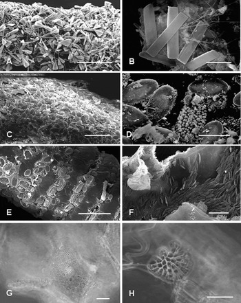
The microassemblages recorded on the hydroid colonies are often very rich. The main epibionts are diatoms (Figure 1) followed by protozoans, both sessile ciliate (Figure 2) and forams (Figure 3). On the branches of Eudendrium racemosum collected in the Ligurian Sea at shallow depths, almost 107 species of diatoms belonging to 52 genera were recorded during an annual cycle (Romagnoli et al., Reference Romagnoli, Bavestrello, Cucchiari, De Stefano, Di Camillo, Pennesi, Puce and Totti2007) (Figure 1A,B). The maximum diatom abundance exceeded 45,000 cells mm−2. This datum is particularly impressive if compared with others recorded on living substrata: 1000 cells mm−2 on gastropod shells (Gillan & Cadée, Reference Gillan and Cadée2000); 120 cells mm−2 on mussel shells (Bodeanu, Reference Bodeanu1988); and 1 cell mm−2 on the carapace of horseshoe crabs (Patil & Anil, Reference Patil and Anil2000). On the bryozoans Electra pilosa, Membranipora membranacea, Flustra foliacea and Alcyonidium gelatinosum from the German Bight, members of 26 diatom genera were found with densities of 71–547, 77–110 and 1–27 cells mm−2 respectively for E. pilosa, M. membranacea and A. gelatinosum while on F. foliacea, diatoms were present with very low abundances (<5 cells mm−2) (Wuchter et al., Reference Wuchter, Marquardt and Krumbein2003).
Algal assemblages associated with marine hydroids. (A–F) Scanning electron microscope (SEM) pictures of diatom assemblages on hydroids. (A) Licmophora spp., erect diatoms that colonize the apical part of Eudendrium racemosum; (B) Cyclophora tenuis forming erect colonies on the E. racemosum skeleton; (C) Cocconeis spp., adnate diatoms covering the basal part of the E. racemosum colony; (D) Cocconeis notata on the inner side of Campanularia hincksii hydrothecae; (E) Cocconeis pseudonotata on the pedicel of Clytia linearis; (F) Navicula sp. inside the theca of Synthecium evansi; (G–H) light microscopy pictures of initial cells of coralline algae epibiontic on marine hydroids; (G) Melobesia membranacea on Sertularella ellisii hydrotheca; (H) Hydrolithon farinosum on Aglaophenia tubiformis hydrotheca. Scale bars: A, 200 μm; B, D, 20 μm; C, E, F–H 50 μm.

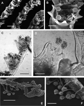
Sessile ciliates. (A) The SEM image of Vorticella sp. on thecae of Aglaophenia kirchenpaueri from Messina Strait; (B) close-up image showing the protozoans attached on the inner side of the marginal teeth; (C) thecae of Aglaophenia octodonta from Conero Riviera hosting numerous cells of Vorticella sp.; (D) living Vorticella on the thecal margin of Aglaophenia tubiformis from Apulian Coasts; (E–F) suctorians of the Ephelota genus on the pedicel of Eudendrium racemosum. Scale bars: A, C, F, 200 μm; B, 50 μm; D, 100 μm; E, 500 μm.

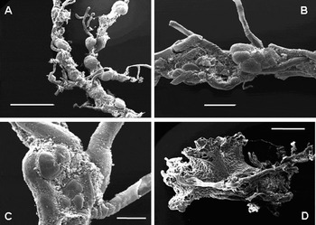
Foraminifera epibiontic on Eudendrium armatum. (A) Several tests of Rosalina globularis on the thin apical pedicels; (B) Lobatula lobatula on a slightly polysiphonic branch; (C) Acervulina inhaerens between two tubes of a polysiphonic branch; (D) Miniacina miniacea growing around the hydroid stem. Scale bars: A, 2 mm; B, 500 μm; C, 200 μm; D, 1 mm.

On rocks, the maximum diatom densities reported range from 2200 to 5600 cells mm−2 (Hudon & Bourget, Reference Hudon and Bourget1981; Brandini et al., Reference Brandini, da Silva, Pellizzari, Fonseca and Fernandes2001; Totti et al., Reference Totti, Cucchiari, De Stefano, Pennesi, Romagnoli and Bavestrello2007). The values observed on hydroids were higher and comparable with those recorded on highly productive soft intertidal sediments (Admiraal et al., Reference Admiraal, Peletier and Zomer1982; Delgado, Reference Delgado1989; Delgado et al., Reference Delgado, De Jonge and Peletier1991; Peletier, Reference Peletier1996).
Together with diatoms, the colonies of Eudendrium racemosum are also rich in assemblages of filamentous cyanobacteria (up to 30,000 cells mm−2) (Romagnoli et al., Reference Romagnoli, Bavestrello, Cucchiari, De Stefano, Di Camillo, Pennesi, Puce and Totti2007).
Also protozoans are often very abundant on the colonies of several hydroid species. On pinnate colonies of Aglaophenia elongata, 7 cm high, living at 25 m depth on the Portofino Promontory (Ligurian Sea), about 3000 cells of Vorticella sp. were recorded, with an average number of about 4 cells per theca (Figure 2A & D) (unpublished).
Few works have focused on the diversity and the abundance of foraminifera on hydroids: Dobson & Haynes (Reference Dobson and Haynes1973) described about 70 species settled on the hydroids dominating the benthic communities of the deep shelf of the Irish and the Malin Seas. Some species, such as Rupertina stabilis, Planulina ariminensis and Cibicioides wuellerstorfi were observed on the hydrocauli of some hydroids (Lutze & Altenbach, Reference Lutze and Altenbach1988; Lutze & Thiel, Reference Lutze and Thiel1989). Recently, we observed on average 25 cells of Rosalina globularis living on Eudendrium colonies (Figure 3A,B) from the northern Sardinia coast (Di Camillo et al., in press), while on specimens from the Portofino Promontory more than 3500 cells per colony were counted (unpublished).
Specificity
At the present state of knowledge, it is difficult to clarify the existence of species-specific relationships between protist epibionts and the hosting hydroids. The existence of host specificity was suggested for diatoms by Sullivan (Reference Sullivan and Mann1984), as certain epiphytic species were distributed only on particular macrophytes, but these have also been interpreted as a response to different physical conditions (Sullivan, Reference Sullivan and Ross1981). Diatoms of the genus Cocconeis and Navicula were frequently observed on thecate hydroids (Figure 1C–F), but these two genera are also widely distributed in many benthic habitats.
A more specific relationship involving hydroids and protists hitherto recorded is that of the filter-feeder ciliates of the genus Vorticella and the hydroids of the genus Aglaophenia and Plumularia. We observed this association in several Aglaophenia species in different localities of the Mediterranean: A. octodonta and A. elongata from Portofino Promontory (Ligurian Sea), A. elongata from the Lebanon coast (eastern Mediterranean), A. harpago and A. octodonta from the Conero Riviera (central Adriatic Sea), A. tubiformis from Otranto (southern Adriatic) and A. kirchenpaueri from the Messina Strait (unpublished). The specificity of this relationship is demonstrated by the spatial position of the cells that are attached through their contractile peduncle exactly on the edge of the hydroid theca (Figure 2C,D). Up to 12 cells may settle on one theca (Figure 2C): if disturbed, the protozoans quickly retract into the inner wall of the theca.
Other relationships are less specific but it is clear that some hydroid groups are particularly suitable as substratum for epibionts. Several species belonging to the family Sertulariidae, both in the Mediterranean and in tropical waters, are covered by a coat composed of coralline algae mixed with benthic diatoms. Encrusting Corallinales were frequently recorded on Sertularella ellisii and S. crassicaulis from several Mediterranean localities (Figure 1G). Although more than 100 hydroid species were examined from Portofino Promontory, Pneophyllum fragile, Hydrolithon cf. farinosum and Melobesia membranacea were found only on these two species and, in one case, also on Aglaophenia tubiformis (Figure 1H) where they form only small spots on the hydrothecae. On the exoskeleton of the two species of Sertularella coralline algae were observed forming a continuous coat completely covering the hydroid perisarc, except for the thecal opercula and gonothecae. During springtime, the coralline coat reaches its maximal development completely covering the colonies which appear pink in colour. The presence of hydranths inside the thecae indicated that the hydroid colonies were still living and probably the host was not damaged by its epibionts (Di Camillo et al., Reference Di Camillo, Puce, Romagnoli, Tazioli, Totti and Bavestrello2006).
Sometimes, epibionts show preferences among different host species belonging to the same genus. On the Sardinia coast the abundance of some foram species (Figure 4) is related to the colonized species of Eudendrium. Rosalina globularis (Figure 4A) was observed on all the Eudendrium species recorded in the locality but its average abundance decreases from 25 tests per colony in E. armatum to 17 in E. glomeratum to 12 in E. racemosum and 8 in E. molouyensis. Lobatula lobatula (Figure 4B), Acervulina inhaerens (Figure 4C) and Miniacina miniacea (Figure 4D) are mainly present on E. armatum, rarely on E. glomeratum, and never on the other species (Di Camillo et al., in press). These data confirm unpublished observations from the Portofino Promontory where the tests of Rosalina globularis present on E. armatum are two orders of magnitude greater than those recorded on E. racemosum. This specificity could be related to the interaction between foraminifera and their host but probably the ecological preferences of the host also affect the epibiont abundances.
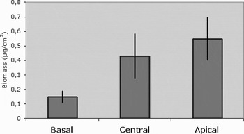
Diatom abundance (means ± SE) on different portions of the colonies of Eudendrium racemosum from the Ligurian Sea.

Spatial distribution
Hydroid colonies represent an amazing mosaic of different microhabitats exploited by a variety of protists. The highly specific position of Vorticella sp. on Aglaophenia theca was described above. On Eudendrium, the suctorian Ephelota was present only on the pedicels, immediately under the polyps (Figure 3E,F) (Di Camillo et al., Reference Di Camillo, Puce, Romagnoli, Tazioli, Totti and Bavestrello2005).
On the branched, polysiphonic colonies of E. armatum, four species of foraminifera—Rosalina globularis, Lobatula lobatula, Acervulina inhaerens and Miniacina miniacea— occupied different positions, due to the shape and the plasticity of their tests and the size of the hydroid branches. In particular, R. globularis settled only at the apex of the thin branches; L. lobatula was found only at the level of branch bifurcation, while A. inhaerens colonized the vertical grooves between two adjacent tubes of a polysiphonic branch. Finally, the large colonies of M. miniacea were observed only in the highly polysiphonic basal region (Di Camillo et al., in press).
Generally, diatoms occupy each part of the hydroid colony, like the base of the stem, the branches, the pedicels and the inner space between the polyp and the theca. At shallow depths, where enough light is available, the stem and branches of Eudendrium racemosum were covered by an extremely rich diatom assemblage (Romagnoli et al., Reference Romagnoli, Bavestrello, Cucchiari, De Stefano, Di Camillo, Pennesi, Puce and Totti2007). The assemblage is mainly composed of motile diatoms (Navicula spp.), followed by erect species such as Licmophora spp. (mainly L. oedipus and L. flabellata), Tabularia tabulata and Cyclophora tenuis, and adnate, such as Amphora spp. and Cocconeis spp. (C. stauroneiformis, C. scutellum and C. neotumensis) and tube-dwelling (Berkeleya rutilans and Parlibellus sp.). As a rule, the erect diatoms colonized the upper part of the hydroid, while the adnate ones colonized the lower part of the stem. Diatom abundance showed a clear relationship with position on the colony. The number of cells increases from the base of the colony towards the apex indicating a direct effect of the polyps on the abundance of the epibionts (Figure 4). The diatom covering stopped at the base of pedicels which are sometimes colonized by Ephelota sp. (Di Camillo et al., Reference Di Camillo, Puce, Romagnoli, Tazioli, Totti and Bavestrello2005). In any case, the abundance of diatoms increases from the base to the apex of the colony, suggesting a certain degree of interaction between algae and polyps (Romagnoli et al., Reference Romagnoli, Bavestrello, Cucchiari, De Stefano, Di Camillo, Pennesi, Puce and Totti2007).
On some hydroids like Campanularia hincksii, diatoms do not settle on the external side of the theca, while a monospecific diatom assemblage of Cocconeis notata living only on the inner side of the theca was recorded (Di Camillo et al., Reference Di Camillo, Puce, Romagnoli, Tazioli, Totti and Bavestrello2005). On the external side of the theca of Clytia linearis, the colonization of Cocconeis pseudonotata starts from the vertical grooves present in each tooth and then continues downwards completely covering the theca and pedicel in a continuous layer. At the same time, inside the theca, a population of the pinnate diatom Cylindrotheca sp. occurs. The green colour and the movement of the cells clearly indicate that they are living and that apparently, their presence did not damage the polyps, which always remained alive. A similar situation was observed on Synthecium evansi where C. neothumensis var. marina colonized the perisarc surrounding stem, branches and thecae, while Navicula sp. was localized exclusively inside the thecae (Di Camillo et al., Reference Di Camillo, Puce, Romagnoli, Tazioli, Totti and Bavestrello2005).
These examples indicate that a number of factors—shape of the host, life-form of epibionts, trophic relationships with the polyps, light exposition, competition between different epibionts—are involved in determining the spatial distribution on the colonies. The narrow space between the internal sides of thecae and polyps is a particularly interesting habitat colonized by diatoms. Probably, the thin perisarc is sufficiently transparent to allow light penetration but on the other hand several mixotrophic diatoms are known in habitats strongly favouring heterotrophic growth, where light is absent or limited and organic substrates are in great supply (Matsuda et al., Reference Matsuda, Ishikawa and Kawaguchi1987). Several diatom species have evolved highly sophisticated and ecologically significant mechanisms for facultative heterotrophy, developing active transport systems for a limited number of organic substrates (glucose, lactate and glutamate) in response to low light or darkness conditions (Myklestad et al., Reference Myklestad, Holm-Hansen, Vårum and Volcani1989).
Temporal distribution
Sometimes the protists epibiontic on hydroid colonies showed a clear seasonal trend. On Aglaophenia the Vorticella cells are present only in summer, with a maximum in May (unpublished). Also the foraminifera of the genus Rosalina on Eudendrium in the Ligurian Sea are present only in summer (unpublished). In these seasonal variations, the cycles of both the epibiont and the host are involved.
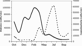
The best described case is the microalgal assemblage on Eudendrium racemosum. On colonies of this species, diatoms and cyanobacteria abundance showed opposite seasonal trends, with diatoms mainly abundant in winter and spring and cyanobacteria in summer (Figure 5). Within the diatom assemblage, the different life-forms—motile, adnate, erect and tube dwelling—showed a different pattern of annual cycle. Among them, adnate diatoms, which live attached to the substratum and have limited motility, showed an annual cycle which paralleled that of hydroid host, suggesting that they can benefit from the host association, probably through the nutrient supply from the host itself (Romagnoli et al., Reference Romagnoli, Bavestrello, Cucchiari, De Stefano, Di Camillo, Pennesi, Puce and Totti2007).
Annual trends of the abundance of diatoms (continuous line) and cyanobacteria (dotted line) on the colonies of Eudendrium racemosum from the Ligurian Sea.

General considerations
The data here summarized clearly indicate that the colonies of large hydroids are not only hot spots of biodiversity hosting numerous, sometimes poorly known species, but also have repertoires of complex life histories co-evolved between epibionts and their hosts, presumably in a short span of time.
In the establishment of these relationships, a pivotal role is played by the perisarc enveloping stem and branches and, in the thecate hydrozoans, also the polyps. In all the revised papers diatoms and protozoans settle on different parts of the perisarc. In athecate hydroids, like Eudendrium, both diatoms and the suctorian Ephelota stop immediately under the polyps. Some diatoms may settle on the hydroid buds that are covered by a thin sheet of perisarc, but when polyps born from the bud, the diatoms disappear (Romagnoli et al., Reference Romagnoli, Bavestrello, Cucchiari, De Stefano, Di Camillo, Pennesi, Puce and Totti2007).
The perisarc is composed of chitin, sometimes linked with different proteins (Jeuniaux, Reference Jeuniaux1963; Holl et al., Reference Holl, Schaefer, Goldberg, Kramer, Morgan and Hopkins1992). However, no comparative studies are available on the perisarc characteristics of different hydroid species. Different kinds of proteins presumably present in different hydroid groups could selectively influence epibiont colonization.
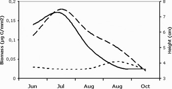
The perisarc acts not only as an inert substratum but in fact it was experimentally demonstrated that on living colonies of E. racemosum, diatom communities showed significantly higher abundance and biomass values than on artificial mimic plastic substrata (Figure 6). When colonies died, on the remaining stems the number of diatom cells become similar to those recorded over the same period on the artificial mimes (Romagnoli et al., Reference Romagnoli, Bavestrello, Cucchiari, De Stefano, Di Camillo, Pennesi, Puce and Totti2007). This evidence demonstrates that the host's metabolism and life style is crucial in affecting the amount and compositions of the epibiontic communities.
Temporal trend of diatom biomass on the colonies of Eudendrium racemosum (continuous line) and on plastic mimes (dotted line) compared with the trend of the height of the hydroid colonies (stacked line).

Several field studies on the effects of habitat structure on species assemblages have indicated that habitat heterogeneity (Pimm, Reference Pimm1984) does not enhance biodiversity as expected theoretically (McGuinness & Underwood, Reference McGuinness and Underwood1986; Beck, Reference Beck and Lunney1998, Reference Beck2000; Guichard & Bourget, Reference Guichard and Bourget1998). This review shows that hydroid colonies can be a driving force in determining levels of biodiversity. This role of hydroid colonies was also demonstrated by Bradshaw et al. (Reference Bradshaw, Collins and Brand2003) in the Irish Sea, where upright sessile epifauna structurally increased habitat heterogeneity leading to a general enrichment of biodiversity.