Case report
A 14-year-old girl with a history of tetralogy of Fallot repair and coil embolisation of major aortopulmonary collateral arteries presented to the clinic with pulmonary regurgitation. She was referred for a cardiac magnetic resonance imaging to assess her pulmonary regurgitation and right ventricular function.
Methods
Magnetic resonance imaging was performed with a cardiac optimised 1.5 T Siemens Symphony (Siemens Medical Solutions, Erlangen, Germany) scanner.
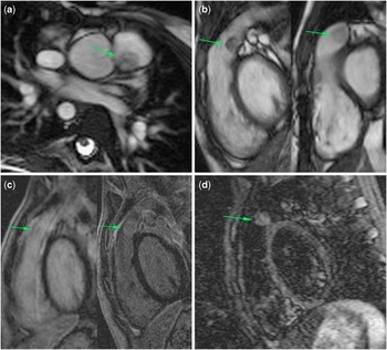
A mobile 13-mm intraluminal mass lesion was demonstrated attached to the floor of the proximal main pulmonary artery, just distal to the pulmonary valve. This mass returned intermediate soft tissue signal on half-fourier acquisition single-shot turbo spin-echo and steady-state free precession sequences (Fig 1a). Subsequent images were acquired post gadolinium, as the mass had not been initially appreciated by the attending physician. Standard fast-angle low-shot and repeat cine steady-state free precession sequences performed at 3–4 minutes showed diffuse enhancement of the lesion (Fig 1b). On late gadolinium enhancement inversion recovery images (11–15 minutes), the lesion had become isointense to blood pool to the point that it was almost imperceptible (Fig 1c). Further delayed late enhancement images were acquired at 26–40 minutes. Diffuse high signal was seen within the lesion on short tau inversion recovery and T1 images, remaining isointense to blood on the steady-state free precession cines and mildly hyperintense to blood pool on inversion recovery images (Fig 1d).
( a ) Axial pre-contrast magnetic resonance images. The pulmonary artery mass (arrow) was of intermediate soft tissue signal on cine SSFP images. ( b ) Pre- (left) and post-gadolinium (right) cine SSFP images of the RVOT showed diffuse enhancement, remaining hypointense to blood pool. ( c ) Delayed inversion recovery late enhancement images of the RVOT acquired at 15 minutes demonstrated isointensity of the mass to blood pool (left). At 26–40 minutes post gadolinium, the mass retained its contrast enhancement, becoming relatively hyperintense to blood pool (right). ( d ) STIR image of the mass at 38 and 40 minutes returned high signal relative to blood pool. RVOT = right ventricular outflow tract; SSFP = steady-state free precession; STIR = short-tau inversion recovery.

In addition, the scan revealed severe pulmonary regurgitation and moderate right ventricular dilatation with restrictive physiology: regurgitant volume 40 ml; regurgitant fraction 41%; right ventricular end-diastolic volume 113 ml/m2; right ventricular end-systolic volume 46 ml/m2; right ventricular stroke volume 92 ml; ejection fraction 59%.
On the basis of these findings, the patient was considered for surgery, before which a computed tomography scan was arranged to characterise the mass further and assess the anatomy of the coronary arteries.
Cardiac and chest computed tomography was performed with a 128-slice Siemens Somatom Definition AS (Siemens Medical Solutions, Erlangen, Germany) scanner. Intravenous β-blocker was administered and a modulated retrospectively gated computed tomography was performed with 4% maximum mAs outside diastolic reconstruction windows. The scan parameters were 80 kV, with a total mAs of 1315. The total dose-length product was 51 Gy cm.
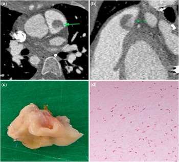
Computed tomography confirmed a 17 × 13 ×15-mm highly mobile, round soft tissue density lesion that was hinged to a small inferolateral attachment in the proximal main pulmonary artery. The base of the lesion enhanced, whereas the bulk showed minor enhancement. Of note, the pulmonary valve was not clearly demonstrated. There was no evidence of mediastinal invasion or secondary disease in the scanned field of view (Fig 2a). The coronary arteries were normally positioned.
Cardiac computed tomography was performed. Retrospective gating with 4% maximum mAs outside diastolic reconstruction windows. ( a ) Axial images demonstrating a 17-mm round, well-defined lesion in the proximal main pulmonary artery (MPA, arrow). ( b ) Sagittal multi-planar reconstructions indicating the basal enhancement seen within the lesion (arrows). ( c ) Photograph of the excised lesion within the MPA, showing a collapsed cyst-like structure. ( d ) Haematoxylin and eosin stain ×400 microscopy of the mass lesion showing myxoid degeneration within the valvular tissue.

These imaging findings were interpreted as a primary tumour of the main pulmonary artery, of which benign myxoma and sarcoma were considered possibilities. Owing to the concurrent severe pulmonary valve regurgitation and the need for tissue diagnosis, surgery was scheduled for a pulmonary valve replacement with wide excision of the main pulmonary artery to ensure complete removal of the mass and any local infiltration.
Pathology
At operation, the mass collapsed and “emptied” serosanguineous fluid on handling. Macroscopy revealed a 12 × 14 mm cystic “empty bag”-like lesion attached to the intima of the pulmonary artery (Fig 2c and d). No normal valvular tissue was seen, and it was initially unclear whether this “cyst” was attached to the valve. Microscopic sections were obtained of the proximal pulmonary trunk, with attached valvular tissue projecting into the lumen. The valvular tissue showed slight myxoid thickening and possible fusion of the leaflets together to form a cystic structure. Overall, the appearances were reported as suggestive of cystic dysplasia of the pulmonary valve.
Discussion
Cardiac valve cysts are a rare finding in adults but have been reported as relatively frequent in post-mortem studies of infants.Reference Zimmerman, Paplanus, Dong and Nagle 1 The majority of these are found on the atrioventricular valves and measure ∼1–2 mm in size.Reference Liese, Brainard and Goto 2 Pulmonary valve cysts often regress spontaneously and are extremely rare beyond infancy; our search of the literature found <20 reported cases.
The exact mechanism by which these lesions form is still unclear. Various aetiologies have been suggested such as haematomas, endocarditis, or vascular malformations. In addition, Minato et alReference Minato, Manabe, Masaki and Kawahara 3 postulated that increased pressure at the valve may cause invagination at the valve crevices and form blood cysts, which may be aggravated in patients with pulmonary stenosis. Dod et alReference Dod, Bhardwaj and Singh 4 reported a case of possible post-traumatic pulmonary valve cyst formation in a patient who had undergone valvuloplasty. Thereafter, these are thought to enlarge over time owing to shear flow or pressure gradients across the valve. The minor contrast enhancement shown on cross-sectional imaging in our case would suggest that there was some degree of blood flow into the cyst, with a relatively slow washout of contrast compared with blood pool.
Pulmonary valve cysts can present with features of functional pulmonary stenosis.Reference Minato, Manabe, Masaki and Kawahara 3 The differential diagnosis includes thromboembolic disease, endocarditis, fibroelastoma, and myxoma. The tricuspid valve has been reported as a location for hydatid cysts.Reference Kurdal, Kahraman, Iskesen and Sirin 5 Typically, the thrombus and the valvular vegetations of the endocarditis can be seen as mobile soft tissue density lesions, but do not show the contrast enhancement pattern demonstrated in our case and are usually subvalvar.
Fibroelastomas are the most common primary cardiac valve tumours,Reference Grebenc, Rosado de Christenson, Burke, Green and Galvin 6 with the majority occurring on the aortic or mitral valves. Myxomas have also been reported to originate from the pulmonary valves, but are relatively less frequent than fibroelastomas.Reference Tansel, El and Gok 7 On cross-sectional imaging, they appear as small, highly mobile pedunculated masses attached to the cardiac valves that enhance post contrast. These imaging features were mimicked by the pulmonary valve cyst in our patient.
Conclusion
This case highlights a rare pulmonary valve cyst that mimicked cardiac valve neoplasia on cross-sectional computed tomography and magnetic resonance imaging. Although these lesions are extremely rare, they should be considered in the differential diagnosis in patients presenting with valvular mass lesions who have conditions conducive to their formation.