Fontan and modified Fontan procedures, which include total cavopulmonary connection, are useful methods for palliation in selected patients with congenital heart disease of univentricular physiology.Reference Kopf, Laks, Stansel, Hellenbrand, Kleinman and Talner1 However, the development of pulmonary arteriovenous fistulas is considered a late complication in patients with cavopulmonary anastomosis and may result in a significant morbidity, such as increased occurrence of cyanosis.Reference McFaul, Tajik, Mair, Danielson and Seward2
Although right heart catheterisation and pulmonary angiography are traditionally used for the diagnosis of pulmonary arteriovenous fistulas,Reference Dines, Seward and Bernatz3 the use of bubble contrast echocardiography with selective injection into both the branch pulmonary arteries has been increasing as it may facilitate earlier diagnosis.Reference Chang, Alejos and Atkinson4 However, the actual efficacy of this diagnostic modality has not been well evaluated, and information about the correlation between contrast echocardiography findings and other clinical parameters obtained via cardiac catheterisation is limited. In this study, we aimed to compare contrast echocardiography findings and other cardiac parameters for the evaluation of pulmonary arteriovenous fistulas and to identify the clinical efficacy of such method in the management of patients with Fontan circulation.
Materials and methods
Patients
The medical records of the patients were retrospectively analysed. Catheter examinations were performed between April 2010 and March 2018. All patients underwent extracardiac total cavopulmonary connection. Moreover, cardiac catheter examinations were conducted for the evaluation of hemodynamic parameters after surgery. Informed consent was obtained from all the patients or their parents.
Cardiac catheter examination
All catheter examinations were performed while the patients were under spontaneous breathing with sedation using intravenous midazolam and pentazocine. At each time point, we measured the pressures and saturations of the inferior vena cava, inferior right pulmonary artery, inferior right pulmonary capillary wedge, inferior left pulmonary artery, inferior left pulmonary capillary wedge, superior vena cava, main ventricle, and ascending aorta.
Data about pulmonary capillary wedge desaturation were graded as mild if saturations were 90–94%, moderate if 85–89%, and severe if <85%.Reference Feinstein, Moore, Rosenthal, Puchalski and Brook5
Bubble contrast echocardiography
A contrast agent was created by agitating solutions manually to form air-filled microbubbles. Two 10-ml syringes were joined by a three-way stopcock. One syringe contained 10 ml of 0.9% saline solution, and the other syringe contained 1 ml of air. The saline solution was agitated by rapidly flushing the solution back and forth between the two syringes via the three-way stopcock ≥10 times. Thereby, a mixture of air and liquid was created. An end-hole balloon catheter was placed, with the tip of the catheter in the left or right pulmonary artery without wedging in the small branches. Then, the agitated saline was rapidly injected.Reference Chang, Alejos and Atkinson4 Patients weighing <20 kg were injected with 5 ml of the agitated saline, and those weighing ≥20 kg were injected with 10 ml of the agitated saline.
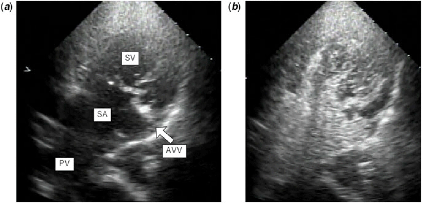
Simultaneous recording of the apical view echocardiogram of the injection was performed to visualise the presence of bubble contrast in the pulmonary venous atrium and systemic ventricle. In a normal individual, after the injection of saline bubble contrast in the pulmonary arteries, the echo contrast quality of the bubbles is lost in the passage via the pulmonary capillary bed; therefore, no bubbles should be observed in the left heart. The presence of bubble contrast in the pulmonary venous atrium and systemic ventricle in three cardiac cycles indicates the lack of passage via the pulmonary capillary bed; therefore, such result indicates the presence of intrapulmonary arteriovenous shunting.Reference Hind and Wong6 Contrast echocardiography opacification of the atrium was graded as follows: 0, no bubbles; 1, occasional filling of the atrium; 2, moderate filling; and 3, complete opacification.Reference Feinstein, Moore, Rosenthal, Puchalski and Brook5 Figure 1 shows the example of bubble contrast echocardiography opacification.
The example of bubble contrast echocardiography opacification before injection of microbubbles. There were no microbubbles in the cardiac chambers. After injection of microbubbles, the microbubbles immediately came into the cardiac chambers. We diagnosed this case as grade 3. AVV = atrioventricular valve; PV = pulmonary vein; SA = single atrium; SV = single ventricle.

Statistics
All values were presented as mean ± standard deviation. Spearman’s correlation coefficient by rank test was used to estimate the correlation between contrast echocardiography grade and other cardiac parameters. p < 0.05 was considered significant.
Results
Patient characteristics
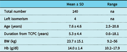
Catheter examination is commonly performed in patients with total cavopulmonary connection 1 year after surgery. Then, such examination is performed every 5–6 years. This study investigated a total of 140 patients. The characteristics of the study population are shown in Table 1. Patients with fenestration were not included. The relative incidence of pulmonary arteriovenous fistula in left isomerism syndrome is reported.Reference Ohuchi, Yasuda and Miyazaki7 However, only four cases were recorded.
Subject characteristics

BW = body weight; Hb = haemoglobin; SD = standard deviation; TCPC = total cavopulmonary connection
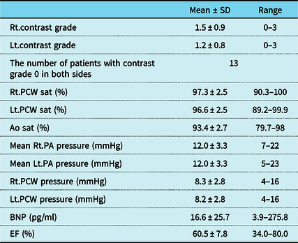
The clinical parameters obtained via cardiac catheter examination are shown in Table 2. Both pulmonary capillary wedge saturations were almost within normal range. The aorta saturation was slightly lower than the pulmonary capillary wedge saturation, but it was within normal range similar to the univentricular circulation. In the univentricular circulation after total cavopulmonary connection, the aorta saturation is not fully saturated because the coronary perfusion cannot circulate the lungs. Only 13 patients had grade 0 both in the right and left contrast echocardiography.
Clinical parameters

Ao = aorta; BNP = brain natriuretic peptide; EF = ejection fraction, Lt = left; PA = pulmonary artery; PCW = pulmonary capillary wedge; Rt; right; Sat = saturation; SD = standard deviation.
Correlation between contrast echocardiography grade and clinical parameters
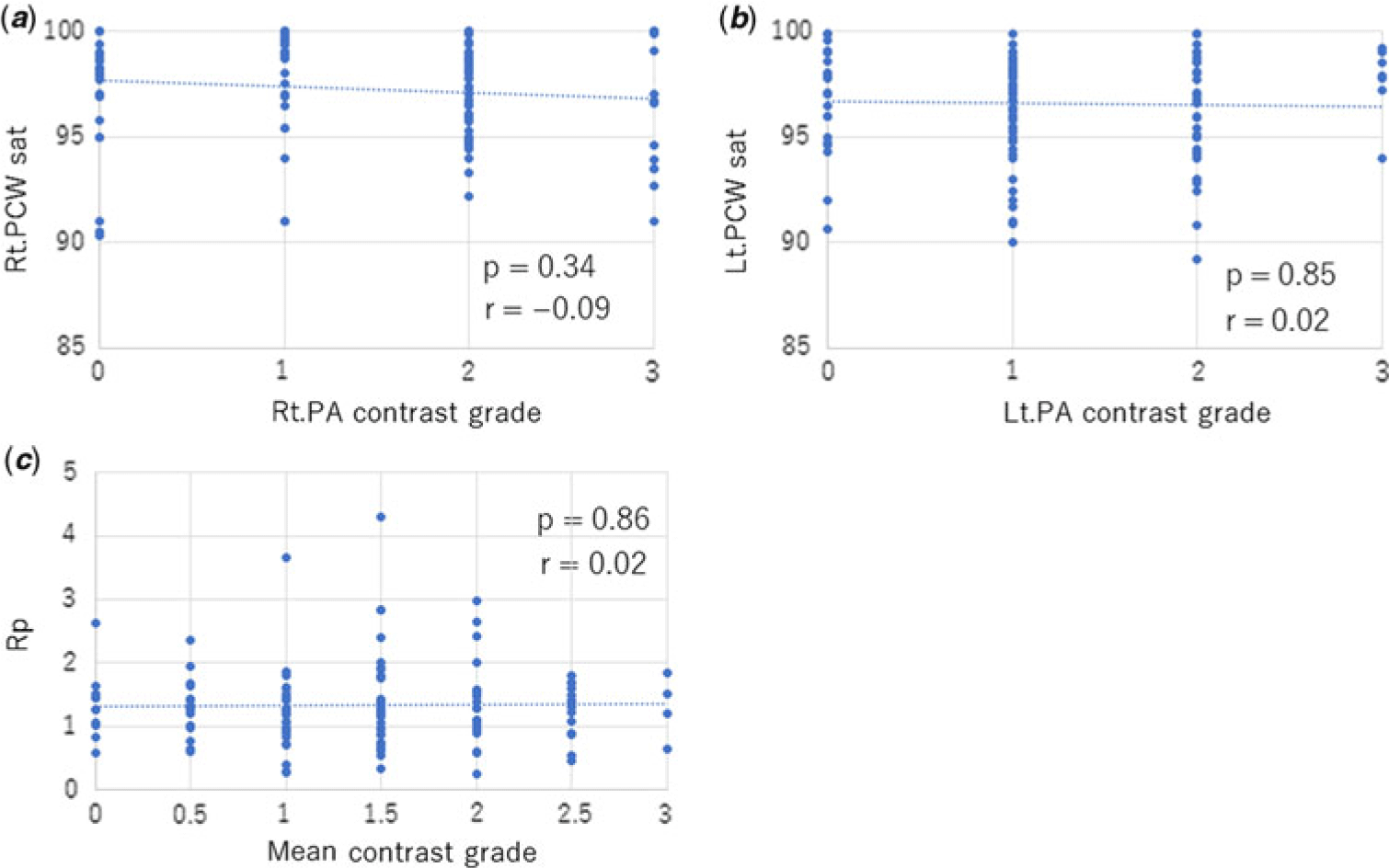
Figure 2 shows the correlation between contrast grade and clinical parameters. Results showed that the clinical parameters were not correlated to the severity of contrast echocardiography grade.
Correlation between bubble contrast grade and cardiac parameters. (a) Rt.PA contrast grade had no correlation with Rt.PCW sat. (b) Lt.PA contrast grade had no correlation with Lt.PCW sat. (c) Rt.PA and Lt.PA mean contrast grade had no correlation with Rp. Lt = left; PA = pulmonary artery; PCW = pulmonary capillary wedge; Rp = pulmonary artery resistance; Rt = right.

Discussion
We investigated the correlation between contrast echocardiography grade and clinical parameters obtained via cardiac catheter examination in 140 patients with total cavopulmonary connection. Results showed no significant correlation between them.
The development of pulmonary arteriovenous fistulas in patients with cavopulmonary anastomosis is a late complication.Reference McFaul, Tajik, Mair, Danielson and Seward2 However, the exact prevalence of this complication is not elucidated.Reference Kopf, Laks, Stansel, Hellenbrand, Kleinman and Talner1 The abnormal distribution of pulmonary blood flow after Glenn shunt or Fontan procedure is considered a contributing factor.Reference Cloutier, Ash and Smallhorn8 Srivastava et al have also reported that the development of this complication is correlated to the exclusion of flow from the liver to the pulmonary circulation.Reference Srivastava, Preminger and Lock9 These findings, along with the assessment results of pulmonary arteriovenous malformations in patients with chronic cirrhosis, have led to the hypothesis that the lack of some hepatic factor(s) contributes to the development of this complication.Reference Barbé, Losay and Grimon10In this study, patients with fenestration were not included. In the univentricular circulation with fenestration, some portion of the blood from the hepatic vein flows directly into the cardiac chamber through fenestration detouring pulmonary circulation. This particular circulation might promote the developing pulmonary arteriovenous fistulas. This supposition made us exclude the patients with fenestration from this study.
Fontan operation can facilitate blood flow from the liver into the pulmonary circulation; however, some patients developed pulmonary arteriovenous malformations after surgery.Reference Moore, Kirby, Madden and Gaither11 Although right heart catheterisation and pulmonary angiography are traditionally performed to diagnose pulmonary arteriovenous fistulas,Reference Dines, Seward and Bernatz3 recently, contrast echocardiography has been used to detect this complication.Reference Chang, Alejos and Atkinson4 The use of hand-agitated saline solution as contrast has been proven safe in echocardiographic studies.Reference Bommer, Shah, Allen, Meltzer and Kisslo12 However, the actual efficacy of this diagnostic modality for the diagnosis of pulmonary arteriovenous fistulas has not been properly evaluated.
Chang et al conducted a study on 14 patients with different forms of cavopulmonary anastomosis and compared the efficacy of bubble contrast echocardiography and pulmonary angiography in detecting fistulas.Reference Chang, Alejos and Atkinson4 The results showed a prevalence rate of 71% via contrast echocardiography. By contrast, the prevalence of pulmonary arteriovenous fistulas was only 21% based on angiographic findings.Reference Chang, Alejos and Atkinson4 The authors have speculated that the difference in the prevalence might be due to the superior sensitivity of contrast echocardiography to traditional angiograms. Moreover, Feinstein et al have shown that contrast echocardiography is extremely sensitive and often obtains positive result even though patients have normal pulmonary vein saturation.Reference Feinstein, Moore, Rosenthal, Puchalski and Brook5 In our study, 127 of the 140 patients showed a positive contrast echocardiography grade. The rate was extremely high and consistent with that of previous reports. In bubble contrast echocardiography, bubbles will only escape entrapment via the pulmonary capillary system if the lungs are overloaded with >20 ml of air.Reference Roelandt13 In this study, we made microbubbles by mixing 10 ml of 0.9% saline solution with 1 ml of air and by injecting 5 or 10 ml of agitated saline based on the patients’ body weight. We believe that no more 20 ml of air was injected. Furthermore, pulmonary arteriovenous fistulas cause hypoxemia due to direct connections between the pulmonary arteries and veins.Reference Dokumcu, Ozcan, Alper and Erdener14 However, our results clarified that >90% of patients showed a positive contrast grade even though they had normal pulmonary capillary wedge saturation and no significant correlation between contrast echocardiography grade and pulmonary capillary wedge saturation. We speculated that bubble contrast echocardiography was an extremely sensitivity method and was likely to obtain false-positive results. Our results also showed that pulmonary artery resistance had no significant correlation with contrast grade. Ohuchi et al indicated that patients with pulmonary arteriovenous fistulas had a lower pulmonary resistance in the Fontan circulation.Reference Ohuchi, Yasuda and Miyazaki15 This also indicated that bubble contrast echocardiography might obtain false-positive results.
Limitations
The present study has several limitations. The main limitation is that the evaluation of contrast echocardiography results was qualitative, not quantitative. In this study, two doctors evaluated the contrast grade and agreed on their evaluation findings. However, this method might not be accurate. Second, the existence of pulmonary arteriovenous fistulas was not evaluated via angiography. Therefore, whether the patients with positive contrast echocardiography grade had pulmonary arteriovenous fistulas was not confirmed. Third, we did not set a control group. There is no evidence if bubble contrast will be completely negative in normal heart. Fourth, we use the capillary wedge saturation as the pulmonary vein saturation. Strictly speaking, we should use the mixed pulmonary venous saturation, combination of each draining different areas of the lungs, to evaluate the diagnosis of pulmonary arteriovenous fistulas. To get this saturation, we need to advance the catheter into left atrium. However, after total cavopulmonary connection with extracardiac conduit without fenestration, it is difficult to advance the catheter into the atrium. We also consider the influence of other factors such as systemic vein to pulmonary vein collaterals or hypoventilation due to sedation when evaluating the capillary wedge saturation, but we did not take these influences into consideration. However, our results showed that both right and left capillary wedge saturation had the normal values. We believe the pulmonary capillary wedge saturation is the good surrogate for pulmonary vein saturation. Finally, this study was conducted after only an average of 5.3 years following total cavopulmonary connection. Kopf et al have reported that the incidence of pulmonary arteriovenous malformations increased with time after cavopulmonary anastomoses.Reference Kopf, Laks, Stansel, Hellenbrand, Kleinman and Talner1 Thus, we need to follow up for a long term to check the frequency of this complication.
Conclusion
We compared contrast echocardiography grade with clinical cardiac parameters, such as pulmonary capillary wedge saturations, to further delineate the role of contrast echocardiography in evaluating pulmonary arteriovenous fistulas after total cavopulmonary connection in univentricular circulation. A significant correlation was not observed between them. Contrast echocardiography has extremely high sensitivity and often obtains false-positive results even though the patients had normal pulmonary capillary wedge saturation. In relation to the numerous factors that may influence the opacity of a contrast echocardiography study, which include injection rate, amount of dissolved contrast, and competitive flow, this study indicates that we have to consider how we make use of the results of contrast echocardiography in detecting pulmonary arteriovenous fistulas in patients with total cavopulmonary connection. Further standardisation of techniques is required.
Acknowledgements
None.
Financial Support
This research received no specific grant from any funding agency, commercial, or not-for-profit sectors.
Conflicts of Interest
None.
Ethical Standards
The authors assert that this work complies with the ethical standards of the relevant national guidelines and with the Helsinki Declaration of 1975, as revised in 2008, and has been approved by the Ethical Review Committee at Kyoto Prefectural University of Medicine, Kyoto, Japan.