




No CrossRef data available.
Tick lectins: structural and functional properties
Published online by Cambridge University Press: 19 April 2005
Abstract
Few papers have been published on tick lectins so far, and therefore more data are needed to complete the mosaic of knowledge of their structural and functional properties. Tissue-specific lectin/haemagglutinin activities of both soft and hard ticks have been investigated. Some tick lectins are proteins with binding affinity for sialic acid, various derivatives of hexosamines and different glycoconjugates. Most tick lectin/haemagglutinin activities are blood meal enhanced, and could serve as molecular factors of self/non-self recognition in defence reactions against bacteria or fungi, as well as in pathogen/parasite transmission. Dorin M, the plasma lectin of Ornithodoros moubata, is the first tick lectin purified so far from tick haemolymph, and the first that has been fully characterized. Partial characterization of other tick lectins/haemagglutinins has been performed mainly with respect to their carbohydrate binding specificities and immunochemical features.
Information
- Type
- Research Article
- Information
- Copyright
- © 2004 Cambridge University Press
INTRODUCTION
The main goal of this review is to provide an overview of our knowledge of tick lectins as tissue-specific carbohydrate-binding proteins of ticks with emphasis on their structural properties and functional role either in defence reactions or pathogen transmission. A few other lectin reviews have been published in the last decade including those dealing with tissue-specific lectins in blood-sucking arthropods (e.g. Ingram & Molyneux, 1991; Grubhoffer, Hypša & Volf, 1997; Grubhoffer & Jindrák, 1998). On the other hand, there are several publications in the last ten years which have drawn attention to plant and animal lectins as well as to lectins as tools in modern glycobiological research (e.g. Jacobson & Doyle, 1996; Rhodes & Milton, 1998). Research on lectins began near the end of 19th century in 1888, with the doctoral thesis of Herman Stilmark at the University of Dorpat, in Estonia on the agglutinins of the seeds of castor bean Ricinus communis (Sharon & Lis, 1988). Further investigations into proteins able to agglutinate red blood cells (RBCs) have been improved significantly by modern biochemical and molecular biological techniques during the past several decades (Sharon & Lis, 1989; Slifkin & Doyle, 1990; Doyle & Slifkin, 1994; Jacobson & Doyle, 1996).
The term lectin comes from latin verb ‘legere’ and reflects an ability of particular proteins (haemagglutinins) to aggregate different kinds of RBCs (Boyd & Shapleigh, 1954). Several authors improved the original definition of lectins as haemagglutinins to make it more exact in terms of modern knowledge of the structural and functional properties of lectin molecules and their sugar binding sites (Goldstein et al. 1980; Kocourek & Hořejší, 1981; Barondes, 1988; Drickamer, 1988; Yoshizaki, 1990; Lee, 1992). According to Peumans & van Damme (1995), lectins are proteins or glycoproteins carrying at least one binding site, possessing a reversible specific interaction with a particular carbohydrate moiety. Lectins are ubiquitous proteins which are likely present in all eukaryotic and many bacterial species as well as in some viruses. This diverse group of proteins is of great importance for living organisms and some of them are often considered to be functional analogues of immunoglobulins in organisms that lack specific immunity (Vasta & Marchalonis, 1984). Lectins as proteins with binding affinity for carbohydrate molecules take part mainly in protein–saccharide interactions. These interactions participate in both effector and regulatory processes in organisms, including those of disease-transmitting blood-sucking arthropods.
The list of identified lectins has grown rapidly in the last two decades with more than ten thousand publications dealing with these molecules. Based on their overall structure, three major types of lectins may be distinguished: mero-, holo-, and chimerolectins (Peumans & van Damme, 1995). Merolectins are proteins that have only carbohydrate-binding domain; hololectins are those comprised of two or more either identical or highly homologous domains; and finally, chimerolectins are those that possess a sugar-binding domain associated with an unrelated one having different biological activity or another carbohydrate-binding specificity. Molecules with such combined functional activities have often been called lectinoids (Gilboa-Garber & Garber, 1989). Combining different binding activities in such molecules offers an effective way to function as regulatory tools for mechanisms of recognition and molecular identification by the receptor/co-receptor systems. On the other hand, proteins having an enzyme activity for the carbohydrate substrates, regulatory proteins (e.g. repressor in the lac operon), or antibodies with idiotypic specificity to carbohydrates/glycoconjugates are not considered as lectins (Kocourek & Hořejší, 1981).
Drickamer (1988) recognized lectins as both soluble and/or membrane-bound post-translationally modified proteins providing often multimolecular complexes built from a number of identical subunits. The author has distinguished two major types of lectins based on their structure: S-type (recently called galectins) with thiol group-dependent activity localized in both intra- and extracellular compartments, and C-type (some of them are called selectins) with Ca2+-dependent activity in the extracellular space. The C-type lectins are more variable in structure and more specific to organs and tissues. However, their sequence data show a common and homologous region responsible for binding of carbohydrates, the carbohydrate region domain (CRD) (Lee, 1992). Collectins represent an important subgroup of C-lectins. They have a collagen domain, a neck region and a globular C-type lectin-binding domain (Epstein et al. 1996; Ezekowitz, Sastry & Reid, 1996).
From a functional point of view, it is important to emphasize that lectins are involved in the innate immunity of arthropods and vertebrates since they provide the primitive/ancient functions, such as aggregation of microorganisms and opsonizing them for phagocytosis (Ratcliffe & Rowley, 1987; Vasta, 1991; Vasta et al. 1994). During evolution, lectins gained more sophisticated functions in defence against pathogens, which is evident from their linkeage to the complement system (Matsushita, 1996; Vasta et al. 1999; Matsushita et al. 2001; Iwanaga, 2002). It has been reported that the lectin activation pathway of the complement system can be triggered not only by the mannose-binding lectin (MBL), a member of the collectins, but also by a group of other lectins called ficolins (Gadjeva, Thiel & Jesenius, 2001; Matsushita et al. 2001). In the lectin activation pathway, MBL with a collagen-like and carbohydrate recognition domains (CRD) (Taylor et al. 1990), activates the complement cascade in cooperation with a MBL-associated serine protease (Hoffmann et al. 1999). Ficolins occur in mammalian sera. Besides a collagen-like domain common in MBL, ficolins contain a fibrinogen-like domain (Lu & Le, 1998). Recent characterization of ficolins present in human, mouse and pig serum/plasma, as well as in the body fluids of ascidians, has revealed that they are lectins with common binding specificity for GlcNAc. Human serum ficolins have been shown to function as opsonins (Matsushita et al. 1996). Most likely, ficolins function as self/nonself recognition lectin molecules in haemolymph (Gokudan et al. 1999). Members of the fibrinogen-related protein family have been identified both from vertebrates and invertebrates (Xu & Doolittle, 1990; Adema et al. 1997; Kurachi et al. 1998; Dimopoulos et al. 2000; Kenjo et al. 2001; Leonard et al. 2001). To complete the list of known lectins, the I-type (belonging to the immunoglobulin superfamily), pentraxins (lectin-like molecules, such as the C-reactive protein) and heparin-binding proteins have to be mentioned (Vasta, Ahmed & Quesenberry, 1996). Lectins that recognize more complex structures at the cell surface, such as C-type lectins and galectins, are found in invertebrate organisms as well as in vertebrates, but the functions of these proteins have evolved separately in different animal lineages (Dodd & Drickamer, 2001).
Moreover, other proteins with carbohydrate-binding domains, which do not fall into the lectin family, can serve as scavenger receptors (Pearson, 1996). They occur also on the surface of haemocytes carrying a binding specificity to anionic polysaccharides (e.g. fucoidan, dextran sulfate, chondroitin sulfate). Scavenger proteins as well as proteins with binding affinity for oligo/polynucleotides seem to play a significant role in the receptor functions over the whole evolution of innate immunity. Together with both lipopolysaccharide (LPS) and β-1,3 glucan-binding proteins, scavenger proteins constitute an important group of molecules engage in non-self recognition in invertebrates.
ARTHROPOD LECTINS
Lectins of general arthropods
Lectins may interact either with ‘self’ glycoconjugates or with glycosylated components of viral, bacterial, protozoan and metazoan pathogens. Soluble or membrane-bound invertebrate lectins take part in the processes of cell adhesion, opsonization, phagocytosis and cytolysis (Vasta & Marchalonis, 1983). They are clearly important molecules involved in humoral and cellular reactions for recognition and defence by opsonization of non-self objects in the inner environment of arthropods. Lectins may also play a part in the transmission of pathogens by vectors, for instance as receptors, homing factors, differentiation factors, etc. A great number of lectins of various invertebrates have been described (Yeaton 1982a,b; Ratcliffe et al. 1985). From the viewpoint of the structure and function, the best known arthropod lectins are those from the horseshoe crabs, Limulus polyphemus and Tachypleus tridentatus, and from the scorpions Centruroides sculpturatus and Parauroctonus (Vasta & Marchalonis, 1984; see Olafsen, 1986, 1996 for reviews). Almost all plasma lectins isolated from the above mentioned group of chelicerates specifically bind N-acetyl neuraminic (sialic) acid and other N-acylamino-carbohydrates. Studies on insect immunity show many molecular aspects common to invertebrate and vertebrate immune responses. For instance, there is a great similarity in the clotting mechanisms of haemolymph and blood. Remarkably, one of the four characterized haemolymph clotting factors of Limulus, factor C (serine protease zymogen), resembles in structure and probable function the selectin family of cell-adhesion molecules. It belongs to C-type (Ca2+ ion dependent) chimerolectins (for review see Iwanaga, 1993; Hoffmann, 1995).
Recently, lectins purified from haemolymph of the Japanese horseshoe crab, T. tridentatus, and called tachylectins 5A/5B, have been characterized and cloned (Gokudan et al. 1999). Both tachylectins (Tl-5A/5B) are proteins of high structural and sequentional similarity to mammalian ficolins. However, Tl-5A/5B have no effector collagen domains in their structure and no lectin-associated serine proteases, as found in mammalian ficolins. Tl-5s, together with other defence molecules released from haemocytes, is employed in effective innate immunity against invading pathogens (Kawabata & Tsuda, 2002).
Lectins of disease vectors
Despite the great influence of vector-borne pathogens on humans and domestic animals, relatively little information is available on lectins of invertebrate vectors, with the exception of reduviid bugs, tsetse flies, mosquitoes and sand flies (Pereira, Andrade & Ribeiro, 1981; Wallbanks, Ingram & Molyneux, 1986; Maudlin & Welburn, 1988; Gomes, Furtado & Coelho, 1991; Volf, 1993; Hypša & Grubhoffer, 1995; Ratcliffe et al. 1996; Chen & Billingsley, 1999).
The best studied vector/parasite system are lectins of various tsetse flies (Glossina spp.) (Maudlin & Welburn, 1988; Welburn & Maudlin, 1990; Ingram & Molyneux, 1991). Lectins have been demonstrated in the gut and haemolymph of tsetse flies that can function as signalling factors of maturation for African trypanosome species or, on the contrary, lytic (killing) factors. In this case, an interaction between the gut lectin of the tsetse fly and symbiotic rickettsia-like organisms, or with products of their saccharide metabolism, is a major factor affecting the life cycle of trypanosomes in tsetse flies (Maudlin & Welburn, 1988). A similar interference between symbiotic rickettsia with protozoa was thought to be involved in ticks without considering lectin activities (Smith et al. 1976). Sand flies, the vectors of Leishmania spp., are another group of vector arthropods that have been well studied with regards to midgut lectins and their receptor function in the process of Leishmania transmission (Volf et al. 1994; Palánová & Volf, 1997; Volf, škařupová & Man, 2002).
Lectin–carbohydrate interactions participate in the vector–pathogen/parasite relationship. Tissue-specific lectins of blood sucking arthropods interact with glycosylated molecules of receptors on the surfaces of pathogens or their cells within appropriate developmental stages and vice versa. For instance, bacteria or other intracellular parasites have often employed their surface lectins to enter into phagocytes by the mechanism of lectinophagocytosis (Ofek & Sharon, 1988).
Tick lectins in overview
The lectins of ticks have not received the same level of attention as those of insects. A role for tick haemolymph lectins in transmission of pathogens by ticks has been suggested (Munderloh & Kurtti, 1995). Lectin/haemagglutinin activites in the haemolymph of four tick species, Ixodes ricinus, Ornithodoros tartakovskyi, O. tholozani (papillipes) and Argas polonicus have been described (Vereš & Grubhoffer, 1990; Grubhoffer & Maťha, 1991; Grubhoffer, Vereš & Dusbábek, 1991). The partially characterized tick lectins have affinity for sialic acid and N-acetyl-D-glucosamine but differ in their binding specificity to other sugars. It is postulated that these lectins can interact with glycosylated structures of transmitted pathogens, but this has not yet been proved. I. ricinus haemolymph lectin was immunolocalized in tick tissues and its distribution supports the idea that the lectin is produced and/or stored in haemocytes and contributes to the immune system by recognition of foreign substances (Kuhn, Uhlíř & Grubhoffer, 1996). Like limulin, the sialic acid-specific lectin of the horseshoe crab, Limulus polyphemus, and other sialic acid-specific lectins of chelicerates, the haemolymph lectin of ixodid ticks might also recognize a wide range of Gram-negative bacteria due to its site specificities for N-acetyl-D-glucosamine, D-galactose and 2-keto-3-deoxyoctonate acid (Vasta & Marchalonis, 1983; Grubhoffer et al. 1991; Kuhn et al. 1996). The haemocytes of I. ricinus phagocytose spirochaetes of Borrelia burgdorferi by the coiling method, and coiling phagocytosis is thought to be a lectin-mediated process (Kuhn et al. 1994).
Kamwendo et al. (1993) reported haemagglutination activities in the haemolymph, gut homogenates and salivary glands in the tick Rhipicephalus appendiculatus, the vector of East Coast fever. This is a serious tick-borne disease of cattle caused by the protozoan parasite Theileria parva (see chapter by Bishop et al. in this Supplement). Bovine red blood cells were found as the most sensitive detection system for all tissue homogenates examined with no significant differences between those in extracts from unfed and fed ticks (Kamwendo et al. 1993). However, human erythrocytes of the ABO system and rabbit red blood cells showed significantly higher values of haemagglutination titres in tissue samples coming from fed than unfed ticks, with the highest increase recorded in the gut extract. Later, Kamwendo et al. (1995) focused particularly on a specific functional role of the tick salivary gland haemagglutinin in terms of the tick's capability to transmit T. parva parasites. R. appendiculatus gut lectin activity also influences transmission of the causative agent of theileriosis in Africa with agglutination of piroplasms (E. Sebitosi, personal communication). This has led to the conclusion that the protein–carbohydrate interactions of piroplasms in the inner environment of tick might be a target for the control of theileriosis. Many functional and morphological aspects of tick tissues are involved in the process of parasite transmission (Friedhoff, 1990). However, little is known about the molecular aspects of cell communication and tick interactions with the envelope components of transmitted pathogens. The specific part of the review which follows consists of two sections devoted to tissue specific lectins of the hard tick, Ixodes ricinus, and the soft tick, Ornithodoros moubata. Due to a lack of published data on tick lectins, most results presented are based on the research of the Tick Lectin Group in the Institute of Parasitology České Budějovice, Czech Republic (see Table 1).

LECTINS OF THE HARD TICK, IXODES RICINUS
Gut
The sheep tick, Ixodes ricinus, which transmits tick-borne encephalitis (TBE) virus and the Lyme disease-causing spirochaete Borrelia burgdorferi sensu lato is the most important tick vector in Europe. Gut haemagglutinating activity of I. ricinus has been found and partially characterized (Uhlíř, Grubhoffer & Volf, 1996). Native mouse erythrocytes were the most sensitive detection cell system in a wide pH range of 6·5–8·0. Binding specificity analysis showed that simple sugars N-acetyl-D-galactosamine, N-acetyl-D-glucosamine, rhamnose, dulcit and glycoconjugates fetuin, hyaluronic acid, laminarin (β-1,3 glucan) and bacterial LPS strongly inhibit haemagglutination. Binding characteristics of the tick gut agglutinin are consistent with LPS-binding agglutinins in the gut tissue of other blood-sucking arthropods (Grubhoffer et al. 1997). It seems that the midgut agglutinin as a potential LPS-binding protein (LPS-BP) could (in co-operation with digestive enzymes) affect Gram-negative spirochaetes B. burgorferi passing through the gut epithelium. Midgut extracts from unfed ticks lack a haemagglutinating activity, which is consistent with observations of Kamwendo et al. (1993) who worked on R. appendiculatus. Mouse polyclonal antibodies raised against midgut haemagglutinating activity (Yeaton, 1982a) recognized, by western blotting, four proteins with molecular weights of 37, 60, 65, and 73 kDa as putative structural components of the lectin(s) (Uhlíř et al. 1996). Whereas the 37 and 60 kDa proteins are glycoproteins modified by both high mannose and complex N-glycans, the 70 kDa subunit is modified by complex type of glycans only (Uhlíř et al. 1994). Using specific mouse polyclonal antibodies, the midgut haemagglutinin/lectin complex was immunohistochemically localized solely in the midgut cells, and not in the gut content or other tick tissues including haemolymph (Uhlíř et al. 1996).
Two agglutinins/lectins of the gut haemagglutinating complex in I. ricinus were isolated from the gut homogenate after delipidation by acetone extraction (Durnová, 1998). Both agglutinins/lectins were purified by affinity chromatography either on immobilized bovine submaxillary mucine (BSM) or laminarin (β-1,3 glucan). Using SDS–PAGE, a 65 kDa protein was found as the main agglutinin with binding affinity to BSM, and a 37 kDa protein showed strong binding specificity for laminarin (β-1,3 glucan). Although crude gut homogenate had no binding affinity for free sialic acid in the haemagglutination inhibition assay, the delipidated proteinaceous ‘acetone powder’ prepared from the crude gut homogenate showed a strong ability to bind sialic acid. It seems to be likely that lipids, including glycolipids abundant in the gut tissue, hide the lectin binding site for sialic acid. The pure 65 kDa agglutinin/lectin also maintained the affinity for free sialic acid. Mouse antibodies raised against the entire haemagglutinating activity of the crude gut homogenate recognized both proteins. Because, the 37 kDa gut agglutinin/lectin functions as a β-1,3 glucan-binding protein, it might be part of the defence machinery protecting the tick against fungal infections.
Factors taking place in processes of pathogen/parasite transmission by ticks were reviewed by Friedhoff (1990). According to Friedhoff (1990), the midgut agglutinin/LPS-binding protein may belong to factors associated with the development of pathogens/parasites in the digestive tract of the tick. The process of blood digestion in ticks differs from that of haematophagous insects, where protein digestion proceeds rapidly and takes place in the lumen of the gut (Gooding, 1972). Instead, digestion in ticks is intracellular (Balashov, 1972). It proceeds slowly, and the pH optimum of proteases is about 3·0, while the pH of the gut content is 6·5 (Akov, Samish & Galun, 1976). In hard ticks, the blood meal is taken up by the digestive cells by means of fluid-phase endocytosis and via receptor-mediated endocytosis. Large particles such as whole cells are phagocytized. The binding activity of the midgut agglutinin/LPS-BP is pH dependent. At pH 3·0, which is an optimum value for the gut proteases, the gut agglutinin does not work at all whereas at pH 6·5, which corresponds to the gut content, a haemagglutinating activity of this binding protein reaches its optimum (Uhlíř et al. 1996). Therefore, the midgut agglutinin/LPS-BP could be involved in the preparatory phase of blood meal digestion, in addition to its role in defence.
Haemolymph (plasma and haemocytes)
Most lectins isolated from arthropods are constituents of the haemolymph, the extracellular circulating fluid that fills the body cavity or haemocoel. The haemolymph, especially haemocytes and large protein complexes, is physically isolated from direct contact with body tissues by basal laminae. However, haemocytes, which are cells within the haemolymph, are in direct contact with this fluid (Mullins, 1985). Haemocytes are immunocytes of arthropods (Gupta, 1991); however, they contribute to nutritional functions, too (Gupta, 1985). Haemolymph lectins and other humoral immune factors in the haemolymph are produced and secreted into the haemolymph mainly by haemocytes and fat body cells. Recently, Gudderra et al. (2002) have published a comprehensive review on haemolymph proteins in ticks emphasizing the structural and functional features of vitellogenin and other tick proteins such as macroglobulins, heme-lipoproteins, antimicrobial proteins and lectins.
Haemagglutinating activity from the haemolymph of I. ricinus was characterized as Ca2+-dependent binding activity of the lectin with a subunit molecular size of 85 kDa by non-reducing SDS–PAGE, and binding affinity to sialic acid as well as N-acetyl-D-glucosamine and D-galactose (Grubhoffer et al. 1991, and unpublished observations). Sialic acid-binding lectins are common in chelicerates (Mandal & Mandal, 1990; Wagner, 1990; Zeng & Gabius, 1992), and recognize Gram-negative bacteria. Mouse polyclonal and monoclonal antibodies prepared toward a lectin haemagglutinating activity (Yeaton, 1982a) of the tick haemolymph identified a protein of 85 kDa as a structural lectin subunit. A semipurified plasma fraction highly enriched with a haemagglutinating activity was obtained by the gel filtration FPLC on Superose 12 (unpublished observations). The lectin was localized to the haemocytes in I. ricinus (Kuhn et al. 1996). Immunoreactivity with poly- and monoclonal antibodies was detected in the granules of both types of granular haemocytes, on the plasma membrane of haemocytes, and on the basal laminae surrounding the haemocoel (Figs. 1, 2). Furthermore, cells attached to the midgut invaginations of Géné's organ and granular inclusions of nephrocytes were labeled. The haemolymph- and haemocyte-associated immunoreactivity supports the idea that this haemolymph lectin may function as a recognition molecule in the immune system of I. ricinus. Therefore, the haemolymph lectin could be involved in transmission of Borrelia burgdorferi as a defence molecule in tick haemolymph/haemocytes. Johns et al. (2001a,b), studying differences in vector capability of various tick species for Borrelia transmission, have suggested a potential role of immune molecules. Several studies have shown that B. burgdorferi migrate from the midgut across the haemocoel to the salivary glands during tick feeding (Ribeiro et al. 1987; Zung et al. 1989). However, less than 5% of midgut population of spirochaetes survived to reach salivary glands (Coleman et al. 1997). Another function could be in the protection of eggs that are coated with secretions by Géné's organ (Kuhn et al. 1996).

Fig. 1. Indirect immunofluorescence detection of the plasma lectin in the haemocytes Ixodes ricinus tick using mouse monoclonal antibody 3/D3/C5 to the plasma lectin activity of I. ricinus (diluted 1[ratio ]100). GII – type II of granular haemocytes; PL – plasmatocyte; T – trachea; M – midgut. Laser confocal microscopy (courtesy Dr K.-H. Kuhn).

Fig. 2. Electron micrographs of immunogold-silver labeling of haemocytes with mouse polyclonal antibodies to the plasma lectin activity of Ixodes ricinus tick. Granular inclusions of type-II granular haemocytes (courtesy Dr K.-H. Kuhn).
The distribution of lectin immunoreactivity, especially the labeling of membranes and granular inclusions of haemocytes and of the basal laminae surrounding the haemocoel, strengthens the assumption that the haemolymph lectin is produced or stored in haemocytes and contributes to the immune system of I. ricinus. Like limulin, the sialic acid-specific lectin of Limulus polyphemus, and other sialic acid-specific lectins of chelicerates, the haemolymph lectin of I. ricinus might also recognize a wide range of Gram-negative bacteria due to its additional specificities for N-acetyl-D-glucosamine, D-galactose, and unrelated molecules such as 2-keto-3-deoxyoctonate (Vasta & Marchalonis, 1983; Grubhoffer et al. 1991). Moreover, it was shown in in vitro assays that B. burgdorferi is a target for the immune system of I. ricinus and is readily ingested by phagocytic haemocytes (Kuhn et al. 1994). Additionally, Kurtti et al. (1988) have reported that B. burgdorferi spirochaetes readily adhere to tick cells in tissue culture. Therefore, the lectin might mediate the interaction of pathogens like B. burgdorferi or tick-borne encephalitis (TBE) virus with the tissues of the vector. The main binding specificity of the haemolymph lectin is for sialic acid (Grubhoffer et al. 1991), which is present in the wall of B. burgdorferi (Hulínská, Volf & Grubhoffer, 1992). In addition, the haemolymph lectin of the tick can bind complex oligosaccharides lacking sialic acid. Therefore, the protein gpE of TBE virus, the only structural glycoprotein that exhibits biantennary complex oligosaccharides (Grubhoffer et al. 1990) could interact with the haemolymph lectin. The localization of lectin immunoreactivity in diverse tissues and cells of the tick does not imply that all labeled proteins are associated with the same function. Probably, polyclonal antibodies recognize common epitopes in functionally diverse proteins with multiple domains. Calcium-dependent globular lectin domains (C-type lectins) (Drickamer, 1993; Drickamer & Taylor, 1993) that are components of vertebrate haemostasis and complement systems (Thiel & Reid, 1989; Sastry & Ezekowitz, 1993; McEver, 1994) and a related protein, factor C (Muta et al. 1991), and an immune protein of the complement–coagulation system of L. polyphemus (Iwanaga, 1993), are examples of this phenomenon.
A cDNA library was constructed from unfed I. ricinus females and screened using a probe obtained from the lectin gene of B. burgdorferi B31 (unpublished observations). Clones showing the strongest hybridization signals were selected and sequenced. The amino acid sequence of a putative carbohydrate recognition domain of the recombinant clone from an I. ricinus cDNA library was compared with those of other lectins (Fig. 3). The predicted amino acid sequence of one clone, termed r58, revealed strong homology (81%) with four lectins from Periplaneta americana cockroach haemolymph (Kawasaki, Kubo & Natori, 1996).

Fig. 3. Comparison of carbohydrate recognition domain of I. ricinus lectin (r58) with those of different lectins. The lectins compared are: r58, clone from I. ricinus cDNA library (unpublished observations); PL21, 31, 9 & 25, four lectins from Periplaneta americana cockroach hemolymph (Kawasaki et al. 1996); HLEC, human hepatic lectin (Halberg et al. 1987); FCE2, mucose immunoglobulin Fc receptor (Bettler et al. 1989); MABC, rat mannose-binding protein A (Drickamer, Dordal & Reynolds, 1986); and MANR, human macrophage mannose receptor (Taylor et al. 1990). Dark grey represents identitical amino acid residues and light grey represents similar residues.
Salivary glands
Kamwendo et al. (1995) were the first to study the role of the tick salivary gland haemagglutinin of the tick R. appendiculatus in relation to the mechanism of transmission of the protozoan parasite Theileria parva. A significant increase was observed in T. parva acinar infection rates in the salivary glands of R. appendicalatus fed on ears of rabbits infused with melibiose and raffinose (haemagglutination inhibiting sugars). In contrast, mannose and turanose (non-inhibitory sugars) did not affect of T. parva acinar infection rates. Haemagglutinating activity in I. ricinus salivary gland (SG) extracts was highest against mouse red blood cells, and was inhibited by sialic acid, D-fructose, L-rhamnose, D-trehalose and several glycoconjugates (unpublished observations). Mouse polyclonal antibodies raised against the haemagglutinating activity were used in western blotting to identify a 70 kDa protein responsible for SG haemagglutinating activity. It is likely that the lectins have a significant role in the entry of pathogens into the salivary gland and, hence, the salivary gland lectin in I. ricinus will be important in the transmission of TBE and B. burgdorferi.
LECTINS OF THE SOFT TICK, ORNITHODOROS MOUBATA
Haemolymph (plasma and haemocytes)
The soft tick Ornithodoros moubata is a vector of African swine fever virus and Borrelia duttoni. Laboratory colonies of this species are valuable experimental models that have been employed to answer questions about the tick physiology and morphogenesis, such as vitellogenin synthesis (Chinzei, Chino & Takahashi, 1983; Chinzei & Yano, 1985; Chinzei, 1988; Gudderra et al. 2002, for review), defensins as immunopeptides (Nakajima et al. 2001), and peritrophic matrix synthesis (Grandjean, 1984). Recently, the immune response of the argasid tick O. moubata induced by infection with the filarial worm Acanthocheilonema viteae has been studied by Hutton, Reid & Towson (2000). A plasma/haemolymph lectin of O. moubata has been thoroughly studied (Grubhoffer & Kovář, 1998; Kovář, Kopáček & Grubhoffer, 2000), though we also have evidence of some lectin activity in the salivary glands and gut of O. moubata (unpublished observations).
A lectin with high haemagglutinating activity, called Dorin M, was identified in the plasma of O. moubata (Grubhoffer & Kovář, 1998; Kovář et al. 2000). The activity of the plasma lectin could be efficiently inhibited by sialic acid, N-acetyl-D-hexosamines and acid (sialo)glycoproteins. Dorin M was purified to homogeneity using two different isolation systems: affinity chromatography on a column of bovine submaxillary mucin conjugated to Sepharose 4B elution with N-acetyl-D-glucosamine (Grubhoffer & Kovář, 1998), or chromatography on Blue-Sepharose followed by anion exchange FPLC on a MonoQ column (Kovář et al. 2000). Dorin M is a glycoprotein with N-type of glycosylation that, in the native state, forms aggregates with molecular mass of about 640 kDa. Non-reducing SDS–PAGE revealed that the lectin consists of two non-covalently bound subunits migrating closely around 37 kDa. After chemical deglycosylation, only one band of about 32 kDa, without haemagglutinating activity, was detected. A tryptic cleavage of purified Dorin M was performed and the amino acid sequence of three resulting internal peptides fragments were determined by Edman degradation. Using degenerate primers and a haemocyte cDNA template, a 290 bp PCR product was cloned and sequenced. The haemocyte origin of the cDNA template demonstrated that haemocytes are a site of Dorin M synthesis (unpublished observations). The sequence of the complete Dorin M cDNA was completed by the method of RACE. The derived amino acid sequence clearly showed that the Dorin M has a fibrinogen-related domain and exhibited the highest significant similarity with tachylectins 5A (38%) and 5B (32%) from the horseshoe crab. Since tachylectins function as non-self recognizing molecules, we believe that Dorin M may play a similar role in innate immunity of the tick and possibly also in pathogen transmission (unpublisahed observations).
More than ten years ago the research on tick haemolymph lectins was initiated with the study of O. tartakovskyi and O. tholozani (papillipes), species closely realated to O. moubata (Vereš & Grubhoffer, 1990; Grubhoffer et al. 1991). Sialic acid-specific lectins are present in Ornithodoros tick plasma/haemolymph with minor differences in binding affinity to other free carbohydrates, such as N-acetyl-D-glucosmine, D-glucosamine and D-galactosamine. Two bands of 30 and 35 kDa in tick haemolymph and a single component of 35 kDa in the haemocyte lysate were distinguished by immunoblotting with polyclonal antibodies. In whole body homogenates, in addition to the 30 kDa protein, a 150 kDa band was identified. Both structural units of haemolymph lectin activity were found to be glycoproteins in the lectin affinoblotting, The 30 kDa protein was modified by the complex type of glycans with terminal D-galactose, whereas the 35 kDa protein was complexing with high mannose type of glycans (Grubhoffer et al. 1991).
FUTURE DIRECTIONS
Protein–carbohydrate interactions are known to participate in vector–pathogen/parasite relationships. One of the most important issues implicating lectins in immune responses to pathogens is the inducibility/upregulation of their binding (haemagglutinating) activities after blood feeding or pathogen invasion. To examine this question, subtractive cDNA libraries of I. ricinus tick were analysed for the occurrence of upregulated clones and a gene with high homology with Jacalin, a plant lectin, was found (unpublished observations). Another important issue of pathogen/parasite transmission by ticks is the reciprocity of lectin–carbohydrate interactions. Here the lectins of transmitted pathogens/parasites (e.g. tick-borne viruses, borrelia spirochaetes, piroplasma, etc.) may recognize carbohydrate moieties of receptors on tick cells. This may facilitate binding of the pathogen to the tissues where they can evade antimicrobial attacks by the host. A lectin/haemagglutinin from the relapsing fever spirochete, Borrelia recurrentis, has been described as well as two different lectins/haemagglutinins from B. burgdorferi spirochaetes (Grubhoffer, Uhlíř & Volf, 1993; Leong et al. 1995; Parveen & Leong, 2000; Rudenko, Golovchenko & Grubhoffer, 2000). With the increasing evidence of the existence of protein–carbohydrate interactions between tick tissues and pathogens, as discussed in this review, the next goal is to elucidate their biological roles and significance in tick–pathogen relationships and, ultimately, to design potential disease transmission-blocking strategies based on our understanding of the tick or tick-borne pathogen lectins.
ACKNOWLEDGEMENTS
We are grateful to Dr K.-H. Kuhn (formerly the University of Regensburg, Germany) for the fruitful collaboration in the past, as well as to Eva Durnová, Vlad'ka Tlustá, Daniela Grubhofferová and Tomáš Klimpera for excellent technical help. This study was supported by the grants of the Grant Agency of the Academy of Sciences of the Czech Republic (No. 62252; No. A6022001), the grant of the Grant Agency of the Czech republic (No. 206/03/1323) and the grants of the Ministry of Education, Youth and Sports of the Czech Republic (VS 96066; MSM 123100003).
References
REFERENCES
ADEMA, C. M., HERTEL, L. A., MILLER, R. D. &
LOKER, E. S. (
1997).
A family of fibrinogen-related proteins that precipitates parasite-derived molecule is produced by an invertebrate after infection.
Proceedings of the National Academy of Sciences, USA
94, 8691–8696.CrossRefGoogle Scholar
AKOV, S., SAMISH, M. &
GALUN, R. (
1976).
Protease activity in female Ornithodoros tholozani ticks.
Acta tropica
33, 36–52.Google Scholar
BALASHOV, Y. S. (
1972).
Bloodsucking ticks (Ixodidae) – vector of disease of man and animals.
Miscellaneous Publications of the Entomological Society of America
8, 161–362.Google Scholar
BARONDES, S. H. (
1988).
Bifunctional properties of lectins: lectins redefined.
Trends in Biochemical Sciences
12, 480–482.CrossRefGoogle Scholar
BETTLER, B., HOFSTETTER, H., YOKOYAMA, W. M., KILCHHERR, F. &
CONRAD, D. H. (
1989).
Molecular structure and expression of the murine lymphocyte low-affinity receptor for IgE (Fc epsilon RII).
Proceedings of the National Academy of Sciences, USA
86, 7566–7570.CrossRefGoogle Scholar
BOYD, W. C. &
SHAPLEIGH, E. (
1954).
Specific precipitating activity of plant agglutinins (lectins).
Science
119, 419.CrossRefGoogle Scholar
CHEN, C. L. &
BILLINGSLEY, P. F. (
1999).
Detection and characterization of a mannan-binding lectin from the mosquito, Anopheles stephensi (Liston).
European Journal of Biochemistry
263, 360–366.CrossRefGoogle Scholar
CHINZEI, Y. (
1988).
A new method for determining vitellogenin in hemolymph of female Ornithodoros moubata (Acari: Argasidae).
Journal of Medical Entomology
25, 548–550.CrossRefGoogle Scholar
CHINZEI, Y., CHINO, H. &
TAKAHASHI, K. (
1983).
Purification and properties of vitellogenin and vitellin from a tick Ornithodoros moubata.
Journal of Comparative Physiology
152B, 13–21.CrossRefGoogle Scholar
CHINZEI, Y. &
YANO, I. (
1985).
Fat body is the site of vitellogenin synthesis in the soft tick, Ornithodoros moubata.
Journal of Comparative Physiology
155B, 671–678.CrossRefGoogle Scholar
COLEMAN, J. E., GEBBIA, J. A., PIESMAN, J., DEGE, J. L., BUGGE, T. H. &
BENACH, J. L. (
1997).
Plasminogen is required for efficient dissemination of Borrelia burgdorferi in ticks and for enhancement of spirochetemia in mice.
Cell
89, 1111–1119.CrossRefGoogle Scholar
DIMOPOULOS, G., CASAVANT, T. L., CHANG, S. R., SCHEETZ, T., ROBERTS, C., DONOHUE, M., SCHULZ, J., BENES, V., BORK, P., ANSORGE, W., SOARES, M. B. &
KAFATOS, F. (
2000).
Anopheles gambiae pilot gene discovery project: identification of mosquito innate immunity genes from expressed sequence tags generated from immune-competent cell lines.
Proceedings of the National Academy of Sciences, USA
97, 6619–6624.CrossRefGoogle Scholar
DODD, R. B. &
DRICKAMER, K. (
2001).
Lectin-like proteins in model organisms: implications for evolution of carbohydrate-binding activity.
Glycobiology
11, 71R–79R.CrossRefGoogle Scholar
doyle, r. j. &
slifkin, m. (ed.) (
1994).
Lectin – Microorganism Interactions.
Marcel Dekker, pp.
1–104.
DRICKAMER, K. (
1988).
Two distinct classes of carbohydrate-recognition domains in animal lectins.
Journal of Biological Chemistry
263, 9557–9560.Google Scholar
DRICKAMER, K. (
1993).
Evolution of Ca2+-dependent animal lectins.
Progress in Nucleic Acid Research Molecular Biology
45, 207–232.CrossRefGoogle Scholar
DRICKAMER, K., DORDAL, M. S. &
REYNOLDS, L. (
1986).
Mannose-specific binding proteins isolated from rat liver contain carbohydrate-recognition domains linked to collagenous tails. Complete primary structures and homlogy with pulmonary surfactant apoprotein.
Journal of Biological Chemistry
261, 6878–6887.Google Scholar
DRICKAMER, K. &
TAYLOR, M. E. (
1993).
Biology of animal lectins.
Annual Review of Cell Biology
1999, 237–264.CrossRefGoogle Scholar
DURNOVÁ, E. (
1998).
Gut agglutinins (lectins) of common sheep tick, Ixodes ricinus L. (Ixodida, Ixodidae): isolation and partial characterization. Master of Science thesis, University of South Bohemia, pp.
3–44. České Budějovice (in Czech).
EPSTEIN, J., EICHBAUM, Q., SHERIFF, S. &
EZEKOWITZ, B. A. R. (
1996).
The collections in inmate immunity.
Current Opinion in Immunology
8, 29–35.CrossRefGoogle Scholar
ezekowitz, r. a. b., sastry, k. n. & reid, k. b. m. (ed.) (
1996).
Collectins and Innate Immunity.
Heidelberg, Springer-Verlag.
FRIEDHOFF, K. T. (
1990).
Interaction between parasite and tick vector.
International Journal for Parasitology
20, 525–535.CrossRefGoogle Scholar
GADJEVA, M., THIEL, S. &
JESENIUS, J. C. (
2001).
The mannan-binding lectin pathway of the innate immune response.
Current Opinion in Immunology
13, 74–78.CrossRefGoogle Scholar
GILBOA-GARBER, N. &
GARBER, N. (
1989).
Microbial lectin cofunction with lytic activities as a model for a general basic lectin role.
FEMS Microbiological Reviews
63, 211–222.CrossRefGoogle Scholar
GOKUDAN, S., MUTA, T., TSUDA, R., KOORI, K., KAWAHARA, T., SEKI, N., MIZUNOE, Y., WAI, S. N., IWANAGA, S. &
KAWABATA, S. I. (
1999).
Horseshoe crab acetyl group-recognizing lectins involved in innate immunity are structurally related to fibrinogen.
Proceedings of the National Academy of Sciences, USA
96, 10086–10091.CrossRefGoogle Scholar
GOLDSTEIN, I. J., HUGHES, R. C., MONSIGNY, M., OSAWA, T. &
SHARON, N. (
1980).
What should be called a lectin?
Nature
285, 66.Google Scholar
GOMES, Y. M., FURTADO, A. F. &
COELHO, L. B. B. (
1991).
Partial purification and some properties of hemolymph lectin from Panstrongylus megistus (Heteroptera: Reduviidae).
Applied Biochemistry and Biotechnology
31, 97–107.CrossRefGoogle Scholar
GOODING, R. H. (
1972).
Digestive processes of haematophagous insects. I. A literature review.
Quaestiones Entomologicae
8, 5–60.Google Scholar
GRANDJEAN, O. (
1984).
Blood digestion in Ornithodoros moubata female.
Acarologia
25, 147–165.Google Scholar
GRUBHOFFER, L., GUIRAKHOO, F., HEINZ, F. X. &
KUNZ, C. H. (
1990).
Interaction of tick-borne encephalitis virus protein E with labelled lectins.
Lectins: Biology, Biochemistry and Clinical Biochemistry
7, 313–319.Google Scholar
GRUBHOFFER, L., HYPšA, V. &
VOLF, P. (
1997).
Lectins (agglutinins) in the gut of the important disease vectors.
Parasite
4, 203–216.CrossRefGoogle Scholar
GRUBHOFFER, L. &
JINDRÁK, L. (
1998).
Lectin and tick–pathogen interactions: a minireview.
Folia Parasitologica
45, 9–13.Google Scholar
GRUBHOFFER, L. &
KOVÁŘ, V. (
1998).
Arthropod lectins: affinity approaches in the analysis and preparation of carbohydrate binding proteins. In
Techniques in Insect Immunology FITC-5 (
ed. Wiesner, A., Dunphy, G. B., Marmaras, V. J., Morishima, I., Sugumaran, M. & Yamakawa, M.), pp.
47–57.
Fair Haven, New Jersey, SOS Publications.
GRUBHOFFER, L. &
MAŤHA, V. (
1991).
New lectins of invertebrates.
Zoological Science (Tokyo)
8, 1001–1003.Google Scholar
GRUBHOFFER, L., UHLÍŘ, J. &
VOLF, P. (
1993).
Functional and structural identification of a new lectin activity of Borrelia recurrentis spirochetes.
Comparative Biochemistry and Physiology
105B, 535–540.CrossRefGoogle Scholar
GRUBHOFFER, L., VEREš, J. &
DUSBÁBEK, F. (
1991).
Lectins as molecular factors of recognition and defence reaction of ticks. In
Modern Acarology Vol. 2 (
ed. Dusbábek, F. & Bukva, V.), pp.
381–388.
The Hague, Academia, Prague and SPB Academic Publishing bv.
GUDDERRA, N. P., SONENSHINE, D. E., APPERSON, C. S. &
ROE, R. M. (
2002).
Hemolymph proteins in ticks.
Journal of Insect Physiology
48, 269–278.CrossRefGoogle Scholar
GUPTA, A. P. (
1985).
Cellular elemments in the hemolymph. In
Comprehensive Insect Physiology, Biochemistry and Pharmacology Vol. 3 (
ed. Kerkut, G. A. & Gilbert, L. I.), pp.
401–451.
Oxford, Pergamon.
GUPTA, A. P. (
1991).
Insect immunocytes and other hemocytes: roles in cellular and humoral immunity. In
Immunology of Insects and Other Arthropods (
ed. Gupta, A. P.), pp.
19–118.
Boca Ratan, CRC.
HALBERG, D. F., WAGER, R. E., FARELL, D. C., HILDRETH, J. 4th, QUESENBERRY, M. S., LOEB, J. A., HOLLAND, E. C. &
DRICKAMER, K. (
1987).
Major and minor forms of the rat liver asialoglycoprotein receptor are independent galactose-binding proteins. Primary structure and glycosylation heterogeneity of minor receptor forms.
Journal of Biological Chemistry
262, 9828–9838.Google Scholar
HOFFMANN, J. A. (
1995).
Innate immunity of insects.
Current Opinion in Immunology
7, 4–10.CrossRefGoogle Scholar
HOFFMANN, J. A., KAFATOS, F. C., JANEWAY, C. A. &
EZEKOWITZ, R. A. (
1999).
Phylogenetic perspectives in innate immunity.
Science
284, 1313–1318.CrossRefGoogle Scholar
HULÍNSKÁ, D., VOLF, P. &
GRUBHOFFER, L. (
1992).
Characterization of Borrelia burgdorferi glycoconjugates and surface carbohydrates.
Zentralblat für Bakteriologie, Microbiologie und Hygiene A
276, 473–480.CrossRefGoogle Scholar
HUTTON, D., REID, A. P. &
TOWSON, S. (
2000).
Immune responses of the argasid tick Ornithodoros moubata induced by infection with the filarial worm Acanthocheilonema viteae.
Journal of Helminthology
74, 233–239.Google Scholar
HYPšA, V. &
GRUBHOFFER, L. (
1995).
An LPS-binding hemagglutinin in the midgut of Triatoma infestans: partial characterization and tissue localization.
Archives of Insect Biochemistry and Physiology
28, 247–255.CrossRefGoogle Scholar
INGRAM, G. A. &
MOLYNEUX, D. H. (
1991).
Insect lectins: role in parasite–vector interactions. In
Lectin Reviews 1 (
ed. Kilpatrick, D. C., Van Driessche, R. E. & Bøg-Hansen, T. C.), pp.
103–127. Sigma Chemical Company, St. Louis, MO, USA.
IWANAGA, S. (
1993).
The Limulus clotting reaction.
Current Opinion in Immunology
5, 74–82.CrossRefGoogle Scholar
IWANAGA, S. (
2002).
The molecular basis of innate immunity in the horseshoe crab.
Current Opinion in Immunology
14, 87–95.CrossRefGoogle Scholar
JACOBSON, R. L. &
DOYLE, R. J. (
1996).
Lectin–parasite interactions.
Parasitology Today
12, 55–60.CrossRefGoogle Scholar
JOHNS, R., CERAUL, S., SONENSHINE, D. E. &
HYNES, W. L. (
2001 a).
Tick immunity to microbial infections: control of representative bacteria in the hard tick Dermacentor variabilis (Acari: Ixodidae). In
Acarology: Proceedings of the 10th International Congress (
ed. Halliday, R. B., Walter, D. E., Proctor, H. C., Norton, R. A. & Colloff, M. J.), pp.
638–644.
Melborne, CSIRO Publishing.
JOHNS, R., OHNISHI, J., BROADWATER, A., SONENSHINE, D. E., DE SILVA, A. M. &
HYNES, W. L. (
2001 b).
Contrasts in tick innate immune response to Borrelia burgdorferi challenge: immunotolerance in Ixodes scapularis versus immunocompetence in Dermacentor variabilis (Acari: Ixodidae).
Journal of Medical Entomology
38, 99–107.Google Scholar
KAMWENDO, S. P., INGRAM, G. A., MUSISI, F. L. &
MOLYNEUX, D. H. (
1993).
Haemagglutinin activity in tick (Rhipicephalus appendiculatus) haemolymph and extracts of gut and salivary glands.
Annals of Tropical Medicine and Parasitology
87, 303–305.CrossRefGoogle Scholar
KAMWENDO, S. P., MUSIS, F. L., TREES, A. J. &
MOLYNEUX, D. H. (
1995).
Effect of haemagglutinin (lectin) inhibitory sugars in Theileria parva infection in Rhipicephalus appendiculatus.
International Journal for Parasitology
25, 29–35.CrossRefGoogle Scholar
KAWABATA, S. &
TSUDA, R. (
2002).
Molecular basis of non-self recognition by the horshoe crab tachylectins.
Biochimica et Biophysica Acta
1572, 414–421.CrossRefGoogle Scholar
KAWASAKI, K., KUBO, T. &
NATORI, S. (
1996).
Presence of the Periplaneta lectin-related protein family in the American cockroach Periplaneta americana.
Insect Biochemistry and Molecular Biology
26, 355–364.CrossRefGoogle Scholar
KENJO, A., TAKAHASHI, M., MATSUSHITA, M., ENDO, Y., NAKATA, M., MIZUOCHI, T. &
FUJITA, T. (
2001).
Cloning and characterization of novel ficolins from the solitary ascidian, Halocynthia roretzi.
Journal of Biological Chemistry
276, 19959–19965.CrossRefGoogle Scholar
KOVÁŘ, V., KOPÁČEK, P. &
GRUBHOFFER, L. (
2000).
Isolation and characterization of Dorin M, a lectin from plasma of the soft tick Ornithodoros moubata.
Insect Biochemistry and Molecular Biology
30, 195–205.CrossRefGoogle Scholar
KUHN, K.-H., RITTING, M., HAUPL, T. &
BURMESTER, G. R. (
1994).
Haemocytes of the tick Ixodes ricinus express coiling phagocytosis of Borrelia burgdorferi.
Developmental Comparative Immunology
18, 115.Google Scholar
KUHN, K.-H., UHLÍŘ, J. &
GRUBHOFFER, L. (
1996).
Ultrastructural localization of a sialic acid – specific hemolymph lectin in the hemocytes and other tissues of the hard tick Ixodes ricinus (Acari: Chelicerata).
Parasitology Research
82, 215–221.CrossRefGoogle Scholar
KURACHI, S., SONG, Z., TAKAGAKI, M., YANG, Q., WINTER, H. C., KURACHI, K. &
GOLDSTEIN, I. J. (
1998).
Sialic acid-binding lectin from the slug Limax flavus: cloning, expression of the polypeptide, and tissue localization.
European Journal of Biochemistry
254, 217–222.CrossRefGoogle Scholar
KURTTI, T. J., MUNDERLOH, U. G., AHLSTRAND, G. G. &
JOHNSON, R. C. (
1988).
Borrelia burgdorferi in tick cell culture: growth and cellular adherence.
Journal of Medical Entomology
25, 256–261.CrossRefGoogle Scholar
LEE, Y. C. (
1992).
Biochemistry of carbohydrate–protein interaction.
FASEB Journal
6, 3193–3200.CrossRefGoogle Scholar
LEONARD, P. M., ADEMA, C. M., ZHANG, S.-M. &
LOKER, E. S. (
2001).
Structure of two FREP genes that combine IgSF and fibrinogen domains, with comments on diversity of the FREP gene family in the snail Biomphalaria glabrata.
Gene
269, 155–165.CrossRefGoogle Scholar
LEONG, J. M., MORRISSEY, P. E., ORTEGA-MARIA, E., PEREIRA, M. E. A. &
COBURN, J. (
1995).
Hemagglutination and proteoglycan binding by the Lyme disease spirochete, Borrelia burgdorferi.
Infection and Immunity
63, 874–883.Google Scholar
LU, J. &
LE, Y. (
1998).
Ficolins and the fibrinogen-like domain.
Immunobiology
199, 190–199.CrossRefGoogle Scholar
MANDAL, C. &
MANDAL, C. (
1990).
Sialic acid-binding lectins.
Experientia
46, 433–441.CrossRefGoogle Scholar
MATSUSHITA, M. (
1996).
The lectin pathway of the complement system.
Microbiology and Immunology
40, 887–893.CrossRefGoogle Scholar
MATSUSHITA, M., ENDO, Y., NONAKA, M. &
FUJITA, T. (
2001).
Activation of the lectin complement pathway by ficolins.
International Immunopharmacology
1, 359–363.CrossRefGoogle Scholar
MATSUSHITA, M., ENDO, Y., TAIRA, S., SATO, Y., FUJITA, T., ICHIKAWA, N. &
MIZUOCHI, T. (
1996).
A novel human serum lectin with collagen- and fibrinogen-like domains that functions as an opsonin.
Journal of Biological Chemistry
271, 2448–2454.CrossRefGoogle Scholar
MAUDLIN, I. &
WELBURN, S. C. (
1988).
The role of lectin and trypanosome genotype in the maturation of midgut infections in Glossina morsitans.
Tropical Medicine and Parasitology
39, 56–58.Google Scholar
MULLINS, D. E. (
1985).
Chemistry and physiology of the hemolymph. In
Comprehensive Insect Physiology, Biochemistry and Pharmacology Vol. 3 (
ed. Kerkut, G. A. & Gilbert, L. I.), pp.
355–400.
Oxford, Pergamon.CrossRef
MUNDERLOH, U. G. &
KURTTI, T. J. (
1995).
Cellular and molecular interrelationships between ticks and prokaryotic tick/borne pathogens.
Annual Review of Entomology
40, 221–243.CrossRefGoogle Scholar
MUTA, T., MIYATA, T., TOKUNAGA, F., NAKAMURA, T., TOH, Y., IKEHARA, Y. &
IWANAGA, S. (
1991).
Limulus factor-C – an endotoxin-sensitive serine protease zymogen with a mosaic structure of complement-like, epidermal growth factor-like, and lectin like domains.
Journal of Biological Chemistry
266, 6554–6561.Google Scholar
NAKAJIMA, Y., VAN DER GOES VAN NATERS-YASUI, A., TAYLOR, D. &
YAMAKAWA, M. (
2001).
Two isoforms of a member of the arthropod defensin family from the soft tick, Ornithodoros moubata (Acari: Argasidae).
Insect Biochemistry and Molecular Biology
31, 747–751.CrossRefGoogle Scholar
OFEK, I. &
SHARON, N. (
1988).
Lectinophagocytosis: a molecular mechanism of recognition between cell surface sugars and lectin in the phagocytosis of bacteria.
Infection and Immunity
56, 539–547.Google Scholar
OLAFSEN, J. A. (
1986).
Invertebrate lectins: biochemical heterogeneity as a possible key to their biological function. In
Immunity in Invertebrates (
ed. Brehélin, M.), pp.
95–111.
Berlin, Heidelberg, Springer Verlag.CrossRef
OLAFSEN, J. A. (
1996).
Lectins: model of natural and induced molecules in invertebrates. In
Advances in Comparative and Environmental Physiology Vol. 24 (
ed. Cooper, E. L.), pp.
49–76.
Heidelberg, Springer Verlag.CrossRef
PALÁNOVÁ, L. &
VOLF, P. (
1997).
Carbohydrate-binding specificities and physico-chemical properties of lectins in various tissues of phlebotominae sandflies.
Folia Parasitologica
44, 71–76.Google Scholar
PARVEEN, N. &
LEONG, J. M. (
2000).
Identification of a candidate glycosaminoglycan-binding adhesin of the Lyme disease spirochete Borrelia burgdorferi.
Molecular Microbiology
35, 1220–1234.CrossRefGoogle Scholar
PEARSON, M. A. (
1996).
Scavenger receptors in innate immunity.
Current Opinion in Immunology
8, 20–28.CrossRefGoogle Scholar
PEREIRA, M. A. E., ANDRADE, A. F. B. &
RIBEIRO, J. M. C. (
1981).
Lectins of distinct specificity in Rhodnius prolixus interact selectively with Trypanosoma cruzi.
Science
211, 597–600.CrossRefGoogle Scholar
PEUMANS, W. J. &
VAN DAMME, E. J. M. (
1995).
Lectins as plant defense proteins.
Plant Physiology
109, 347–352.CrossRefGoogle Scholar
RATCLIFFE, N. A., NIGAM, Y., MELLO, C. B., GARCIA, E. S. &
AZAMBUJA, P. (
1996).
Trypanosoma cruzi and erythrocyte agglutinins: a comparative study of occurrence and properties in the gut and hemolymph of Rhodnius prolixus.
Experimental Parasitology
83, 83–93.CrossRefGoogle Scholar
RATCLIFFE, N. A. &
ROWLEY, A. F. (
1987).
Insect responses to parasites and other pathogens. In
Immune Responses in Parasitic Infections Vol. 4 (
ed. Soulsby, E. J. L.), pp.
271–332.
Boca Raton, CRC Press Inc.
RATCLIFFE, N. A., ROWLEY, A. F., FITZGERALD, S. W. &
RHODES, C. P. (
1985).
Invertebrate immunity: basic concepts and recent advances.
International Review of Cytology
97, 183–349.CrossRefGoogle Scholar
RHODES, J. M. &
MILTON, J. D. (
1998).
Lectin Methods and Protocols.
Totowa, New Jersey, Humana Press.
RIBEIRO, J. M. C., MATHER, T. N., PIESMAN, J. &
SPIELMAN, A. (
1987).
Dissemination and salivary delivery of Lyme disease spirochetes in vector ticks (Acari: Ixodiae).
Journal of Medical Entomology
24, 201–205.Google Scholar
RUDENKO, N., GOLOVCHENKO, N. &
GRUBHOFFER, L. (
2000).
Lectin-like sequences in genome of Borrelia burgdorferi.
Folia Parasitologica
46, 81–90.Google Scholar
SASTRY, K. &
EZEKOWITZ, R. A. (
1993).
Collectins pattern recognition molecules involved in first line host defense.
Current Opinion in Immunology
5, 59–66.CrossRefGoogle Scholar
SHARON, N. &
LIS, H. (
1988).
A century of lectin research (1888–1988).
Trends in Biochemical Sciences
12, 488–491.Google Scholar
SLIFKIN, M. &
DOYLE, R. J. (
1990).
Lectins and their application to clinical microbiology.
Clinical Microbiology Reviews
3, 197–217.CrossRefGoogle Scholar
SMITH, R. D., SELLS, D. M., STEPHENSON, E. M., RISTIC, M. &
HUXDOLL, D. L. (
1976).
Development of Ehrlichia canis, causative richettsia.
American Journal of Veterinary Research
37, 119–126.Google Scholar
TAYLOR, M. E., CONARY, J. T., LENNERTZ, M. R., STAHL, P. D. &
DRICKAMER, K. (
1990).
Primary structure of the mannose receptor contains multiple motifs resembling carbohydrate-recognition domains.
Journal of Biological Chemistry
265, 12156–12162.Google Scholar
THIEL, S. &
REID, K. B. M. (
1989).
Structures and functions associated with the group of mammalian lectins containing collagen-like sequences.
FEBS Letters
250, 78–84.CrossRefGoogle Scholar
UHLÍŘ, J., GRUBHOFFER, L., BORSKÝ, I. &
DUSBÁBEK, F. (
1994).
Antigens and glycoproteins of larvae, nymphs and adults of the tick Ixodes ricinus.
Medical Veterinary Entomology
8, 141–150.CrossRefGoogle Scholar
UHLÍŘ, J., GRUBHOFFER, L. &
VOLF, P. (
1996).
Novel agglutinin in the midgut of the tick Ixodes ricinus.
Folia Parasitologica
43, 233–239.Google Scholar
VASTA, G. R. (
1991).
The multiple biological roles of invertebrate lectins: their participation in nonself recognition mechanisms. In
Phylogenesis of Immune Functions (
ed. Warr, G. W. & Cohen, N.), pp.
183–199.
Boca Raton, CRC Press Inc.
VASTA, G. R., AHMED, H., FINK, N. E., ELOLA, M. T., MARSH, A. G., SNOWDEN, A. &
ODOM, E. W. (
1994).
Animal lectins as self/non-self recognition molecules. Biochemical and genetic approaches to understanding their roles and evolution.
Annals of the New York Academy of Sciences
712, 55–73.CrossRefGoogle Scholar
VASTA, G. R., AHMED, H. &
QUESENBERRY, M. S. (
1996).
Invertebrate C-type lectins and pentraxins as non-self recognition molecules. In
New Directions in Invertebrate Immunology (
ed. Söderhäll, K., Iwanaga, S. & Vasta, G. R.), pp.
189–227.
Fair Haven, New Jersey, SOS Publications.
VASTA, G. R. &
MARCHALONIS, J. J. (
1983).
Humoral recognition factors in the arthropoda. The specificity of chelicerate serum lectins.
American Zoology
23, 157–171.Google Scholar
VASTA, G. R. &
MARCHALONIS, J. J. (
1984).
Summation: immunobiological significance of invertebrate lectins.
Progress in Clinical Biological Research
154, 177–191.Google Scholar
VASTA, G. R., QUESENBERRY, M. S., AHMED, H. A. &
O'LEARY, N. (
1999).
C-type lectins and galectins mediate innate and adaptive immune functions: their roles in the complement activation pathway.
Developmental Comparative Immunology
23, 401–420.CrossRefGoogle Scholar
VEREš, J. &
GRUBHOFFER, L. (
1990).
Detection and partial characterization of a new plasma lectin in the hemolymph of the tick Ornithodoros tartakovskyi.
Microbios Letters
45, 61–64.Google Scholar
VOLF, P. (
1993).
Lectin activity in the gut extract of sandfly Lutzomyia longipalpis.
Folia Parasitologica
40, 155–156.Google Scholar
VOLF, P., KILLICK-KENDRICK, R., BATES, P. &
MOLYNEUX, D. H. (
1994).
Comparison of the haemagglutination activities in the gut and head extracts of various species and geographical populations of phlebotominae sandflies.
Annals of Tropical Medicine and Parasitology
88, 337–340.CrossRefGoogle Scholar
VOLF, P., šKAŘUPOVÁ, S. &
MAN, P. (
2002).
Characterization of the lectin from females of Phlebotomus duboscqi sandflies.
European Journal of Biochemistry
269, 6294–6301.CrossRefGoogle Scholar
WAGNER, M. (
1990).
Sialic acid specific lectins.
Advances in Lectin Research
3, 36–82.Google Scholar
WALLBANKS, K. R., INGRAM, G. A. &
MOLYNEUX, D. H. (
1986).
The agglutination of erythrocytes and Leishmania parasites by sandfly gut extracts: evidence for lectin activity.
Tropical Medicine and Parasitology
37, 409–413.Google Scholar
WELBURN, S. C. &
MAUDLIN, I. (
1990).
Haemolymph lectin and the maturation of trypanosome infections in tsetse.
Medical and Veterinary Entomology
4, 43–48.CrossRefGoogle Scholar
XU, X. &
DOOLITTLE, R. F. (
1990).
Presence of a vertebrate fibrinogen-like sequence in an echinoderm.
Proceedings of the National Academy of Sciences, USA
87, 2097–2101.CrossRefGoogle Scholar
YEATON, R. W. (
1982 a).
Invertebrate lectins: I. Occurrence.
Developmental Comparative Immunology
5, 391–402.Google Scholar
YEATON, R. W. (
1982 b).
Invertebrate lectins: II. Diversity of specificity, biological synthesis and function in recognition.
Developmental Comparative Immunology
5, 535–545.Google Scholar
YOSHIZAKI, N. (
1990).
Functions and properties of animal lectins.
Zoological Science (Tokyo)
7, 581–591.Google Scholar
ZENG, F. Y. &
GABIUS, H. J. (
1992).
Sialic acid-binding proteins: characteriazation, biological function and application.
Zeitschrift für Naturforschung C
47, 641–653.Google Scholar
ZUNG, J. L., LEWENGRUB, S., RUDZINSKA, M. A., SPIELMAN, A., TELFORD, S. R. &
PIESMAN, J. (
1989).
Fine structural evidence for the penetration of the Lyme disease spirochete Borrelia burgdorferi through the gut and salivary tissues of Ixodes dammini.
Canadian Journal of Zoology
67, 1737–1748.CrossRefGoogle Scholar

Table 1. Tick lectins/agglutinins and their basic characteristics

Fig. 1. Indirect immunofluorescence detection of the plasma lectin in the haemocytes Ixodes ricinus tick using mouse monoclonal antibody 3/D3/C5 to the plasma lectin activity of I. ricinus (diluted 1[ratio ]100). GII – type II of granular haemocytes; PL – plasmatocyte; T – trachea; M – midgut. Laser confocal microscopy (courtesy Dr K.-H. Kuhn).

Fig. 2. Electron micrographs of immunogold-silver labeling of haemocytes with mouse polyclonal antibodies to the plasma lectin activity of Ixodes ricinus tick. Granular inclusions of type-II granular haemocytes (courtesy Dr K.-H. Kuhn).
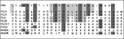
Fig. 3. Comparison of carbohydrate recognition domain of I. ricinus lectin (r58) with those of different lectins. The lectins compared are: r58, clone from I. ricinus cDNA library (unpublished observations); PL21, 31, 9 & 25, four lectins from Periplaneta americana cockroach hemolymph (Kawasaki et al. 1996); HLEC, human hepatic lectin (Halberg et al. 1987); FCE2, mucose immunoglobulin Fc receptor (Bettler et al. 1989); MABC, rat mannose-binding protein A (Drickamer, Dordal & Reynolds, 1986); and MANR, human macrophage mannose receptor (Taylor et al. 1990). Dark grey represents identitical amino acid residues and light grey represents similar residues.
 You have
Access
- 24
- Cited by