
Introduction
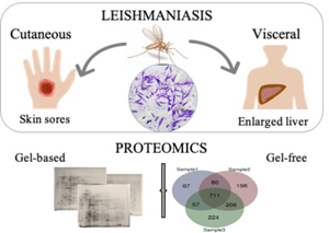
Leishmaniasis is an infectious disease mostly affecting people who live in developing countries with a lack of sanitation. The disease rises from parasites of the genus Leishmania; however, sandflies can be accused of contraction since a bite suffices to transmit these obligate intracellular protozoans to vertebrates. Three main clinical forms, cutaneous (CL), mucocutaneous and visceral (VL) leishmaniases emerge regarding body location where parasites in amastigote form are accumulated. The life-threatening form, VL, affects the inner organs, especially the lymph nodes, spleen and liver. On the contrary, CL which may leave behind unpleasant permanent scars is diagnosed by skin lesions on the exposed part of the body. Muco-cutaneous form is more severe than cutaneous form since it may result in the mutilation of the nose, mouth or throat (Chappuis et al., Reference Chappuis, Sundar, Hailu, Ghalib, Rijal, Peeling, Alvar and Boelaert2007; Steverding, Reference Steverding2017; Garrido-Jareño et al., Reference Garrido-Jareño, Sahuquillo-Torralba, Chouman-Arcas, Castro-Hernández, Molina-Moreno, Llavador-Ros, Gómez-Ruiz, López-Hontangas, Botella-Estrada, Salavert-Lleti and Pemán-García2020). Among more than 50 Leishmania species defined, approximately 20 are pathogenic for humans and the disease forms are associated with those species. In this context, only Leishmania braziliensis is seen in the cases of muco-cutaneous leishmaniasis, Leishmania donovani and Leishmania infantum are primarily causative agents of VL and finally Leishmania major, Leishmania mexicana, Leishmania tropica and the others are generally responsible for CL. Apart from that, L. infantum can cause CL to a lesser extent and the same goes for L. tropica about VL (Akhoundi et al., Reference Akhoundi, Downing, Votýpka, Kuhls, Lukeš, Cannet, Ravel, Marty, Delaunay, Kasbari, Granouillac, Gradoni and Sereno2017).
Atypical case is quite a broad term and refers to abnormalities including the unusual appearance of lesions (Guimarães et al., Reference Guimarães, Queiroz, Silva, Silva, Magalhães, Lago, Machado, Bacellar, Wilson, Beverley, Carvalho and Schriefer2016; Espinoza-Morales et al., Reference Espinoza-Morales, Lucchetti Rodríguez, Silva-Caso, Suarez-Ognio, Pons and del Valle Mendoza2017; Meireles et al., Reference Meireles, Maia, Soares, Teodoro, Gadelha, da Silva and de Lima2017) and unfamiliar clinical indications due to the coinfections and concurrent occurrence of other diseases (Calvopina et al., Reference Calvopina, Gomez, Uezato, Kato, Nonaka and Hashiguchi2005; Da-Cruz et al., Reference Da-Cruz, Filgueiras, Coutinho, Mayrink, Grimaldi, De Luca, Mendonça and Coutinho1999; Kouyialis et al., Reference Kouyialis, Archontakis, Bilinis, Nikolaou, Stavropoulou, Samaras, Sarafoglou, Nicolaou, Parasi and Minadaki2005; Celesia et al., Reference Celesia, Cacopardo, Massimino, Gussio, Tosto, Nunnari and Pinzone2014; Hashemi et al., Reference Hashemi, Badirzadeh, Sabzevari, Nouri and Seyyedin2018; Martínez et al., Reference Martínez, Verdonck, Kaye, Adaui, Polman, Llanos-Cuentas, Dujardin and Boelaert2018). Apart from that, clinical manifestations caused by unexpected Leishmania spp. which has become widespread around the world are also considered as atypical cases (Mebrahtu et al., Reference Mebrahtu, Van Eys, Guizani, Lawyer, Pamba, Koech, Roberts, Perkins, Were and Hendricks1993; Siriwardana et al., Reference Siriwardana, Noyes, Beeching, Chance, Karunaweera and Bates2007; Elamin et al., Reference Elamin, Guizani, Guerbouj, Gramiccia, El Hassan, Di Muccio, Taha and Mukhtar2008; Lopes et al., Reference Lopes, Vasconcelos, Borges-Costa, Soares-Almeida, Campino and Filipe2013; Özbilgin et al., Reference Özbilgin, Harman, Karakuş, Bart, Töz, Kurt, Çavuş, Polat, Gündüz, Van Gool and Özbel2017; Thakur et al., Reference Thakur, Singh, Shanker, Negi, Jain, Matlashewski and Jain2018). Genomic studies demonstrated that mixed Leishmania infections and interspecies Leishmania hybrids which were promoted by climate change, mass migrations and travels (Dereure et al., Reference Dereure, Vanwambeke, Malé, Martinez, Pratlong, Balard and Dedet2009; González et al., Reference González, Wang, Strutz, González-Salazar, Sánchez-Cordero and Sarkar2010; Alawieh et al., Reference Alawieh, Musharrafieh, Jaber, Berry, Ghosn and Bizri2014; King et al., Reference King, Stelkens, Webster, Smith and Brockhurst2015; Karakuş et al., Reference Karakuş, Çizmeci, Karabela, Erdoğan and Güleç2019; Lypaczewski and Matlashewski, Reference Lypaczewski and Matlashewski2021), render the disease more detrimental for this reason aforementioned complications supposedly have emerged under these factors (Akopyants et al., Reference Akopyants, Kimblin, Secundino, Patrick, Peters, Lawyer, Dobson, Beverley and Sacks2009; Romano et al., Reference Romano, Inbar, Debrabant, Charmoy, Lawyer, Ribeiro-Gomes, Barhoumi, Grigg, Shaik, Dobson, Beverley and Sacks2014; Ferreira et al., Reference Ferreira, Cruz, Cañavate, de Melo, Pereira, Madeira, Valério, Cunha, Paglia and Gontijo2015; Alves Souza et al., Reference Alves Souza, Souza Leite, de Oliveira Silva, Groenner Penna, Figueiredo Felicori Vilela, Melo and de Andrade2019). Since these alterations somehow jeopardize disease control and prevention, to keep up with the biological alterations and to update our knowledge, Leishmania parasites should be investigated from all perspectives, especially involving the ones isolated from patients.
Differential proteomics enables scientists to reveal the alterations between two or more different conditions by comparing their overall protein expression. Two methodical approaches namely gel-based and gel-free proteomics have gained wide acceptance for the comparison. While the first is based on protein separation in a porous gel, the latter is built on the separation of complex peptide mixtures with liquid chromatography after single-shot digestion of the protein mixture (Lambert et al., Reference Lambert, Ethier, Smith and Figeys2005). Two-dimensional polyacrylamide gel electrophoresis (2D-PAGE) was formerly the leading method, in which proteins are separated on thin square polyacrylamide gels, however, shotgun proteomics has become the method of choice today due to accurate and high-throughput data accumulation in a short time (Zhang et al., Reference Zhang, Fonslow, Shan, Baek and Yates2013). Since only a limited number of proteins can be detected with the gel-based method, gel-free or shotgun proteomics techniques which keep evolving with technological advancements and improvements in auxiliary materials, dominate over gel-based techniques nevertheless seeing proteins isolated on the gel visible to the naked eye enables scientists a reassurance for the protein expression while providing simplicity during database search. Although these two approaches can give results at a completely different levels and perspectives, both of them can be used as complementary to each other. Therefore, to determine differential proteins on Leishmania isolates obtained from VL and CL patients, both gel-based and gel-free methods were applied to gain a broader insight into the Leishmania parasite tropism. The comparison was built on the species, L. infantum and L. tropica were partially incorporated since these two are the prevalent species in Europe and the Mediterranean region including our country (Ready, Reference Ready2010).
Materials and methods
Parasite cultivation and total protein extraction
The Leishmania isolates obtained from patients who were admitted to dermatology clinics in different provinces of Turkey were genetically identified and cryopreserved as described in the previous study (Özbilgin et al., Reference Özbilgin, Harman, Karakuş, Bart, Töz, Kurt, Çavuş, Polat, Gündüz, Van Gool and Özbel2017). Real-time ITS1-polymerase chain reaction (PCR) and PCR-based cysteine protease B methods were used for the species discrimination (Hide and Bañuls, Reference Hide and Bañuls2006; Toz et al., Reference Toz, Culha, Zeyrek, Ertabaklar, Alkan, Vardarlı, Gunduz and Ozbel2013). Parasite Bank in Manisa Celal Bayar University supplied these isolate samples which were cryopreserved in liquid nitrogen after cell cultivation and labelling. VL and CL promastigote cells were cultivated in enriched NNN medium at 25 °C and then transferred to RPMI-1640 medium containing 10% foetal calf serum (FCS), 1% penicillin/streptomycin and 1% gentamycin. When the population reached approximately 108 cells mL−1, the growth medium was centrifuged at 4400 rpm for 10 min at 4 °C. Pellets were incubated overnight in RPMI-1640 medium without FCS and harvested again by centrifugation at 4400 rpm (Özbilgin et al., Reference Özbilgin, Çulha, Uzun, Harman, Topal, Okudan, Zeyrek, Gündüz, Östan, Karakuş, Töz, Kurt, Akyar, Erat, Güngör, Kayabaşı, Çavuş, Bastien, Pratlong, Kocagöz and Özbel2016). After washing gently with phosphate-buffered saline three times, 1 mL of Mammalian Cell Lysis Reagent (Fermentas Life Sciences; Thermo Fisher) was added for the cell lysis. The resuspended solution was incubated at room temperature on a shaker at 900 rpm and centrifuged at 14 500 rpm for 15 min. Finally, the supernatant was decanted gently to a new tube and stored at −20 °C for further processing. The protein solution was concentrated with centrifugal filters (Amicon Ultra 10K molecular weight cut-off 0.5 mL; Merck Millipore) and the slurry was resuspended in the rehydration buffer. Protein concentration was measured with Bradford protein assay (Coomassie Plus Bradford Assay Reagent; Thermo Scientific). Both gel-based and gel-free approaches were employed for differential protein expression analysis by using different mass spectrometers.
Gel-based proteomics: two-dimensional gel electrophoresis and in-gel tryptic digestion
Protein was dissolved in the rehydration buffer containing 7 m urea, 2 m thiourea, 4% v/v CHAPS, 65 mm dithiothreitol (DTT) and 2.5% ampholyte solution pH 3–10 (40%, w/v). A total of 400 μg protein in 350 μL solution was loaded onto 17 cm, pH 3–10 nonlinear strip (ReadyStrip™ IPG Strips; BioRad). In addition, two pH 4–7 strips and one pH 5–8 strip were run with remaining samples to evaluate the degree of resolution in narrower ranges. After passive rehydration for 2 h and subsequent active rehydration at 50 V for 16 h, the first stage of two-dimensional sodium dodecyl sulphate polyacrylamide gel electrophoresis (2D-SDS PAGE) (O'Farrell, Reference O'Farrell1975), isoelectric focusing (IEF) was started to run until a total of 60 000 Vh electrophoresis was completed (Protean IEF cell; Bio-Rad). Before mounting onto the polyacrylamide gels, strips were first equilibrated in a solution containing 6 m urea, 50 mm Tris-HCl pH 8.8, 2% w/v SDS, 30% w/v glycerol and 65 mm DTT for 15 min. Following this, they were kept in another equilibration solution in which 4% w/v 2-iodoacetamide (IAA) was substituted with DTT for 15 min more, this time in the dark. After the equilibration step, the strips were washed by immersing into electrophoresis running buffer Tris-glycine-SDS. The second stage, SDS PAGE, was performed placing the strips onto 12% polyacrylamide gels and applying 180 V for approximately 9 h. Gels were sealed with overlay agarose solution containing 0.5% agarose and 0.003% bromophenol blue in the running buffer to monitor the reaching of the dye to the bottom of the gel to terminate the current. After rinsing the gel with water, proteins were detected with silver staining (Gromova and Celis, Reference Gromova, Celis and Celis2006) and selected spots were subjected to in-gel digestion (Shevchenko et al., Reference Shevchenko, Tomas, Havlis, Olsen and Mann2006) with trypsin (proteomic grade porcine pancreas, Sigma Aldrich). No computational tool was utilized for the comparison and differential protein assignment therefore only explicit and repeating changes were taken into consideration. Peptide solution was desalted with the C18 ZipTip™ pipette tip (Millipore) and mixed with alpha cyano-4-hydroxycinnamic acid for the thin layer deposition onto a matrix-assisted laser desorption/ionization (MALDI) plate (Dai et al., Reference Dai, Whittal and Li1999). MALDI mass spectrometry (MS) measurements were performed on an Autoflex III Smartbeam MALDI TOF/TOF MS (Bruker).
Gel-free proteomics: off-line high-performance liquid chromatography (HPLC) and liquid chromatography–mass spectrometry/mass spectrometry (LC-MS/MS)
For in-solution tryptic digestion, the aforementioned 10 kDa MWCO filter tube was used to remove excess urea and detergent which interferes with trypsin and causes denaturation (Wiśniewski et al., Reference Wiśniewski, Zougman, Nagaraj and Mann2009). Before trypsin addition, 5 μL of 0.2 m reducing agent DTT has been added to 400 μg protein solutions and incubated for 1 h. Later, 20 μL of 0.2 m alkylating agent IAA was added and incubated for 1 h in the dark. Next, 20 μL of 0.2 m reducing agent DTT was added again and incubated for 1 h to neutralize the excessive IAA. Samples were centrifuged at 14 000 rpm for 10 min for the solvent exchange with Tris-HCl. Two washing steps were performed by adding 200 μL of 100 mm Tris-HCl buffer followed by centrifugation. Finally, 4 μg trypsin (enzyme to protein 1:100) was mixed in a solution containing 50 mm, pH 8.5 Tris-HCl and 10% acetonitrile. An enzyme solution of 200 μL was added to the concentrated protein mixture and incubated at 37 °C overnight. Since hundreds of thousands of peptides are produced from protein mixtures by a single tryptic digestion step, this method is also called shotgun proteomics. Although shotgun proteomics seems labourless due to the absence of protein separation, this time the peptide mixture needs to be reduced to a measurable degree before the LC-MS/MS analysis. This task can be achieved with 2D-HPLC systems so-called online analysis or by collecting fractions from another HPLC, the off-line separation. For the off-line peptide separation, high pH reverse-phase chromatography (Shimadzu Prominence UFLC, LC Solution Version 1.25 SP3) with a fraction concatenation strategy was applied at a 0.5 mL min−1 flow rate (Wang et al., Reference Wang, Yang, Gritsenko, Wang, Clauss, Liu, Shen, Monroe, Lopez-Ferrer, Reno, Moore, Klemke, Camp and Smith2011). Peptide solution was evaporated with a speed-vac vacuum concentrator and 100 μL solution was injected to C18, 5 μm, 250 × 4.6 mm analytical column (C18-Teknokroma Mediterranea sea18) which was equipped with a 10 × 4.6 mm column protection cartridge (uniguard; Thermo). Eluent was collected in a 96-well plate with 35-s intervals (SunCollect micro fraction collector MALDI spotter; SunChrom). To decrease the 96 fractions, six wells were pooled into a new 1.5 mL tube by skipping 15 fractions each time and then all 16 fractions were completely evaporated. All 16 fractions belonging to one sample were resuspended, cleaned with the C18 ZipTip™ pipette tip and analysed by using capillary HPLC (DIONEX UltiMate 3000) coupled with an ESI linear ion trap MS (LTQ XL mass spectrometer, Thermo Scientific) twice. The outlet of the reversed-phase capillary column (15 cm × 500 μm; 2.7 μm; Sigma Supelco Ascentis) having a 5 μL min−1 flow rate was connected to the ESI source whose spray voltage and the capillary temperature was set to 5 kV and 200 °C, respectively. MS/MS data were collected through data-dependent acquisition (Xcalibur software 2.0.7) that allows the fragmentation of the nine most abundant ions in positive mode unless the ion count is below 10 000, using 35% collision energy. Full spectra were collected from m/z 400–1800 and dynamic exclusion was 30 s.
Protein identification with peptide database search
In the gel-based part, MS and MS/MS spectra were collected with software flexControl 3.0 and transferred from software flexControl 3.0 to Biotools 3.0 for database search with the Mascot search engine (version 2.3; Matrix Science) (Perkins et al., Reference Perkins, Pappin, Creasy and Cottrell1999). For the database search, carbamidomethylation (C) as fixed global modification and oxidation (M) as variable modification and allowance for up to one tryptic cleavage were set. The peptide mass tolerance was either 200 ppm or 1.0 Da and fragment ion mass tolerance was 0.5 Da. Charge state was 1+ and monoisotopic option mass was chosen. The mass range of the analyses was between 700 and 3500 Da. Data were searched against the NCBI database (NCBI Resource Coordinators, 2017) of Leishmania genus and alternatively UniProt (The UniProt Consortium, 2016) protein database of L. infantum.
In the gel-free part, 32 MS/MS raw files were converted to a mascot generic file (mgf) by Proteome Discoverer 1.4. for each sample. The combined data of all fractions were searched against the L. infantum proteome database obtained from UniProt using an in-house Mascot server version 2.3 as the first option. After post-processing with Mascot percolator (Brosch et al., Reference Brosch, Yu, Hubbard and Choudhary2009), the results were exported and further analysed in MS Excel. Apart from that, all mgf files were submitted to software SearchGUI (Vaudel et al., Reference Vaudel, Barsnes, Berven, Sickmann and Martens2011) for multiple-search using the search engines Comet (Eng et al., Reference Eng, Jahan and Hoopmann2013), MS Amanda (Dorfer et al., Reference Dorfer, Pichler, Stranzl, Stadlmann, Taus, Winkler and Mechtler2014), MS-GF+ (Kim and Pevzner, Reference Kim and Pevzner2014) and xTandem (Craig and Beavis, Reference Craig and Beavis2004). Results were displayed and exported to excel files by PeptideShaker (Vaudel et al., Reference Vaudel, Burkhart, Zahedi, Oveland, Berven, Sickmann, Martens and Barsnes2015). The following set-up parameters were entered to SearchGUI for all searches: enzyme: trypsin, fixed modifications: carbamidomethyl, variable modification: oxidation, peptide tolerance: 1.0 Da, MS/MS tolerance: 0.5 Da, peptide charge: 2+ and 3+, instrument: ESI-TRAP and experimental mass values: monoisotopic. The same settings were used for Mascot searches except for the peptide tolerance and MS/MS tolerance which were changed to default values 1.2 and 0.6 Da, respectively. Protein false discovery rates (FDR) of all searches were <1%. To determine the differential proteins, emPAI values (Ishihama et al., Reference Ishihama, Oda, Tabata, Sato, Nagasu, Rappsilber and Mann2005) of Mascot results and spectrum counting values obtained from PeptideShaker (Powell et al., Reference Powell, Weaver, Jennings, McAfee, He, Weil and Link2004) were transferred to Microsoft Excel spread sheet and normalized values (Shinoda et al., Reference Shinoda, Tomita and Ishihama2010) were imported to Perseus software (Tyanova et al., Reference Tyanova, Temu, Sinitcyn, Carlson, Hein, Geiger, Mann and Cox2016) for the statistical analysis. Web-based Venn diagram tools were utilized when necessary (VIB/UGent, 2021; Bardou et al., Reference Bardou, Mariette, Escudié, Djemiel and Klopp2014).
Results
Promastigote parasites isolated from different patients were examined in three groups which were designated with CLi for the CL group caused by L. infantum, CLt for the CL group caused by L. tropica and VLi or just VL for the VL group caused by L. infantum. To compare the protein expression of promastigote parasites caused to different disease forms such as VL and CL, both gel-based and gel-free proteomic approaches were applied. The first was studied on 15 samples while the latter were contained nine out of those 15 samples.
Gel-based approach: 2D-SDS PAGE results
From a broad perspective, gel images of CL isolates, belonging to either L. infantum or L. tropica group, were quite similar whereas those of VL isolates were distinguishable from them, especially in particular samples. Main differentiation between CL and VL isolates was observed as downregulation in several proteins on the gels of VL samples.
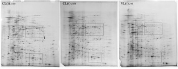
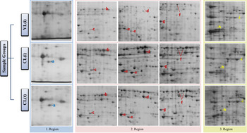
Whole gel images of all samples and the list of identified protein spots are available in the Supplementary material_1; however, one gel from each group is given in Fig. 1 for representation purposes. For ease of viewing, specific regions of interest (ROIs) whose locations are shown by dashed rectangles in Fig. 1. Aligned version of all these gel sections for each ROI is given in Supplementary material_1. In those figures, all VL samples are laid on the top row while CL(i) samples follow them in the middle and CL(t) samples are aligned down on the third row. Gels of three samples are missing in the first ROI due to a technical problem during the electrophoresis, therefore sections from two pH 4–7 and one pH 5–8 gels are added on the left column for this region. Protein spots only identified with in-gel digestion and mass spectrometric analysis by MALDI-TOF/TOF MS were indicated with a number. Some of these spots which were identified more than once were like anchor points helping us to ensure about the positions during the visual inspection. A total of 40 protein spots were identified by using peptide mass fingerprinting and peptide fragmentation or both. Among them, nine protein spots seemed deserve special attention therefore they are flagged with a lower-case letter and gathered in Fig. 2. Our first observation was the absence of a spot in the VL group in the first ROI. As it is seen on the left side of Fig. 2, this protein was pinned with a blue -a letter and defined as superoxide dismutase. In this region, we have also determined another antioxidant protein namely peroxidoxin corresponding to a quite dark and large spot. As we verified with extra runs using narrow pH ranges (pH 4–7 to two samples and pH 5–8 to one sample), peroxidoxin and superoxide dismutase have protein variations whose molecular weight and pI values are too close.
One whole gel image from each group and locations of ROIs as rectangular frames.

Differentiated spots between the groups.

Since the second ROI which was nearer to alkaline side of the gel was expanded by the addition of aligned sections from the other samples (Fig. 2) to facilitate the tracking. Herein, letters from -b to -g are corresponding to pyruvate kinase, aspartate aminotransferase, putative succinyl-coA:3-ketoacid-coenzyme A transferase, putative aldose 1-epimerase, putative dihydrolipoamide dehydrogenase and putative eukaryotic initiation factor 4a, respectively. Except for the latter, all of these proteins seemed downregulated mostly in the samples of the VL group (top row).
In the third region, two collateral spots which were represented with the letter -h was identified as putative N-acyl-l-amino acid amidohydrolase and distinguished from a very close other spots below namely metallopeptidase (spots 28 and 31; Supplementary material_1). The other protein spot in this region which were downregulated in all VL samples and represented with the letter -i, was identified as enolase.
Gel-free approach: shotgun proteomic results
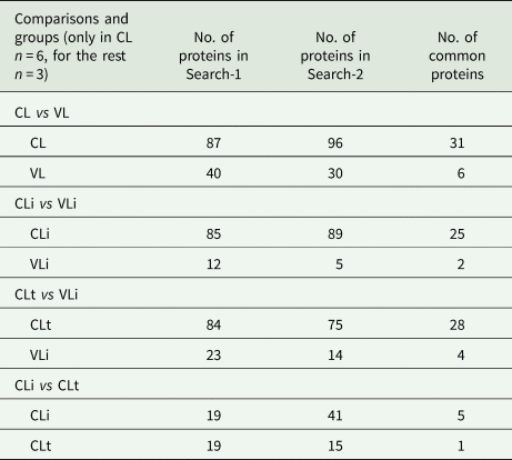
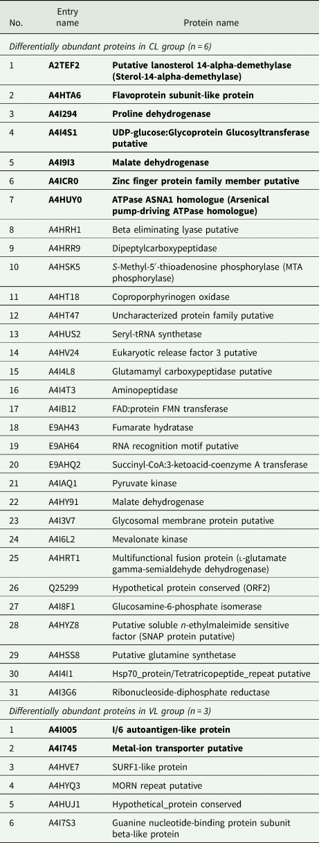
All shotgun proteomic data were searched against the L. infantum proteome database using two different bioinformatic search engines for the cross-check therefore we did not apply the two-peptide rule (Gupta and Pevzner, Reference Gupta and Pevzner2009) for the reliable protein identification. After the elimination of contaminant proteins, both searches resulted in more than 1900 Leishmania proteins in total. Samples were grouped based on clinical disease forms (CL and VL) they caused, and moreover, samples of CL were divided into two namely CLi and CLt representing L. infantum and L. tropica to be able to make an interspecific and intraspecific comparison. Tables containing both search findings (the Mascot Search and the Search via Search GUI) and the results of statistical comparisons of the groups are given in Supplementary material_2 and Supplementary material_3, respectively. Table 1 shows the number of differentially expressed proteins obtained from all comparisons and the last column contains those of common proteins between the searches which are considered more reliable. To sum up, according to t-test with P < 0.05 in which the spectrum counting of clinical forms (CL and VL) was compared, a total of 127 proteins differentially abundant were detected in the first search by mascot and 126 proteins were in the second search run by searchgui. Thirty-seven proteins common in these outputs are given in Table 2. Exceptionally, one protein called malate dehydrogenase was appeared twice with different entry names. In conclusion, among 36 proteins, six proteins were determined differentially abundant in the VL samples, and the rest 30 proteins were more expressed in the CL samples. Finally, a peptide centric comparison was carried out between the CL and VL groups which are provided in Supplementary material_4, in case a specific peptide sequence peculiar to a group might come out.
Number of differentially expressed proteins between the clinical forms and the species

Differentially abundant proteins obtained from the comparison of the gel-free analysis (Bold text indicates a significance level of p < 0.01)

Discussion
In the early days of proteomics, 2D gel electrophoresis was the leading method since protein separation or protein isolation was inevitable for the further analysis. With the advent of automatic LC-MS/MS data acquisition, it became possible to detect and identify thousands of protein in a complex mixture thus shotgun proteomic analysis has taken over the lead role gradually in association with the contributions of technological developments. Undoubtedly, the latter is superior to a gel-based method with regards to the number of proteins attained and keeps pushing the limits via expensive high-end mass spectrometers and powerful computers. On the contrary, highly abundant soluble proteins mostly get the chance to be examined in a gel-based method; therefore, it addresses to proteoform investigation rather than proteome-level scanning. Apart from that, visualized proteins in the gel with conserved integrity have an idiosyncratic persuasive effect on the expression of that protein.
Herein, proteomic comparison between the two clinical forms of leishmaniasis disease namely visceral and cutaneous has been performed using the abovementioned both gel-based and gel-free proteomic approaches. Although L. donovani and L. infantum are the main causative agents of the clinic form, VL, they can cause the cutaneous form to a lesser extent. Since L. infantum is more common than L. donovani in the Mediterranean region including our country, intra-species alteration between the clinical forms was investigated on L. infantum. In addition, samples of another common species, L. tropica which corresponds to the typical cases for CL in Turkey, was included to obtain an intra-species comparison. Gel-based and gel-free proteomic methods were applied to the promastigote parasites which were isolated from different patients. The first was studied on 15 samples while the latter were contained nine out of those 15 samples.
The gel images of CL samples exhibited more similar picture regardless of the species while the gel images of VL samples were discriminable from them based on three specific locations according to our visual inspection. Among these regions, one distinct spot which was identified as superoxide dismutase protein was only detected in CL samples. In the gel-free results, we observed that VL samples shared a specific peptide sequence of superoxide dismutase while none of the CL samples were containing it. These findings from two different methods might indicate a structural regulation on this antioxidant enzyme regarding the tissue tropism in the Leishmania; therefore, targeted analysis focusing on this protein will provide a deeper understanding. In the second ROI, especially three spots which seemed faded and smaller in the VL samples, repetitively, were considered as downregulation. Two of them, pyruvate kinase and putative succinyl-coA:3-ketoacid-coenzyme A transferase were confirmed by a gel-free method; however, the third one, putative dihydrolipoamide dehydrogenase which, in fact, consists of two lateral spots, did not show up in any comparison results of the gel-free method. Apart from them, two spots identified as aspartate aminotransferase and putative aldose 1-epimerase which can be said partially supported by the gel-free method, looked as if they were downregulated in VL samples too. These two proteins emerged in the list of comparison result of the gel-free method which was based on Mascot database searching. Finally, a protein identified as putative N-acyl-l-amino acid amidohydrolase was prominently seen in the CL samples. This protein was observed in the third ROI and encountered in the list of comparison result of the gel-free method which was based on the other database searching with Search GUI.
In a comparative proteomic study which was conducted on promastigote L. tropica parasites caused to CL and VL in a dog (Hajjaran et al., Reference Hajjaran, Mousavi, Burchmore, Mohebali, Mohammadi Bazargani, Salekdeh, Kazemi-Rad and Khoramizadeh2015), general downregulation in the proteins of energy metabolism and protein synthesis was observed in the VL sample too. This correlation does not rely on the same proteins nevertheless a particular hypothetical protein with the entry name Q4QFN8 was noted. This uncharacterized protein which corresponds to A4HW39 for L. infantum with 95% similarity is present in five of the six CL samples according to our shotgun proteomic results.
In the gel-free method, shotgun proteomic data of each sample were analysed by using two different bioinformatic search tools; thus, an ultimate protein list containing more dependable proteins was generated from the output in common. Since we acquired the LC-MS/MS data from quite an outmoded low-resolution mass spectrometer with microlitre LC flow, two-peptide rule would not be efficient for our data; therefore, we aimed to compensate these drawbacks partially by confirming the search results with another one. Numerically searching outcomes of the both bioinformatic tools were quite close to each other. Both searchings were resulted in total protein between 1900 and 1930 cumulatively and more than 125 of them were potentially related to clinical forms. The ultimate list of differentially abundant proteins which were constituted from the two searches contained 37 components with specific entry name. As an exception, malate dehydrogenase protein was emerged twice with two different entry names. Consequently, six proteins associated with VL and 30 proteins associated with CL were determined as more reliable differentially expressed proteins. Finally, this list was created in the order of importance by taking into account the P value of the t-test which means significance level for the first nine proteins was less than 0.01% and similarly P was less than 0.03% for the next 14 proteins (2 of them belong to VL).
There might be remarkable findings in this list judging by the studies in the literature of Leishmania research. For example, putative lanosterol 14-alpha-demethylase, a protein that participates in steroid biosynthesis deserves special attention due to its connection with the protozoan counterpart of cholesterol, ergosterol. In our results of the gel-free method, this protein was not detected in any of the VL samples while all the CL samples were containing it. In addition, another enzyme related to ergosterol biosynthesis namely mevalonate kinase which protects cells against oxidative stress (Shafi et al., Reference Shafi, Bamra, Das, Kumar, Abhishek, Kumar, Kumar, Kumar, Mukherjee, Sen and Das2021) was observed downregulated in VL samples. Since major antifungal and antileishmanial drugs target the sterol metabolism to destroy the membrane integrity through ergosterol (Emami et al., Reference Emami, Tavangar and Keighobadi2017), drug-resistant parasites seemingly figured out to avoid from this issue by being less predictable on the membrane content. Reduction of ergosterol coupled with alternative sterol formation as well as a mutation on the gene of sterol 14-alpha-demethylase in the amphotericin B-resistant cells supports this notion (Mwenechanya et al., Reference Mwenechanya, Kovářová, Dickens, Mudaliar, Herzyk, Vincent, Weidt, Burgess, Burchmore, Pountain, Smith, Creek, Kim, Lepesheva and Barrett2017). Moreover, lipid profile changes between L. infantum and L. amazonensis (Negrão et al., Reference Negrão, Abánades, Jaeeger, Rocha, Belaz, Giorgio, Eberlin and Angolini2017) indicate that alterations on cell membrane enable the parasite to escape from the host's immune system and treatments. A similar mechanism pertaining to ergosterol content can be anticipated between the viscerotropic and dermotropic strains of L. infantum.
Flavoprotein subunit-like protein and proline dehydrogenase proteins were never detected in any VL samples while expressed in all six CL samples. Up-regulation of the first was shown in some drug-resistant L. infantum strains (Vincent et al., Reference Vincent, Racine, Légaré and Ouellette2015). In addition to proline dehydrogenase, a more expression of a second proline degradation protein, multifunctional fusion protein (A4HRT1) might be related to the high proline content in the skin collagen (Shoulders and Raines, Reference Shoulders and Raines2009). Similar to these, I/6 autoantigen-like protein was like a determinant protein for the VL group since none of the CL samples contained it while it was expressed in all VL samples. Considering the importance of glycosylation on protein recognition and cell adhesion (Mule et al., Reference Mule, Saad, Fernandes, Stolf, Cortez and Palmisano2020), UDP-glucose: glycoprotein glucosyltransferase putative (A4I4S1) might be noteworthy to understand the dermotropic strains in L. infantum. Similarly, ion transporters are crucial membrane proteins for nutrient uptake; therefore, occurrence of metal-ion transporter protein (A4ICR0) only in viscerotropic strains must have vital implications for the organism survival in an inner organ. Indeed, more evidence is needed to claim their relevance to clinical forms per se. Apart from the protein-centric comparison, a qualitative peptide-centric comparison was provided in the case of being used in an immunoassay or targeted proteomics someday.
To conclude, proteomic comparison in which capacity and perspectives of gel-based and gel-free proteomic analyses can be evaluated is presented in this study to gain more insight about Leishmania parasites once caused to VL and CL in people.
Supplementary material
The supplementary material for this article can be found at https://doi.org/10.1017/S0031182021001967
Data
All mass spectrometric data have been deposited at the ProteomeXchange Consortium via the PRIDE partner repository with the dataset identifier PXD016333 and 10.6019/PXD016333.
Acknowledgements
The authors acknowledge National Mass Spectrometry Application and Research Center (NMSC) in Izmir Institute of Technology (IzTech).
Author contributions
TY and AÖ conceived and designed the study. İÇ and AÖ provided biological samples. İÇ performed cell cultivation and protein extraction. MD conducted sample preparation and mass spectrometric data gathering. MD performed bioinformatic analyses. MD and TY wrote the article.
Financial support
The authors gratefully acknowledge financial support for doctoral research by the Scientific and Technological Research Council of Turkey, TUBITAK (project no. 215Z257).
Conflict of interest
None
Ethical standards
This study was approved by the Institutional Review Board of Manisa Celal Bayar University, Faculty of Medicine and written informed consent was obtained from all patients.