Introduction
Cutaneous haemangiomas are the most common skin tumour of infancy, affecting 12 per cent of children. There is a greater incidence in females, Caucasians and premature infants. Haemangiomas consist of proliferating endothelial cells, in contrast to vascular malformations which consist of dysplastic microvascular proliferation.Reference Mulliken and Glowacki1 Most cutaneous haemangiomas develop within months of birth (the proliferative phase) and usually involute spontaneously months to years later (the involutional phase) without specific treatment.Reference Chang, Haggstrom, Drolet, Baselga, Chamlin and Garzon2
The established treatment modalities for cutaneous haemangioma, both surgical and non-surgical, have significant potential co-morbidity. For example, steroid therapy may cause Cushing's syndrome, with adrenal suppression; endoscopic laser treatment and open excision carry a small risk of inducing subglottic stenosis; and an obstructed tracheostomy may result in hypoxic brain injury.
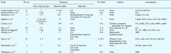
In 2008, Leaute-Labreze et al. described the novel use of propranolol therapy in 11 children with disfiguring cutaneous haemangiomas.Reference Leaute-Labreze, de la Roque, Hubiche, Boralevi, Thambo and Taieb3 Subsequently, several cases of such treatment have been reported, with promising results (Table I), and propranolol has been recommended as a first-line treatment for infantile haemangioma when intervention is required.Reference Zimmermann, Wiegand, Werner and Eivazi14
Previous reports of propranolol therapy for infantile haemangioma

*Mean average for group. Pts = patients; FU = follow up; mth = months; += some patients’ treatment ongoing at time of writing; CS = corticosteroids; US = ultrasonography; NA = not advised; Rx = therapy; inc = increments to the treatment dose; wk = weeks; cardio = paediatric cardiology review; obs = general observations (i.e. heart rate, blood pressure, respiration rate, oxygen saturation and blood glucose); ECG = electrocardiography; echo = echocardiography; AUS = abdominal ultrasonography; RBTs = routine blood tests (i.e. full blood count, urea and electrolyte concentration, liver function tests, glucose tests, and thyroid function tests); ophth = ophthalmology review (because of location of lesion); FL = flexible laryngoscopy; CT = computed tomography; MRI = magnetic resonance imaging; DLTB = direct laryngotracheobronchoscopy
We present our experience of propranolol therapy for childhood haemangiomas of the head and neck, and we describe an important case in which propranolol therapy was unsuccessful.
Case reports
Case one
A three-month-old girl presented with a two-week history of a barking cough and biphasic stridor.
Clinical examination confirmed biphasic stridor, with evidence of respiratory distress. There was no history of previous endotracheal intubation, and no cutaneous haemangiomas were evident.
The child was commenced on oral dexamethasone. Rigid airway endoscopy demonstrated a left-sided subglottic haemangioma causing 80 per cent airway narrowing (Figure 1).
Rigid airway endoscopy views for case one, showing subglottic haemangioma (a) prior to treatment and (b) 3 days, (c) 3 weeks and (d) 13 months after initiation of propranolol treatment.

Following a satisfactory paediatric cardiology assessment, including electrocardiography and echocardiography, the child was commenced on a course of propranolol at 1 mg/kg/day in three divided doses for one week.
Marked clinical improvement was noted during the child's two-week in-patient stay, and this was verified objectively with repeated rigid airway endoscopies (Figure 1). After the first week, the child's propranolol dose was increased to 2 mg/kg/day in three divided doses. Her blood pressure and glucose were monitored regularly throughout this period.
On discharge from hospital, there was complete resolution of the child's stridor, and no other signs of respiratory distress.
A further rigid airway endoscopy was performed two weeks following discharge, showing almost complete resolution of the subglottic haemangioma.
The child continued to experience no symptoms or complications resulting from propranolol treatment, (and this continued to the time of writing (child aged 18 months).
After five months of treatment, she was commenced on a reducing dose of propranolol. Rigid airway endoscopy was planned in the near future, on completion of withdrawal of propranolol treatment.
Case two
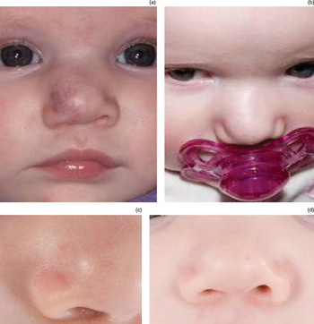
An 11-week-old girl presented with an enlarging haemangioma involving the right side of her nasal tip, distorting the contour of the alar margin (Figure 2). There was no evidence of stridor or respiratory distress.
Clinical photographs of case two, showing nasal tip haemangioma (a) prior to treatment and (b) one, (c) two and (d) eight weeks after initiation of propranolol treatment.

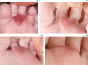
On secondary clinical examination, a haemangioma was noted on the sole of her right foot (Figure 3).
Clinical photographs of case two, showing pedal haemangioma (a) prior to treatment and (b) one, (c) two and (d) eight weeks after initiation of propranolol treatment.

The child was commenced on a course of prednisolone (1 mg/kg once daily).
After two weeks of treatment, there had been no clinical improvement.
An ultrasound scan of the craniofacial soft tissues was performed, showing a 17 × 18 × 6 mm nasal lesion which was heterogeneous, hypoechoic and moderately hypervascular, consistent with a haemangioma.
Following these initial assessments, the child was commenced on a new regime of propranolol at 1 mg/kg/day in three divided doses, increasing to 2 mg/kg/day after one week.
In response to this treatment, the child's nasal tip and foot haemangiomas both showed marked improvement in terms of pigmentation and local mass effect. There was no residual asymmetry of the nasal tip, and the foot lesion was less pigmented.
At the time of writing, the child was eight months of age and the improvement in the appearance of both lesions had been maintained. At this stage, it was planned to commence stepwise reduction in propranolol dosage when the child was one year old.
Case three
A nine-week-old boy was seen in clinic with a swelling in the right parotid region (Figure 4a). Shortly after birth, he had contracted an Escherichia coli infection which had caused sepsis requiring ventilation for three days. On recovery, a swelling in the region of the right parotid had been noted and empirical antibiotic treatment commenced. The swelling had been found to cause narrowing of the external auditory meatus, but had displayed no erythema or tenderness, and there was no related lymphadenopathy to suggest acute parotitis. Soon after, a tiny vascular area had been seen on the surface of the lesion, prompting referral to our unit.
Clinical photographs of case three, showing facial appearance prior to treatment (a, b) and after four months’ propranolol treatment (c, d).

Ultrasound scanning suggested a right parotid haemangioma, and magnetic resonance imaging confirmed an infiltrating haemangioma involving the right parotid gland (Figure 5).
Coronal magnetic resonance imaging scan of case three, prior to treatment, showing diffuse right parotid enlargement.

The child underwent a series of investigations prior to commencing propranolol treatment. Electrocardiography was normal, as was an ultrasound of the abdomen (apart from demonstrating mild left hydronephrosis). A full blood count, renal screen and glucose blood tests were all within normal limits.
After 48 hours’ observation in hospital, the child was discharged on a maintenance dose of propranolol (1 mg/kg/day in three divided doses), increased to 2 mg/kg/day after one week, during a second admission to hospital.
After four months of treatment, the size of the lesion had decreased markedly, and there were no signs of skin pigmentation (Figure 4b).
At the time of writing, the child remained under regular follow up. At each hospital appointment, his blood pressure and blood glucose were checked and his dose of propranolol was increased according to his weight.
Case four
A four-month-old boy was referred to our department with large cutaneous haemangiomas in a beard-like distribution. At this stage, there was no evidence of airway compromise. The referring hospital had already commenced him on oral corticosteroids.
Magnetic resonance imaging demonstrated features consistent with extensive multiple haemangiomas bilaterally, involving the parotid and the submandibular glands.
Following a satisfactory cardiac assessment and blood tests, it was considered that the child had no features to suggest PHACES syndrome (i.e. posterior fossa structural brain abnormalities, haemangiomas of the head and neck, arterial lesions, cardiac anomalies, and eye abnormalities), and he was commenced on propranolol (1 mg/kg/day).
During an in-patient stay of two weeks, a reduction in the extent of the cervical haemangiomas was seen.
The boy was subsequently discharged home on a maintenance dose of propranolol (2 mg/kg/day).
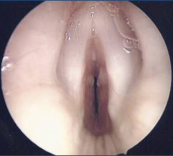
Four weeks later, he presented to the emergency department with a history of progressive, biphasic stridor with significant respiratory distress. This failed to respond to conservative measures, and the child underwent an urgent rigid airway endoscopy (Figure 6). A significant, circumferential subglottic haemangioma was found, and a tracheotomy was performed to secure the airway. A biopsy was also taken of the neck swelling via the tracheotomy incision.
Rigid airway endoscopy view of case four, showing a circumferential subglottic haemangioma.

Biopsy results confirmed the presence of features consistent with an infantile haemangioma. The tissue was also positive for GLUT-1 (glucose transporter 1) immunoreactivity. Such immunoreactivity is a useful marker of juvenile haemangioma, and has been shown to accurately distinguish haemangiomas from vascular malformations (i.e. arterio-venous, venous, lymphatic and port wine malformations).Reference Leon-Villapalos, Wolfe and Kangesu15, Reference North, Waner, Mizeracki and Mihm16
The child made a good post-operative recovery and was eventually discharged home with the tracheostomy. As the child had developed a significant subglottic haemangioma whilst taking propranolol, this treatment was stopped. He was maintained on the standard medical treatment of oral steroids, with a good clinical response of the cervical haemangiomas.
Endocrinology input was arranged, as was our usual practice for any patient receiving a prolonged course of steroids. The child subsequently developed a Cushingoid appearance, and he was commenced on a reducing regime of prednisolone under endocrinologist review.
At the time of writing, the child was six months of age, and his cervical haemangiomas had reduced significantly, especially on the left side. He had a 3.5 PED Shiley tracheostomy in situ, and vocalised around it. Surveillance rigid airway endoscopy was planned, including attempted tracheostomy decannulation.
Discussion
The exact mechanism of action of propranolol on haemangiomas is not fully understood. The natural course of infantile haemangiomas is characterised by a growth phase followed by an involutional phase. It has been observed that, when treatment with propranolol is commenced, there is a rapid decrease in pigmentation and a palpable softening of the lesion. It has been proposed that this may be due to vasoconstriction.Reference Leaute-Labreze, de la Roque, Hubiche, Boralevi, Thambo and Taieb3 There then follows a progressive reduction in haemangioma volume.
The two main pro-angiogenic factors involved in haemangioma growth are basic fibroblast growth factor and vascular endothelial growth factor. Following commencement of propranolol treatment, the resulting gradual reduction in haemangioma volume may be due to down-regulation of the RAF protein kinase mitogen activated pathway, with triggering of apoptosis.Reference D'Angelo, Lee and Weiner17, Reference Sommers Smith and Smith18 The RAF kinase family is involved in the regulation of endothelial apoptosis and therefore angiogenesis. It is also known that β-2 receptors for propranolol are expressed in haemangioma endothelial cells.Reference Denoyelle, Leboulanger, Enjolras, Harris, Roger and Garabedian5
The side effects of propranolol include bradycardia, hypotension and hypoglycaemia.Reference Hoffmann and Katzung19 Patients with diffuse haemangiomas, or syndromes with associated cardiac abnormalities, are at risk of high-output cardiac compromise; not only does propranolol have the potential to conceal the tachycardia, tachypnoea and hyperhidrosis of cardiac failure, it may also exacerbate them.Reference D'Angelo, Lee and Weiner17 Irritation and unsettled behaviour are signs associated with hypoglycaemia, which would be masked by propranolol. Prolonged hypoglycaemia in infancy has been associated with long-term neurological morbidity.Reference Inder20, Reference Burns, Rutherford, Boardman and Cowan21
Propranolol is contraindicated in patients with coexisting anomalous brain vasculature and insufficient collateral arterial supply (e.g. the PHACE syndrome), due to the theoretical risk of infarction of brain tissue resulting from propranolol-induced hypotension.Reference Lawley, Siegfried and Todd7
For cases of proliferating haemangiomas requiring intervention, until the introduction of propranolol there had been no available treatment without potentially serious short- and long-term adverse effects.
To date, corticosteroids have been the first-line treatment for infantile haemangiomas, and many of the case reports cited in this article have used corticosteroids as an adjunct to propranolol treatment. However, the doses required are often high, the response rate ranges from 30–60 per cent, and the resulting regression is often incomplete.Reference Sans, Dumas de la Roque, Berge, Grenier, Boralevi and Mazereeuw-Hautier11 Furthermore, the side effect profile associated with corticosteroids is extensive and potentially clinically unacceptable, and includes Cushing's syndrome (as seen in our fourth case), hirsutism, hypertrophic cardiomyopathy, delayed wound healing, immunosuppression and hypertension.
Medical treatments used as second- and third-line therapy for infantile haemangiomas include interferon-α and vincristine.
Interferon-α has led to complete regression in 40–50 per cent of complicated cases, but is associated with fever and myalgia at the onset of treatment, and haematological and hepatic toxicity throughout treatment; furthermore, neurotoxicity with developmental delay may occur in up to 30 per cent of cases.Reference Sans, Dumas de la Roque, Berge, Grenier, Boralevi and Mazereeuw-Hautier11
Vincristine has almost 100 per cent efficacy when given in weekly infusions, with involution usually beginning after three weeks of treatment. However, treatment may be associated with constipation, jaw pain, neuropathy, haemotoxicity and inappropriate secretion of antidiuretic hormone.Reference Sans, Dumas de la Roque, Berge, Grenier, Boralevi and Mazereeuw-Hautier11 Thus, vincristine is no longer used by most units.
• Infantile haemangiomas typically proliferate rapidly, then involute slowly
• Those with the potential to cause disfigurement or morbidity require intervention
• All previous treatment modalities have had significant associated morbidities
• Propranolol has become the first-line treatment for such haemangiomas
• However, in a presented case, haemangioma development occurred despite propranolol treatment; regular patient review is thus advised
Surgical treatment of subglottic haemangioma, via a laryngofissure external approach and submucous resection, carries a risk of damage to surrounding vital structures during the procedure, and a further risk secondary to post-operative fibrosis and tissue contraction.Reference Bitar, Moukarbel and Zalzal22 Tracheostomy to secure the airway, whilst awaiting natural involution, is a treatment option; however, tracheotomy is associated with mortality rates as high as 4 per cent in some studies, and requires appropriate parental training prior to eventual hospital discharge.Reference Bitar, Moukarbel and Zalzal22
Three of the four cases described above showed a reduction in haemangioma size following propranolol treatment. All three had previously failed to respond to medical intervention. Despite the heterogeneity of their presentations, the initial results of these three cases were dramatic, and no adverse effects of propranolol treatment were seen.
Our institution's infantile haemangioma treatment protocol and pre-treatment investigations are based on the practice followed at the Great Ormond Street Hospital.Reference Jephson, Manunza, Syed, Mills, Harper and Hartley6 There is currently no consensus regarding the duration of propranolol treatment; however, we believe that the propranolol dose can be tapered off after the expected proliferation phase has elapsed, after one to two years, depending upon the response to treatment.
Conclusion
The fourth case in our infantile haemangioma series is an important addition to the expanding literature on the use of propranolol for infantile haemangioma. The literature suggests that propranolol is universally effective in the treatment of infantile haemangioma. To our knowledge, our fourth case represents the first report of failure of initial propranolol therapy for this condition. This child's subsequent development of airway compromise suggests that his subglottic haemangioma developed, or continued to enlarge, despite treatment with propranolol at what is considered to be an acceptable treatment dose for haemangiomas.
The possibility of such non-response to treatment is important, and highlights the need for regular review of such children receiving propranolol treatment, and not just to monitor the side effects of treatment.
Propranolol is an important new treatment modality, but the clinician may still need to employ more established techniques in certain cases.