Introduction
Paratestis is the anatomical region around testis which includes spermatic cord and its contents, vas deferens, testicular tunics, epididymis, efferent ductules and rete testis. Reference Bostwick, Bostwick and Cheng1 Soft tissue sarcomas are accounts for approximately 1% of all malignant tumours Reference Clark, Fisher, Judson and Meirion Thomas2 and the paratestis is an extremely rare site accounting for less than 1% of sarcomas. Reference Stiller, Trama and Serraino3 So, primary paratesticular soft tissue sarcomas are rarest of rare tumours. Due to the rarity of cases, a consensus on the best treatment has yet to be reached, thus presenting a diagnostic and therapeutic challenge for clinicians. Presently, treatment recommendations are based on case reports, small case series and literature reviews, resulting in a number of unresolved issues. The standard therapeutic approach for these tumours has been complete excision of the tumour and radical inguinal orchiectomy with resection of surrounding tissue as well as high ligation of the spermatic cord at the external inguinal ring. Reference Rodríguez and Olumi4 The reported local relapse rate in the scrotum and groin after orchidectomy is 25–37%. Risk factors for local recurrence are a large tumour, an inguinal location, narrow or positive margins after resection and previous intralesional surgery. Nodal failure in the retroperitoneum has been reported in 14–29% of paratesticular tumours. Reference Khoubehi, Mishra, Ali, Motiwala and Karim5 The high rate of local-regional recurrence advocates the use of adjuvant treatment such as radiotherapy.
Conventional 2D technique is the most widely used technique for adjuvant radiotherapy treatment of paratesticular sarcoma. It has potential for higher gastrointestinal and genitourinary toxicities as compared to conformal techniques such as intensity-modulated radiotherapy as seen for pelvic tumours. Reference Gandhi, Sharma and Rath6 So far one study has illustrated conformal intensity-modulated radiotherapy technique in paratesticular sarcoma, but in that study only the tumour bed is irradiated without addressing regional lymph nodes treatment. Reference Cerda, Martin, Truc, Créhange and Maingon7
We are presenting a case report of paratesticular leiomyosarcoma along with technical illustration of conformal planning, target volume delineation and dosimetric comparison between different conformal field set-ups.
Case Presentation
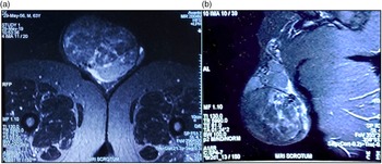
A 62-year-old obese male presented with history of painless right scrotal swelling which was insidious in onset and slowly increasing in size for the last 4 years. Patient was evaluated, clinically the mass was about 15 cm in size. Based on clinical examination, scrotal swelling was diagnosed as hydrocele for which surgical exploration was done, which was abandoned on table due to suspicion of right testicular mass. Ultrasound of bilateral scrotum showed grossly enlarged right testes with heterogeneously echotexture suggestive of solid cystic lesion with internal vascularity and enlarged veins in the region of right spermatic cord. Left testes was normal in size, shape, echotexture and vascularity. MRI perineum including scrotum showed large heterogenous lobulated soft tissue mass lesion sized 13 × 9·5 × 13 cm (Figure 1) within the right scrotum, arising from right epididymis, displacing the right testis antero-superiorly. Patient underwent right high inguinal orchidectomy with ligation of the spermatic cord. Large encapsulated 15 × 15 cm, solid cystic mass removed. Histopathological evaluation of the specimen showed encapsulated spindle cell tumour with features of well-differentiated leiomyosarcoma, Fédération Nationale des Centres de Lutte Contre Le Cancer (FNCLCC) grade 1, immunopositive for desmin and smooth muscle actin, no capsular invasion seen. A PET-CT scan post-operatively showed no definite evidence of metabolically activelesion.
Magnetic Resonance Imaging T1 Weighted (MRI T1W) sequence showing a) axial section and b) sagittal section of scrotum. Arrow depicting large heterogenous lobulated soft tissue mass lesion within the right scrotum, displacing the right testis.

Radiotherapy Planning
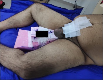
In multidisciplinary sarcoma clinic based on size of tumour >5 cm, it was decided to deliver adjuvant radiotherapy. A BlueBAG™ (Elekta, Stockholm, Sweden) vacuum cushion shaped in treatment position where patient is lying in frog leg position with hands on chest and scrotum is kept flat to couch using styrofoam support (Figure 2). Bladder protocol of voiding followed by sipping 250-ml water 20 mins prior to planning was used. A planning CT was taken (Philips Brilliance Big Bore 16 slice CT scanner) with foot first (slice thickness of 3 mm). The CT scan image dataset imported to the Monaco 5.11 Planning system (Elekta, Stockholm, Sweden). Organs at risk (OARs) included bilateral femoral head, urinary bladder, rectum, bowel, penis and bilateral kidney.
Showing use of vacuum cushion and styrofoam as positioning device to maintain frog leg position.

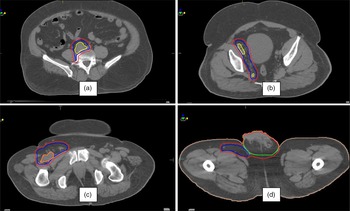
There are no contouring guidelines for paratesticular sarcoma but target volume delineation done based on review by Enoch et al. the ‘The field for radiotherapy should include the inguinal canal, ipsilateral pelvic tissue and the scrotum’. Lower pelvic nodes delineation started from bifurcation of common iliac vessel, ipsilateral external and internal iliac vessels were delineated down till the appearance of femoral head. Reference Taylor, Rockall, Reznek and Powell8 Addition of 7-mm margin to iliac vessels given with the upper border maintained at common iliac vessel bifurcation and contour is extended around common iliac vessels posteriorly and laterally so as to include connective tissue between iliopsoas muscles and lateral surface of vertebral body. Reference Bansal, Patel, Rai, Gulia, Dhanireddy and Sharma9 To cover obturator nodes, a strip of 18-mm wide is created medial to the pelvic side wall, by joining the contour of external iliac vessels with internal iliac vessels. Contouring of obturator nodes continued lower down along pelvic side wall, till superior part of obturator foramen. Reference Taylor, Rockall, Reznek and Powell8 Inguinal nodal was delineated by adding isotropic margin of 2 cm around the femoral vessels and tailored to not involve muscles as explained by Kim et al. Reference Kim, Olson, Kim and Beriwal10 Tumour bed included scrotum and incision site (Figure 3).
Showing target volume delineation of a) pelvic node, b) obturator node, c) inguinal node and d) tumour bed. The yellow structure depicts iliac vessels, orange structure depicts inguinal vessels, blue structure depicts clinical target volume and red structure depicts the planning target volume.

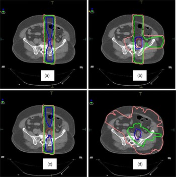
Lower pelvic nodal expansion, inguinal nodal expansion and tumour bed combined to form clinical target volume (CTV); a margin of 5 mm given to CTV to form planning target volume (PTV). Dose of 50·4 Gy in 28 fractions at 1·8 Gy per fraction prescribed to PTV and QUANTEC dose constraints followed for OARs. Multiple field set-up planes of a) 3D conformal 2-field, b) 3D conformal 3-field, c) 3D conformal field-in-field and d) Volumetric Modulated Arc Technique beam (VMAT) were made (Figure 4). Plan was evaluated following ICRU 83 report. Reference Menzel11
Showing isodose lines of 107% (blue), 95% (green) and 10% (peach) in axial section of a) 3D conformal 2-field, b) 3D conformal 3-field, c) 3D conformal field-in-field and d) volumetric arc technique. The red structure describes the planning target volume.

Results
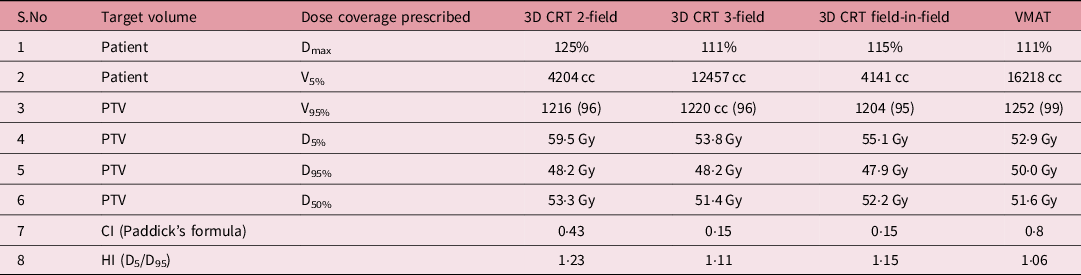
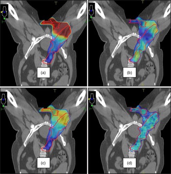
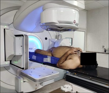
All the plans were dosimetrically compared (Table 1) and OAR dose constraints (Table 2). The VMAT plan with 6-MV beam energy was chosen for treatment which had most conformal plan and meeting OAR constraints (Figure 5). Treatment was done in Versa HD™ (Elekta, Stockholm, Sweden) linear accelerator under daily cone beam CT image guidance to minimise set-up errors (Figure 6). Same bladder protocol followed before daily set-up to mimic planning CT. Patient developed grade II skin reactions post 15 fractions of radiotherapy, for which moisturiser and emollients given, post 20 fractions radiotherapy patient developed CTCAE V5.0 grade III scrotal dermatitis radiation and dysuria, a treatment gap of 10 days given, after which reactions subsided to grade I. On completion of 25 fractions patient again started complaining of burning micturition and developed CTCAE V5.0 grade III dermatitis radiation. Treatment was halted due to poor tolerance. One of the reasons for poor tolerance was the presence of multiple skin folds on patients’ groin and pelvis due to obesity. Patient had skin ulcerations and skin reactions, which was managed symptomatically and skin began to heal by second-week post-radiotherapy and completely healed by fifth-week post-radiotherapy. Patient is kept on 3-month follow-up with clinical examination and ultrasound pelvis and scrotum done in in each follow-up. After 12 months of follow-up, patient had no complaints and no evidence of recurrence. Patient will be kept on long-term follow-up and will be observed for recurrence and toxicity.
Showing dosimetry analysis of a) 3D conformal 2-field, b) 3D conformal 3-field, c) 3D conformal field-in-field and d) volumetric arc technique

PTV, planning target volume; CI, conformity index; HI, homogeneity index; 3D CRT, 3 Dimensional conformal radiotherapy.
Showing OAR dose constraints analysis of a) 3D conformal 2-field, b) 3D conformal 3-field, c) 3D conformal field-in-field and d) volumetric arc technique

OAR, organ at risk.
Showing dose colour wash of 95% of the prescribed dose in coronal section of a). 3D conformal 2-field, b) 3D conformal 3-field, c) 3D conformal field-in-field and d) volumetric arc technique. The red structure describes the planning target volume.

Showing treatment position of patient on linear accelerator.

Discussion
Radiotherapy for sarcomas is well established to achieve a local control benefit of up to 90% in extremities and up to 60% in retroperitoneum. Reference Tiong, Dickie, Haas and O’Sullivan12 For paratesticular sarcoma, Catton et al. suggested that all patients should undergo surgical dissection and adjuvant radiotherapy of the ipsilateral pelvic and groin nodes, and scrotum based on their observations of failure patterns post-surgery, local recurrence being the main failure pattern. Reference Catton, Jewett, O’Sullivan and Kandel13 The majority of high-grade tumours have been seen to fail, mainly in retroperitoneal nodes or systemically.
A prospective study suggested that there is a significant correlation between recurrence and size of tumour. With no adjuvant treatment, there was local recurrence in 5 out of 23 patients (21·7%). Reference Bozzini, Albersen and Romero Otero14 Some authors recommend that adjuvant radiotherapy should be instituted for all types and grades of paratesticular leiomyosarcoma and treatment volume should cover inguinal canal, ipsilateral pelvic tissue and scrotum. Reference Enoch, Wharton and Murray15 Largest analysis till date of 362 paratesticular sarcoma patients is done by Rodriguez et al., based on SEER database they identified 27·4% lymph node positive in leiomyosarcoma. They identified positive lymph nodes and leiomyosarcoma histology as independent predictors of decreased disease-specific survival. Reference Rodríguez, Barrisford, Sanchez, Preston, Kreydin and Olumi16 Due to rarity of disease, current evidence is not enough to accurately identify which patient needs adjuvant radiotherapy and define target volumes. Evidence of other sites of soft tissue sarcoma cannot be extrapolated to paratesticular site as sarcomas in paratestis tend to be lower grade and have higher propensity of lymphatic spread.
Historically, anterior–posterior oblique portals were used to treat scrotal area, inguinal region and ipsilateral lower pelvic nodes doses of 40 to 60 Gy. Radiation dermatitis, genitourinary and gastrointestinal toxicities dissuade us to use adjuvant radiotherapy. Good dosimetry may permit higher therapeutic ratio and may thus improve local tumour control with decreased exposure to surrounding normal tissues, subsequently reducing acute and late radiation toxicities.
In our patient on VMAT treatment, we had CTCAE V 5.0 grade III radiation dermatitis and dysuria. Treatment gap of 10 days was given and treatment stopped after 45 Gy in 25 fractions. There was no gastrointestinal toxicity.
Conclusion
Our case study demonstrated technical illustration of conformal planning, target volume delineation and dosimetric comparison of different field set-ups. Our patient treated by VMAT had acute radiation dermatitis and genitourinary toxicities, which were managed conservatively. Adjuvant radiotherapy by advanced delivery technique such as VMAT can provide good dose distribution with good sparing of organ at risk. The downside of conformal radiation delivery is that it is resource-intensive and has no consensus target volume delineation guidelines.
Acknowledgements
None.
Ethical Approval
Ethical approval is not required at our institution for publishing an anonymous case.
Statement of Human and Animal Rights
All procedures followed were in accordance with the ethical standards of responsible committee on human experimentation (institutional and national) and with the Helsinki Declaration of 1975, as revised in 2008.
Statement of Informed Consent
Informed consent was obtained from patients for being included in the study. Additional informed consent was obtained for identifying information included in the article.
Declaration of Conflicting Interests
The author(s) declared no potential conflicts of interest with respect to the research, authorship and/or publication of this article.
Funding
The author(s) received no financial support for the research, authorship and/or publication of this article.